. 2000 May;20(9):2970-83.
doi: 10.1128/MCB.20.9.2970-2983.2000.

Domains in the SPT5 protein that modulate its transcriptional regulatory properties

Affiliations

Domains in the SPT5 protein that modulate its transcriptional regulatory properties

D Ivanov et al. Mol Cell Biol. 2000 May.

Abstract

SPT5 and its binding partner SPT4 regulate transcriptional elongation by RNA polymerase II. SPT4 and SPT5 are involved in both 5,6-dichloro-1-beta-D-ribofuranosylbenzimidazole (DRB)-mediated transcriptional inhibition and the activation of transcriptional elongation by the human immunodeficiency virus type 1 (HIV-1) Tat protein. Recent data suggest that P-TEFb, which is composed of CDK9 and cyclin T1, is also critical in regulating transcriptional elongation by SPT4 and SPT5. In this study, we analyze the domains of SPT5 that regulate transcriptional elongation in the presence of either DRB or the HIV-1 Tat protein. We demonstrate that SPT5 domains that bind SPT4 and RNA polymerase II, in addition to a region in the C terminus of SPT5 that contains multiple heptad repeats and is designated CTR1, are critical for in vitro transcriptional repression by DRB and activation by the Tat protein. Furthermore, the SPT5 CTR1 domain is a substrate for P-TEFb phosphorylation. These results suggest that C-terminal repeats in SPT5, like those in the RNA polymerase II C-terminal domain, are sites for P-TEFb phosphorylation and function in modulating its transcriptional elongation properties.


Figures
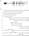
FIG. 1
FIG. 1
Schematic of the SPT5 protein. (A) A schematic of the SPT5 protein with the positions of the acidic domain, the four KOW domains that have homology to the E. coli NusG protein, and two domains, CTR1 and CTR2, each of which has multiple amino acid repeats, is shown. (B) A variety of contructs with mutations in different domains of the SPT5 protein are shown. These mutants were analyzed for their function using both in vivo interaction and in vitro transcription assays.
FIG. 2
FIG. 2
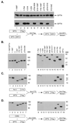
SPT5 association with SPT4, RNA polymerase II, and CDK9. (A) Expression vectors containing either a Myc-tagged SPT4 construct or the indicated Flag-tagged SPT5 constructs were cotransfected into COS cells. Extracts were prepared from each of these transfections, and the SPT4 levels were analyzed by Western blot analysis with antibody directed against the Myc epitope (upper panel). Extracts prepared from the SPT4-SPT5-cotransfected cells were immunoprecipitated with M2 monoclonal antibody to isolate the Flag-tagged SPT5 proteins, and then Western blot analysis was performed with monoclonal antibody directed against the Myc epitope to detect the epitope-tagged SPT4 protein (lower panel). In lane 2, an expression vector containing the SPT4 cDNA was transfected into COS cells alone without cotransfection of the SPT5 cDNA. (B) Western blot analysis of COS cell extracts prepared following transfection of expression vectors containing Flag-tagged SPT5 cDNA constructs (lanes 1 to 17) or a Flag-tagged SPT4 cDNA (lane 18) was performed using the M2 monoclonal antibody. In lane 1, an expression vector containing SPT4 was cotransfected with the wild-type SPT5 construct. MW, molecular weight in thousands. (C and D) Flag-tagged SPT5 constructs were each transfected into COS cells, and extracts were prepared. Immunoprecipitation of these extracts was performed with antibodies directed against either the RNA polymerase II CTD (C) or CDK9 (D). Western blot analysis with the M2 monoclonal antibody was then performed to detect the Flag-tagged SPT5.
FIG. 3
FIG. 3
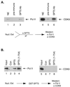
Association of SPT5 with RNA polymerase II and CDK9 in vivo and in vitro. (A) HeLa nuclear extract was immunoprecipitated with either preimmune sera (lanes 2 and 5) or rabbit polyclonal antibody directed against SPT5 (lanes 3 and 6). A total of 20% of the HeLa nuclear extract that was subjected to immunoprecipitation is shown (lanes 1 and 4). Following immunoprecipitation, Western blot analysis was performed with antibody directed against the RNA polymerase II (Pol II) CTD (lanes 1 to 3) or CDK9 (lanes 4 to 6). (B) Purified RNA polymerase II (lanes 1 to 4) or baculovirus-purified CDK9/cyclin T1 (lanes 5 to 8) was either untreated (30% of the input is shown in lanes 1 and 5) or incubated with glutathione-agarose beads containing GST (lanes 2 and 6), GST-SPT5 (lanes 3 and 7), or a GST-SPT5 fusion protein extending from aa 1 to 754 (lanes 4 and 8). Following extensive washing, Western blot analysis was performed with antibodies directed against the RNA polymerase II CTD (lanes 1 to 4) or CDK9 (lanes 5 to 8).
FIG. 4
FIG. 4
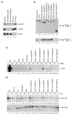
Reconstitution of DRB inhibition and Tat activation by recombinant SPT5 proteins. (A) Western blot analysis was performed with antibodies directed against cyclin T1, CDK9, and SPT5 using unfractionated HeLa nuclear extract (Nuc Ext) (lane 1), affinity-purified SPT4-SPT5 complex produced following transfection of epitope-tagged SPT4 and SPT5 cDNAs into COS cells (lane 2), baculovirus-expressed and purified SPT4-SPT5 (lane 3), or 10-μl (lane 4) or 50-μl (lane 5) aliquots of a 1.0 M potassium acetate fraction of HeLa nuclear extract obtained following phosphocellulose (PC) chromatography. (B) Western blot analysis of affinity-purified SPT4 and SPT5 proteins. Expression vectors containing the different Flag-tagged SPT5 cDNA constructs and a Myc-tagged SPT4 cDNA construct were cotransfected into COS cells. The Flag epitope-tagged SPT5 and associated SPT4 protein in these extracts were purified by binding to protein G beads containing M2 monoclonal antibody followed by elution with Flag peptide. Western blot analysis was performed with anti-Flag monoclonal antibody to detect SPT5 proteins (top panel) or anti-Myc monoclonal antibody to detect the associated SPT4 (lower panel). (C) In vitro transcription analysis was performed with the pTF3-6C2AT template using unfractionated HeLa nuclear extract (NE) (lanes 1 and 2), or the 1.0 M potassium acetate fraction of HeLa nuclear extract eluted from the phosphocellulose (PC) column (lanes 3 to 24). The 1.0 M potassium acetate fraction was assayed either alone (lanes 3 and 4), with affinity-purified SPT4 added alone (lanes 5 and 6), with affinity-purified SPT5 added alone (lanes 7 and 8), or with both SPT4 and the different affinity-purified SPT5 proteins shown in panel B added as indicated (lanes 9 to 24). DRB was added to the even-numbered lanes in the in vitro transcription assays. (D) In vitro transcription analysis was performed with the HIV-1 LTR wild-type (top panel) or the HIV-1 LTR loop mutant (lower panel) templates using unfractionated HeLa nuclear extract (lanes 1 to 4), the 1.0 M potassium acetate fraction of HeLa nuclear extract eluted from the phosphocellulose (PC) column alone (lanes 5 and 6), or the 1.0 M potassium acetate fraction in the presence of affinity-purified SPT4 and SPT5 proteins (lanes 7 and 8), SPT5 alone (lane 9 and 10), or SPT4 and affinity-purified SPT5 mutants (lanes 11 to 24) as indicated. Tat (25 ng) was added to the odd-numbered lanes and GST (25 ng) was added to the even-numbered lanes in the in vitro transcription assays. Following the in vitro transcription analysis with these templates containing G-less cassettes, the labeled RNAs were digested with RNase T1 and gel electrophoresis and autoradiography were performed.
FIG. 5
FIG. 5
P-TEFb phosphorylation of SPT5. (A) Baculovirus-produced and purified proteins including CDK9 (lanes 1 and 6), CDK9 and cyclin T1 (lanes 2 and 7), CDK9 and cyclin T2a (lanes 3 and 8), and CDK9 and cyclin T2b (lanes 4 and 8) were assayed in in vitro kinase assays in the absence of the GST-SPT5 substrate (lanes 1 to 4), the GST-SPT5 substrate was assayed alone (lane 5), or the GST-SPT5 substrate was added along with the different CDK9 preparations (lanes 6 to 9). (B) The baculovirus-produced and purified preparations of CDK9 (lane 1), CDK9 and cyclin T1 (lane 2), CDK9 and cyclin T2a (lane 3), and CDK9 and cyclin T2b (lane 4) were assayed in Western blot analysis using antibodies directed against CDK9 (top panel), cyclin T1 (middle panel), or cyclin T2 (lower panel).
FIG. 6
FIG. 6
Comparison of P-TEFb and CAK phosphorylation of SPT5 constructs. (A) Purified RNA polymerase II (Pol II) was assayed in in vitro kinase assays using RNA polymerase II alone (lane 1) or in the presence of baculovirus-produced and purified CAK including CDK7, cyclin H, and MAT1 (lane 2) or CDK9 and cyclin T1 (lane 3). MW, molecular weight in thousands. (B) In vitro kinase assays were performed using baculovirus-produced preparations of CDK9/cyclin T1 (lanes 1 to 6) or CAK (lanes 7 to 12) with either no added substrate (lanes 1 and 7), GST-SPT5 (lanes 2 and 8), GST-SPT5 (aa 1 to 754) (lanes 3 and 9), GST-SPT5 (aa 760 to 852) (lanes 4 and 10), GST-SPT5 (aa 814 to 1087) (lanes 5 and 11), or GST-SPT5 (aa 760 to 1087) (lanes 6 and 12). Phosphorylation was assayed following SDS-PAGE and autoradiography. (C) A Coomassie blue-stained gel of the various GST-SPT5 fusion proteins used in the in vitro kinase assays in part B is shown.
FIG. 7
FIG. 7
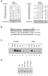
Comparison of P-TEFb and CAK phosphorylation of SPT5 CTR1 repeats. (A) Sequences of CTR1 and CTR2 repeats. The positions of the C-terminal repeat motifs in the CTR1 and CTR2 domains of SPT5 are indicated, as is the consensus sequence for each domain, as previously described (49). (B) The sequences of the two CTR1 repeats that were fused to GST and used as substrates in in vitro kinase reactions are shown. The boxes indicate the positions of the repeats, and the mutated amino acids are shown in boldface type. (C) In vitro kinase reactions were performed using baculovirus-produced and purified CDK9-cyclin T1 (lanes 1 to 6) or the baculovirus-produced and purified CAK components including CDK7, cyclin H, and MAT1 (lanes 7 to 12). The kinase reactions were performed without the addition of substrate (lanes 1 and 7) or following the addition of GST (lanes 2 and 8), a GST-CTD fusion protein containing two repeats of the RNA polymerase II CTD (lanes 3 and 9), or GST fusion proteins containing two CTR1 repeats with either the wild-type (WT) sequences (lanes 4 and 10), serine residues substituted for alanine (lanes 5 and 11), or threonine residues substituted for alanine (lanes 6 and 12). (D) A Coomassie blue-stained SDS-polyacrylamide gel of the GST fusion proteins that were used as substrates for kinase assays in panel B is shown.
FIG. 8
FIG. 8
Role of P-TEFb phosphorylation on SPT4-SPT5 function in mediating DRB repression. (A) In vitro transcription assays were performed with the pTF3-6C2AT template in the presence (even-numbered lanes) or absence (odd-numbered lanes) of DRB. Untreated HeLa nuclear extract (lanes 1 and 2), HeLa nuclear extract immunodepleted with GST antibody (lanes 3 and 4), or HeLa nuclear extract immunodepleted with SPT5 antibody (lanes 6 to 20) were used in the in vitro transcription assays. In vitro transcription assay mixtures with the SPT5-immunodepleted HeLa nuclear extracts were supplemented with the SPT4-SPT5 complex purified following baculovirus expression in the absence of other treatment (lanes 7 and 8), the SPT4-SPT5 complex preincubated with P-TEFb (lanes 9 and 10 and lanes 15 and 16), the SPT4-SPT5 complex preincubated without P-TEFb (lanes 11 and 12 and lanes 17 and 18), or P-TEFb alone preincubated in the absence of SPT4-SPT5 (lanes 13 and 14 and lanes 19 and 20). The preincubation procedure was performed in the absence of ATP (lanes 9 to 14) or in the presence of 5 mM ATP (lanes 15 to 20), which was subsequently removed by dialysis. (B) A schematic of the functional domains in the SPT5 protein that were analyzed in this study is shown.

Similar articles

Cited by

References

    1. Basrai M A, Kingsbury J, Koshland D, Spencer F, Hieter P. Faithful chromosome transmission requires Spt4p, a putative regulator of chromatin structure in Saccharomyces cerevisiae. Mol Cell Biol. 1996;16:2838–2847. - PMC - PubMed
    1. Bengal E, Flores O, Krauskopf A, Reinberg D, Aloni Y. Role of the mammalian transcription factors IIF, IIS, and IIX during elongation by RNA polymerase II. Mol Cell Biol. 1991;11:1195–1206. - PMC - PubMed
    1. Bentley D L. Regulation of transcriptional elongation by RNA polymerase II. Curr Opin Genet Dev. 1995;5:210–216. - PubMed
    1. Bieniasz P D, Grdina T A, Bogerd H P, Cullen B R. Recruitment of a protein complex containing Tat and cyclin T1 to TAR governs the species specificity of HIV-1 Tat. EMBO J. 1998;17:7056–65. - PMC - PubMed
    1. Bortvin A, Winston F. Evidence that Spt6p controls chromatin structure by a direct interaction with histones. Science. 1996;272:1473–1476. - PubMed

Publication types

MeSH terms

Substances