

Genetic and biochemical characterization of the pathway in Pantoea citrea leading to pink disease of pineapple
- PMID: 10735866
- PMCID: PMC111272
- DOI: 10.1128/JB.182.8.2230-2237.2000
Genetic and biochemical characterization of the pathway in Pantoea citrea leading to pink disease of pineapple
Abstract
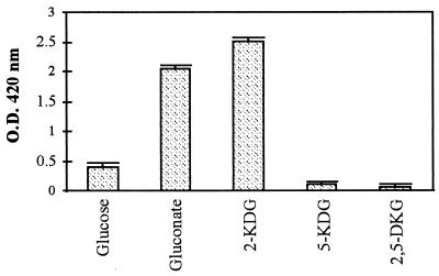
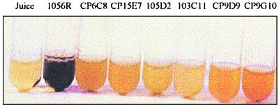
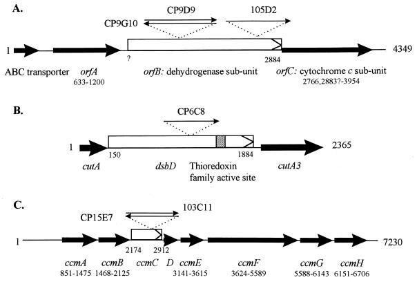
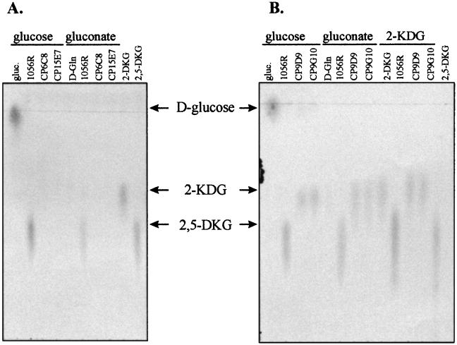
Pink disease of pineapple, caused by Pantoea citrea, is characterized by a dark coloration on fruit slices after autoclaving. This coloration is initiated by the oxidation of glucose to gluconate, which is followed by further oxidation of gluconate to as yet unknown chromogenic compounds. To elucidate the biochemical pathway leading to pink disease, we generated six coloration-defective mutants of P. citrea that were still able to oxidize glucose into gluconate. Three mutants were found to be affected in genes involved in the biogenesis of c-type cytochromes, which are known for their role as specific electron acceptors linked to dehydrogenase activities. Three additional mutants were affected in different genes within an operon that probably encodes a 2-ketogluconate dehydrogenase protein. These six mutants were found to be unable to oxidize gluconate or 2-ketogluconate, resulting in an inability to produce the compound 2,5-diketogluconate (2,5-DKG). Thus, the production of 2,5-DKG by P. citrea appears to be responsible for the dark color characteristic of the pink disease of pineapple.
Figures
Similar articles
-
Identification and characterization of a Pantoea citrea gene encoding glucose dehydrogenase that is essential for causing pink disease of pineapple.Appl Environ Microbiol. 1997 Jan;63(1):71-6. doi: 10.1128/aem.63.1.71-76.1997. Appl Environ Microbiol. 1997. PMID: 8979341 Free PMC article.
-
gdhB, a gene encoding a second quinoprotein glucose dehydrogenase in Pantoea citrea, is required for pink disease of pineapple.Microbiology (Reading). 1999 May;145 ( Pt 5):1217-1226. doi: 10.1099/13500872-145-5-1217. Microbiology (Reading). 1999. PMID: 10376838
-
Characterization of pUCD5000 involved in pink disease color formation by Pantoea citrea.Plasmid. 1998 Sep;40(2):169-73. doi: 10.1006/plas.1998.1355. Plasmid. 1998. PMID: 9735319
-
New developments in oxidative fermentation.Appl Microbiol Biotechnol. 2003 Feb;60(6):643-53. doi: 10.1007/s00253-002-1155-9. Epub 2002 Dec 18. Appl Microbiol Biotechnol. 2003. PMID: 12664142 Review.
-
Pantoea ananatis: an unconventional plant pathogen.Mol Plant Pathol. 2009 May;10(3):325-35. doi: 10.1111/j.1364-3703.2009.00542.x. Mol Plant Pathol. 2009. PMID: 19400836 Free PMC article. Review.
Cited by
-
The cytochrome c maturation locus of Legionella pneumophila promotes iron assimilation and intracellular infection and contains a strain-specific insertion sequence element.Infect Immun. 2002 Apr;70(4):1842-52. doi: 10.1128/IAI.70.4.1842-1852.2002. Infect Immun. 2002. PMID: 11895946 Free PMC article.
-
Uncovering lipopolysaccharide regulation in bacteria via the critical lipid binding tunnel of YciS/YciM.iScience. 2022 Aug 20;25(9):104988. doi: 10.1016/j.isci.2022.104988. eCollection 2022 Sep 16. iScience. 2022. PMID: 36093049 Free PMC article.
-
Metabolic engineering of Escherichia coli BL21 (DE3) for de novo production of L-DOPA from D-glucose.Microb Cell Fact. 2019 Apr 25;18(1):74. doi: 10.1186/s12934-019-1122-0. Microb Cell Fact. 2019. PMID: 31023316 Free PMC article.
-
Transcriptional modulation of squalene synthase genes in barley treated with PGPR.Front Plant Sci. 2015 Sep 1;6:672. doi: 10.3389/fpls.2015.00672. eCollection 2015. Front Plant Sci. 2015. PMID: 26388880 Free PMC article.
-
2,5-Diketo-D-Gluconate Hyperproducing Gluconobacter sphaericus SJF2-1 with Reporting Multiple Genes Encoding the Membrane-Associated Flavoprotein-Cytochrome c Complexed Dehydrogenases.Microorganisms. 2022 Oct 27;10(11):2130. doi: 10.3390/microorganisms10112130. Microorganisms. 2022. PMID: 36363722 Free PMC article.
References
-
- Alexeyev M F, Shokolenko I N. Mini-Tn10 transposon derivatives for insertion mutagenesis and gene delivery into the chromosome of gram-negative bacteria. Gene. 1995;160:59–62. - PubMed
-
- Ameyama M, Adachi O. 2-Keto-d-gluconate reductase from acetic acid bacteria. Methods Enzymol. 1982;89:203–209. - PubMed
-
- Anderson S, Marks C B, Lazarus R, Miller J, Stafford K, Seymour J, Light D, Rastetter W, Estell D. Production of 2-keto-l-gulonate, an intermediate in l-ascorbate synthesis, by a genetically modified Erwinia herbicola. Science. 1985;230:144–149. - PubMed
-
- Birnboim H C. A rapid alkaline extraction method for the isolation of plasmid DNA. Methods Enzymol. 1983;100:243–255. - PubMed
-
- Bouvet O M, Lenormand P, Grimont P A. Taxonomic diversity of the d-glucose oxidation pathway in the enterobacteriaceae. Int J Syst Bacteriol. 1989;39:61–67.
Publication types
MeSH terms
Substances
Associated data
- Actions
- Actions
- Actions
LinkOut - more resources
Full Text Sources
Other Literature Sources
