Synaptotagmin VII regulates Ca(2+)-dependent exocytosis of lysosomes in fibroblasts
- PMID: 10725327
- PMCID: PMC2174306
- DOI: 10.1083/jcb.148.6.1141
Synaptotagmin VII regulates Ca(2+)-dependent exocytosis of lysosomes in fibroblasts
Abstract
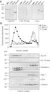
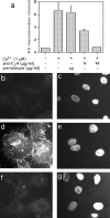
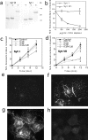
Synaptotagmins (Syts) are transmembrane proteins with two Ca(2+)-binding C(2) domains in their cytosolic region. Syt I, the most widely studied isoform, has been proposed to function as a Ca(2+) sensor in synaptic vesicle exocytosis. Several of the twelve known Syts are expressed primarily in brain, while a few are ubiquitous (Sudhof, T.C., and J. Rizo. 1996. Neuron. 17: 379-388; Butz, S., R. Fernandez-Chacon, F. Schmitz, R. Jahn, and T.C. Sudhof. 1999. J. Biol. Chem. 274:18290-18296). The ubiquitously expressed Syt VII binds syntaxin at free Ca(2+) concentrations ([Ca(2+)]) below 10 microM, whereas other isoforms require 200-500 microM [Ca(2+)] or show no Ca(2+)-dependent syntaxin binding (Li, C., B. Ullrich, Z. Zhang, R.G.W. Anderson, N. Brose, and T.C. Sudhof. 1995. Nature. 375:594-599). We investigated the involvement of Syt VII in the exocytosis of lysosomes, which is triggered in several cell types at 1-5 microM [Ca(2+)] (Rodríguez, A., P. Webster, J. Ortego, and N.W. Andrews. 1997. J. Cell Biol. 137:93-104). Here, we show that Syt VII is localized on dense lysosomes in normal rat kidney (NRK) fibroblasts, and that GFP-tagged Syt VII is targeted to lysosomes after transfection. Recombinant fragments containing the C(2)A domain of Syt VII inhibit Ca(2+)-triggered secretion of beta-hexosaminidase and surface translocation of Lgp120, whereas the C(2)A domain of the neuronal- specific isoform, Syt I, has no effect. Antibodies against the Syt VII C(2)A domain are also inhibitory in both assays, indicating that Syt VII plays a key role in the regulation of Ca(2+)-dependent lysosome exocytosis.
Figures


Similar articles
-
Ca(2+)-dependent and -independent activities of neural and non-neural synaptotagmins.Nature. 1995 Jun 15;375(6532):594-9. doi: 10.1038/375594a0. Nature. 1995. PMID: 7791877
-
Synaptotagmin VII is targeted to dense-core vesicles and regulates their Ca2+ -dependent exocytosis in PC12 cells.J Biol Chem. 2004 Dec 10;279(50):52677-84. doi: 10.1074/jbc.M409241200. Epub 2004 Sep 28. J Biol Chem. 2004. PMID: 15456748
-
Synaptotagmin V is targeted to dense-core vesicles that undergo calcium-dependent exocytosis in PC12 cells.J Biol Chem. 2002 Jul 5;277(27):24499-505. doi: 10.1074/jbc.M202767200. Epub 2002 May 2. J Biol Chem. 2002. PMID: 12006594
-
Synaptotagmin regulates mast cell functions.Immunol Rev. 2001 Feb;179:25-34. doi: 10.1034/j.1600-065x.2001.790103.x. Immunol Rev. 2001. PMID: 11292024 Review.
-
There's more to life than neurotransmission: the regulation of exocytosis by synaptotagmin VII.Trends Cell Biol. 2005 Nov;15(11):626-31. doi: 10.1016/j.tcb.2005.09.001. Epub 2005 Sep 15. Trends Cell Biol. 2005. PMID: 16168654 Review.
Cited by
-
The 8-oxoguanine DNA glycosylase-synaptotagmin 7 pathway increases extracellular vesicle release and promotes tumour metastasis during oxidative stress.J Extracell Vesicles. 2024 Sep;13(9):e12505. doi: 10.1002/jev2.12505. J Extracell Vesicles. 2024. PMID: 39235072 Free PMC article.
-
Host cell invasion by Trypanosoma cruzi: a unique strategy that promotes persistence.FEMS Microbiol Rev. 2012 May;36(3):734-47. doi: 10.1111/j.1574-6976.2012.00333.x. Epub 2012 Mar 13. FEMS Microbiol Rev. 2012. PMID: 22339763 Free PMC article. Review.
-
FM dyes enter via a store-operated calcium channel and modify calcium signaling of cultured astrocytes.Proc Natl Acad Sci U S A. 2009 Dec 22;106(51):21960-5. doi: 10.1073/pnas.0909109106. Epub 2009 Dec 9. Proc Natl Acad Sci U S A. 2009. PMID: 20007370 Free PMC article.
-
MicroRNA profiles of MS gray matter lesions identify modulators of the synaptic protein synaptotagmin-7.Brain Pathol. 2020 May;30(3):524-540. doi: 10.1111/bpa.12800. Epub 2019 Nov 17. Brain Pathol. 2020. PMID: 31663645 Free PMC article.
-
Role of calcium-sensor proteins in cell membrane repair.Biosci Rep. 2023 Feb 27;43(2):BSR20220765. doi: 10.1042/BSR20220765. Biosci Rep. 2023. PMID: 36728029 Free PMC article. Review.
References
-
- Black D.L. Splicing in the inner eara common tune, but what are the instruments? Neuron. 1998;20:165–168. - PubMed
-
- Bommert K., Charlton M.P., DeBello W.M., Chin G.J., Betz H., Augustine G.J. Inhibition of neurotransmitter release by C2-domain peptides implicates synaptotagmin in exocytosis. Nature. 1993;363:163–165. - PubMed
-
- Burgoyne R.D., Morgan A. Calcium sensors in regulated exocytosis. Cell Calcium. 1998;24:367–376. - PubMed
-
- Butz S., Fernandez-Chacon R., Schmitz F., Jahn R., Sudhof T.C. The subcellular localizations of atypical synaptotagmins III and VI. Synaptotagmin III is enriched in synapses and synaptic plasma membranes but not in synaptic vesicles. J. Biol. Chem. 1999;274:18290–18296. - PubMed
Publication types
MeSH terms
Substances
Grants and funding
LinkOut - more resources
Full Text Sources
Other Literature Sources
Molecular Biology Databases
Miscellaneous

