. 2000 Apr;20(7):2475-87.
doi: 10.1128/MCB.20.7.2475-2487.2000.

H-ras but not K-ras traffics to the plasma membrane through the exocytic pathway

Affiliations

H-ras but not K-ras traffics to the plasma membrane through the exocytic pathway

A Apolloni et al. Mol Cell Biol. 2000 Apr.

Abstract

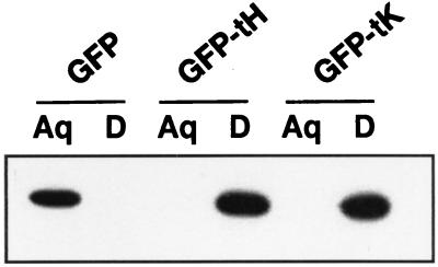
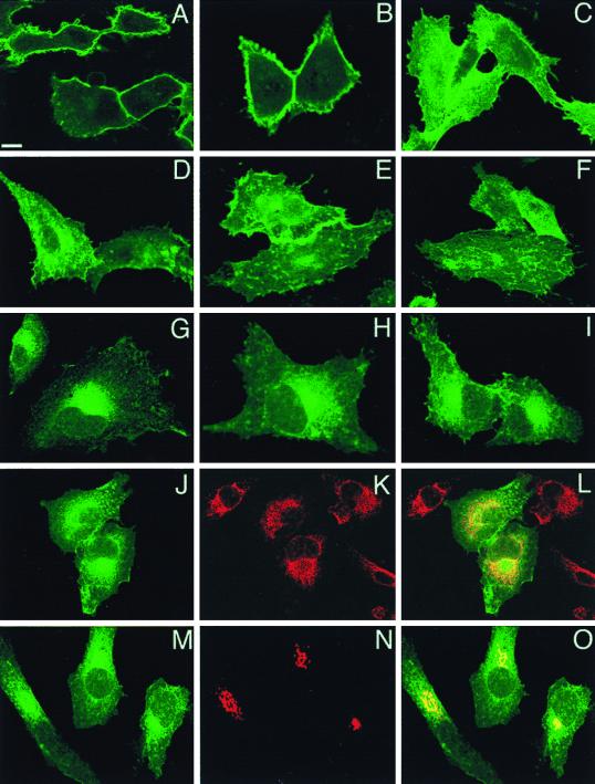
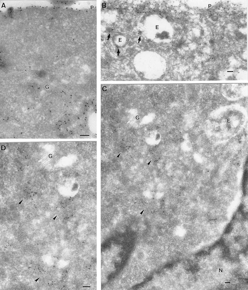
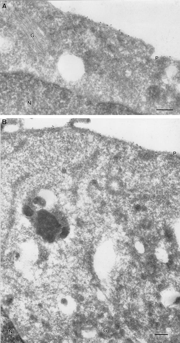
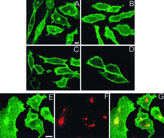
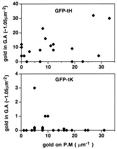
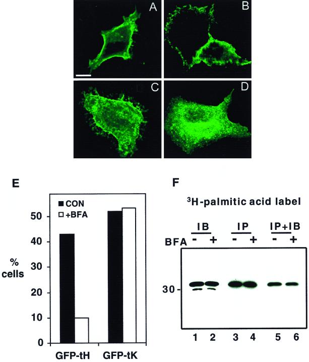
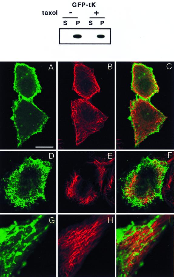
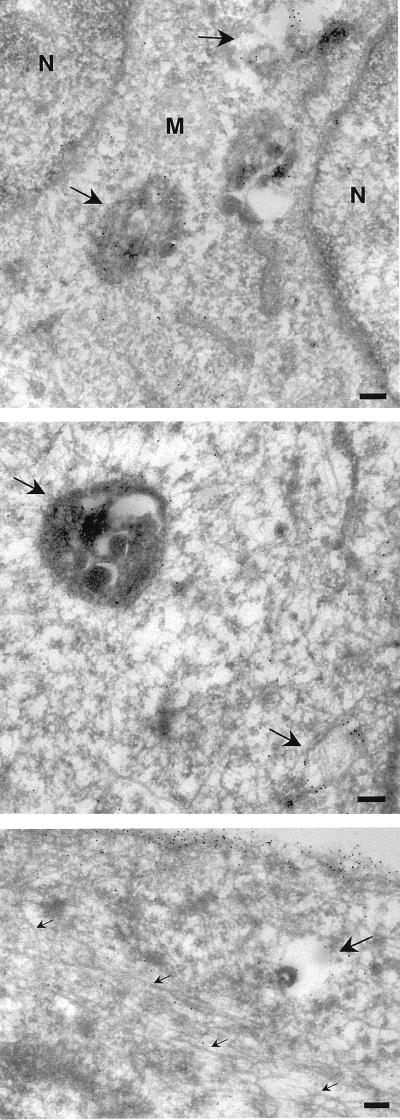
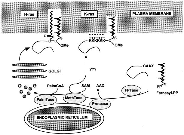
Ras proteins must be localized to the inner surface of the plasma membrane to be biologically active. The motifs that effect Ras plasma membrane targeting consist of a C-terminal CAAX motif plus a second signal comprising palmitoylation of adjacent cysteine residues or the presence of a polybasic domain. In this study, we examined how Ras proteins access the cell surface after processing of the CAAX motif is completed in the endoplasmic reticulum (ER). We show that palmitoylated CAAX proteins, in addition to being localized at the plasma membrane, are found throughout the exocytic pathway and accumulate in the Golgi region when cells are incubated at 15 degrees C. In contrast, polybasic CAAX proteins are found only at the cell surface and not in the exocytic pathway. CAAX proteins which lack a second signal for plasma membrane targeting accumulate in the ER and Golgi. Brefeldin A (BFA) significantly inhibits the plasma membrane accumulation of newly synthesized, palmitoylated CAAX proteins without inhibiting their palmitoylation. BFA has no effect on the trafficking of polybasic CAAX proteins. We conclude that H-ras and K-ras traffic to the cell surface through different routes and that the polybasic domain is a sorting signal diverting K-Ras out of the classical exocytic pathway proximal to the Golgi. Farnesylated Ras proteins that lack a polybasic domain reach the Golgi but require palmitoylation in order to traffic further to the cell surface. These data also indicate that a Ras palmitoyltransferase is present in an early compartment of the exocytic pathway.


Figures

FIG. 1
FIG. 1
Prenylation of GFP-Ras constructs. BHK cells expressing GFP, GFP-tH, or GFP-tK were lysed in Triton X-114. The lysates, normalized for protein content, were warmed to 37°C, and the detergent (D) and aqueous (Aq) phases were separated by centrifugation exactly as previously described (15, 17). Equal proportions of each fraction were resolved by sodium dodecyl sulfate-polyacrylamide gel electrophoresis and immunoblotted using anti-GFP serum. Wild-type GFP is hydrophilic and partitions exclusively into the aqueous phase, whereas prenylation of the CAAX motifs of GFP-tH and GFP-tK imparts sufficient hydrophobicity to partition the proteins into the detergent phase. Prenylation is complete, since all of the expressed GFP-tH and GFP-tK has shifted into the detergent phase.
FIG. 2
FIG. 2
Confocal localization of GFP-Ras constructs. BHK cells were transfected with GFP-tH, GFP-tK, or GFP-6QtK and incubated at 37°C for 18 h prior to fixation for confocal microscopy. Scale bars, 10 μm. (A to F) Panels A, B, D, and E show cuts through cells at the level of the nucleus, whereas those in panels C and F are higher-level cuts showing the cell surface. In cells expressing GFP-tK (A to C), the protein is almost completely localized to the plasma membrane. In cells expressing GFP-tH (D to F), there is staining of the plasma membrane and intracellular structures. The intensity of plasma membrane fluorescence is less with GFP-tH than with GFP-tK. (G to I) Representative cells expressing GFP-6QtK. There is substantial intracellular staining adjacent to and contiguous with the nuclear membrane, with minimal staining of the plasma membrane. (J to L) Cells expressing GFP-6QtK were costained for PDI, an ER marker (red channel), with anti-KDDD antibody (K and L). The overlay (L) shows extensive localization of GFP-6QtK to the ER. (M to O) GFP-6QtK was cotransfected with VSV G-tagged sialyltransferase, a Golgi-resident protein, and the cells were stained for the G epitope tag. The overlay in panel O shows that GFP-6QtK is present in the Golgi, a result confirmed by immunoelectron microscopy.
FIG. 3
FIG. 3
Ultrastructural localization of GFP-tH at 37 and 15°C. BHK cells were transfected with GFP-tH and incubated for 18 h at 37°C (A and B) or for 16 h at 37°C, followed by 2 h at 15°C (C and D). The cells were then labeled with antibodies to GFP, followed by 10-nm protein A-gold particles. Scale bars, 100 nm. (A and B) Specific labeling for GFP-tH is evident on the plasma membrane (P). Despite the high expression level, there is negligible cytosolic labeling. Specific labeling is also apparent on the Golgi complex (G). Putative endosomes (E) show low labeling, whereas small vesicles nearby show higher labeling (arrows). (C and D) At 15°C, GFP-tH accumulates in groups of small vesicular structures (arrowheads) close to the Golgi complex (G), as shown at higher magnification in panel D. Again, note the lack of significant cytosolic labeling and the absence of labeling on the ER surrounding the nucleus (N).
FIG. 4
FIG. 4
Ultrastructural localization of GFP-tK at 37 and 15°C. BHK cells were transfected with GFP-tK and incubated for 18 h at 37°C (A) or for 16 h at 37°C, followed by 2 h at 15°C (B). The cells were then labeled with antibodies to GFP, followed by 10-nm protein A-gold particles. Scale bars, 100 nm. (A) Specific labeling for GFP-tK is evident on the plasma membrane (P). Negligible labeling is apparent on the Golgi complex (G). (B) Again, specific labeling for GFP-tK is evident on the plasma membrane (P). Negligible labeling is apparent on the Golgi complex (G), in contrast to the results obtained with GFP-tH (cf. Fig. 3C and D). N, nucleus.
FIG. 5
FIG. 5
Newly synthesized GFP-tH accumulates in the Golgi complex at 15°C. BHK cells were transfected with GFP-tH (A, B, and E to G) or GFP-tK (C and D) or cotransfected with GFP-tH and VSV G-tagged sialyltransferase (E to G). After incubation at 37°C for 24 h, cells were incubated for a further 2 h at 15°C prior to fixation. Scale bars, 10 μm. (A and B) GFP-tH accumulates in perinuclear structures after incubation at 15°C (A); this intracellular accumulation of GFP-tH is blocked by incubation of the cells in cycloheximide for 2 h before and during the temperature block (B). (C and D) GFP-tK does not show any evidence of intracellular accumulation at 15°C (C and D). Cells shown in panel D were incubated in cycloheximide. (E to G) All cells were incubated at 15°C prior to fixation. Sialyltransferase (red channel), a Golgi marker, shows extensive colocalization with the intracellular accumulation of GFP-tH.
FIG. 6
FIG. 6
Semiquantitative comparison of the subcellular distribution of GFP-ras constructs. Cells expressing GFP-tK and GFP-tH were fixed after a 2-h incubation at 15°C, cryosectioned, and examined by electron microscopy after immunogold labeling for GFP. Cells positively expressing GFPtH or GFPtK were assayed for the number of gold particles present on randomly chosen fixed-unit lengths of plasma membrane (P.M) or in fixed-unit Golgi complex areas (G.A). These data are, in part, arrayed as two scatter diagrams; note the log difference in the y axis scales. The plasma membrane pool, which represents the largest subcellular pool of Ras, contains similar amounts of labeling for GFPtH and GFPtK, indicating that levels of expression were comparable for the two peptides (means, 13.75 and 13.21 gold particles/μm, respectively). GFP-tH was present on and in the vicinity of the Golgi apparatus (mean, 28.62 gold particles/μm2), whereas GFPtK was almost completely excluded from this region (mean, 0.25 gold particles/μm2).
FIG. 7
FIG. 7
BFA inhibits the plasma membrane accumulation of GFP-tH but not GFP-tK. BHK cells were transfected with GFP-tH or GFP-tK. At 2 h after transfection, BFA (5 μg/ml) was added to half of the cultures. After 5 h, coverslips were examined by confocal microscopy. Bar, 10 μm. (A to D) Representative cells from control cultures (A and C) and BFA-treated cultures (B and D). The localization of GFP-tK (A and B) is unaffected, while GFP-tH (C and D) is prevented from trafficking to the plasma membrane. (E) Greater than 450 cells (462 to 524) per experimental condition were scored for intensity of plasma membrane fluorescence by an observer blind to the transfection and treatment conditions. The graph shows the percentages cells with clear plasma membrane staining. It is a representative experiment that was repeated three times with similar results. CON, control. (F) BHK cells plated in 10-cm-diameter dishes were transfected with GFP-tH. At 2 h after transfection, cells were switched to labeling medium containing [3H]palmitic acid; simultaneously, BFA (10 μg/ml) was added to half of the cultures (+BFA) and ethanol carrier was added to the remainder (−BFA). After a further 3 h of incubation, coverslips were quantified as for panel E; the remainder of the cells were harvested into lysis buffer. Lysates were immunoblotted (IB, lanes 1 and 2) or immunoprecipitated (IP, lanes 3 to 6) using monoclonal anti-GFP serum. Radiolabeled proteins in the immunoprecipitates were visualized by fluorography (lanes 3 and 4). Immunoprecipitates were also probed with polyclonal anti-GFP serum to confirm that equal amounts of GFP-tH had been captured (lanes 5 and 6). Note that there is no effect of BFA on GFP-tH expression (lanes 1 and 2) or on palmitic acid incorporation (lanes 3 and 4). BFA did, however, decrease the number of cells with strong plasma membrane staining to 8% from the 48% in the untreated control.
FIG. 8
FIG. 8
Taxol causes redistribution of GFP-tK. BHK cells were incubated in taxol for 2 h prior to and 12 h after lipofection and then harvested for subcellular fractionation or fixed for confocal or electron microscopy. The cytosolic (S = S100) and membrane (P = P100) fractions were prepared from cells expressing GFP-tK as previously described (45), normalized for protein content, and immunoblotted with anti-GFP serum (upper panel). GFP-tK remained fully associated with the P100 fraction in taxol-treated cells. The lower panel shows confocal images of BHK cells from identical cultures that have been costained for α-tubulin. Cells in panels A to F have been cut at the level of the nucleus. The images in panels G to I show the edge of a cell magnified 2.5× relative to the other images. Bar, 10 μm for panels A to F and 4 μm for panels G to I. (A to C) Untreated control cells showing normal microtubules (red channel) and plasma membrane-localized GFP-tK (green channel). (D to I) Taxol-treated cells show some thickening and disruption of the microtubules, accompanied by accumulation of intracellular GFP-tK and a reduction in the amount of GFP-tK seen at the plasma membrane. GFP-tK is seen decorating vesicular and tubulovesicular structures, but there is very little actual colocalization of GFP-tK with tubulin. Some of the tubulovesicular structures are apparently aligned alongside microtubules; this is seen particularly well in panels G to I.
FIG. 9
FIG. 9
Electron microscopic analysis of taxol-treated cells. BHK cells were taxol treated and transfected as described in the legend to Fig. 8. Frozen sections were then labeled with antibodies to GFP, followed by protein A-gold particles. Specific labeling is associated with the plasma membrane and endosomal structures (arrows), but negligible labeling is associated with microtubules (small arrows, bottom panel). N, nucleus; M, mitochondria; bars, 200 nm.
FIG. 10
FIG. 10
Model of Ras trafficking. After the common processing steps of farnesylation, AAX proteolysis, and methylation which are completed on the cytosolic surface of the ER, the trafficking routes of the different Ras proteins diverge. K-ras, by virtue of its C-terminal polybasic domain, is sorted out of the conventional exocytic pathway and takes an undefined pathway to the cell surface that bypasses the Golgi. H- and N-ras are palmitoylated by an ER-localized palmitoyltransferase and enter the exocytic pathway; they traffic to the cell surface via the Golgi. OMe, carboxymethyl; PalmCoA, palmitoyl coenzyme A; SAM, S-adenosyl methionine; PalmTase, palmitoyltransferase; MethTase, methyltransferase; FPTase, farnesyl protein transferase; Farnesyl-PP, farnesyl pyrophosphate.

Similar articles

Cited by

References

    1. Alexandrov K, Horiuchi H, Steele Mortimer O, Seabra M C, Zerial M. Rab escort protein-1 is a multifunctional protein that accompanies newly prenylated Rab proteins to their target membranes. EMBO J. 1994;13:5262–5273. - PMC - PubMed
    1. Aronheim A, Engelberg D, Li N, al Alawi N, Schlessinger J, Karin M. Membrane targeting of the nucleotide exchange factor Sos is sufficient for activating the Ras signaling pathway. Cell. 1994;78:949–961. - PubMed
    1. Bijlmakers M-J J E, Marsh M. Trafficking of an acylated cytosolic protein: newly synthesized p56lck travels to the plasma membrane via the exocytic pathway. J Cell Biol. 1999;145:457–468. - PMC - PubMed
    1. Booden M A, Baker T C, Solski P A, Der C J, Parker S G, Buss J E. A non-farnesylated Ha-Ras protein can be palmitylated and trigger potent differentiation and transformation. J Biol Chem. 1999;274:1423–1431. - PubMed
    1. Bos J L. ras oncogenes in human cancer: a review. Cancer Res. 1989;49:4682–4689. - PubMed

Publication types