. 2000 Mar;11(3):969-82.
doi: 10.1091/mbc.11.3.969.

Apg5p functions in the sequestration step in the cytoplasm-to-vacuole targeting and macroautophagy pathways

Affiliations
Free PMC article

Apg5p functions in the sequestration step in the cytoplasm-to-vacuole targeting and macroautophagy pathways

M D George et al. Mol Biol Cell. 2000 Mar.
Free PMC article

Abstract

The cytoplasm-to-vacuole targeting (Cvt) pathway and macroautophagy are dynamic events involving the rearrangement of membrane to form a sequestering vesicle in the cytosol, which subsequently delivers its cargo to the vacuole. This process requires the concerted action of various proteins, including Apg5p. Recently, it was shown that another protein required for the import of aminopeptidase I (API) and autophagy, Apg12p, is covalently attached to Apg5p through the action of an E1-like enzyme, Apg7p. We have undertaken an analysis of Apg5p function to gain a better understanding of the role of this novel nonubiquitin conjugation reaction in these import pathways. We have generated the first temperature-sensitive mutant in the Cvt pathway, designated apg5(ts). Biochemical analysis of API import in the apg5(ts) strain confirmed that Apg5p is directly required for the import of API via the Cvt pathway. By analyzing the stage of API import that is blocked in the apg5(ts) mutant, we have determined that Apg5p is involved in the sequestration step and is required for vesicle formation and/or completion.


Figures
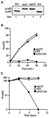
Figure 1
Figure 1
Epitope-tagged Apg5p complements defects in prAPI import and macroautophagy in an apg5Δ strain. (A) API maturation in steady-state cultures. Cells from strains SEY6210 (WT, lane 1), apg5 (lane 2), apg5Δ (lane 3), and apg5Δ expressing Apg5HAp from a CEN plasmid (HA, lane 4) were grown to log phase, and protein extracts were prepared and immunoblotted with antiserum to API, as described in MATERIALS AND METHODS. mAPI, mature API. (B) Kinetic analysis of prAPI import. Cells from strains SEY6210 (WT), apg5Δ, and apg5Δ expressing Apg5HAp from a CEN plasmid (APG5-HA) were pulse labeled for 10 min at 30°C and subjected to a nonradioactive chase for the indicated times. API was recovered by immunoprecipitation and detected with a STORM PhosphorImager. The percentage of mature API (%mAPI) was calculated by dividing mature API by mature API plus prAPI at each time point. (C) Viability during nitrogen starvation. Cells from SEY6210 (WT), apg5Δ, and apg5Δ expressing Apg5HAp from a CEN plasmid (APG5-HA) were analyzed for sensitivity to starvation, as described in MATERIALS AND METHODS.
Figure 2
Figure 2
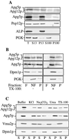
Subcellular fractionation and characterization of membrane association. (A) Apg5HAp is pelletable. Spheroplasts from the apg5Δ strain expressing Apg5HAp were lysed osmotically, as described in MATERIALS AND METHODS. After centrifugation at 500 × g for 5 min to remove debris and unlysed spheroplasts, the supernatant (T, total lysate) was fractionated by 13,000 × g centrifugation for 10 min, generating a pellet (P13) and a supernatant (S13) fraction. The supernatant fraction was further centrifuged at 100,000 × g for 30 min to generate a pellet (P100) and a supernatant (S100) fraction. Each fraction was subjected to SDS-PAGE and immunoblotting with the use of antibodies to the HA epitope (Apg5p), Pep12p (endosomal marker; P13 and P100), ALP (vacuolar marker; P13), or PGK (cytosolic marker; S13 or S100). (B) Apg5p is membrane associated. Spheroplasts expressing Apg5HAp were subjected to lysis with PS200 buffer containing 5 mM MgCl2. The lysate was loaded on the bottom of a Ficoll step gradient, as described in MATERIALS AND METHODS, in the absence or presence of 1% Triton X-100 (TX-100). The resulting gradients were centrifuged at 13,000 × g for 10 min. Membrane-containing float (F), nonfloat (NF), and pellet (P) fractions were collected and subjected to immunoblotting with antibodies to HA or Dpm1p (membrane protein marker) or antiserum to PGK (soluble protein marker), as indicated. A portion of Apg5HAp and the Apg12p-Apg5HAp conjugate are found in the membrane-associated float fraction. (C) Biochemicalcharacterization of pelletable Apg5p. Spheroplasts expressing Apg5HAp were lysed in PS200 buffer containing 5 mM MgCl2, as described in MATERIALS AND METHODS. The pellet fractions were resuspended in buffer alone or buffer containing 1 M KCl, 0.1 M Na2CO3, pH 10.5, 3 M urea, or 1% Triton X-100 (TX-100) and separated into supernatant (S) and pellet (P) fractions, as described in MATERIALS AND METHODS. Western blotting was performed with antibodies to HA or Dpm1p (integral membrane marker) or with antiserum to Vma2p (peripheral membrane marker).
Figure 3
Figure 3
Apg5pGFP is localized to punctate staining structures. Cells from the apg5Δ strain (WT) and from the mutant strains apg8, apg12Δ, and apg16Δ expressing Apg5pGFP from a 2μ plasmid were grown in SMD or shifted to SD(−N), as described in MATERIALS AND METHODS, and observed on a Leica DM IRB confocal microscope with the use of 510- to 525-nm filter settings. The fluorescent punctate pattern of Apg5pGFP in apg12Δ was essentially the same as that observed for apg7Δ.
Figure 4
Figure 4
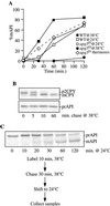
The apg5ts mutant displays rapid inactivation kinetics for prAPI import. (A) Strains apg5Δ expressing apg5ts from a centromeric plasmid (apg5ts) and SEY6210 (WT) were preincubated for 5 min at 24 and 38°C, labeled for 10 min, and subjected to a nonradioactive chase. Aliquots were removed at the indicated times and precipitated with trichloroacetic acid. The samples were then immunoprecipitated with antiserum to API, as described in MATERIALS AND METHODS. After resolution of the immunoprecipitated samples by SDS-PAGE, the amounts of both prAPI and mature API were quantified with the use of a STORM PhosphorImager. The percentage of mature API (%mAPI) was calculated as mature API divided by prAPI plus mature API at each time point. The dotted line corresponds to apg5ts cells shifted from nonpermissive to permissive temperatures, as described in C below. (B) CPY sorting is unaffected at the nonpermissive temperature in the apg5ts mutant. apg5ts cells were preincubated and labeled as described for A at 38°C and subjected to nonradioactive chase for the indicated times. Samples were then immunoprecipitated with antiserum to API or CPY, resolved by SDS-PAGE, and visualized with the use of a STORM PhosphorImager. (C) The apg5ts mutant is thermally reversible. apg5ts cells were preincubated at 38°C for 5 min, labeled for 10 min, subjected to a 30-min nonradioactive chase to accumulate prAPI, and then shifted to 24°C for 120 min of additional chase. Aliquots were removed during the chase at the permissive temperature at the indicated times and immunoprecipitated with antiserum to API, and the proteins were resolved by SDS-PAGE. API was detected with a STORM PhosphorImager. Quantification of the import kinetics is presented in A for comparison (dotted line).
Figure 5
Figure 5
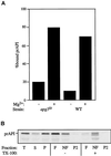
(A) prAPI assembles into higher-order, pelletable Cvt complexes in apg5ts. Spheroplasts were shifted to 38°C for 5 min, pulse labeled for 5 min, and subjected to a cold chase for 20 min. The labeled spheroplasts were lysed by resuspension in PS200 with or without 5 mM MgCl2 and then separated into a soluble fraction and a membrane-containing pellet fraction, as described in MATERIALS AND METHODS. The percentage of prAPI recovered in the pellet fraction is represented. (B) prAPI is membrane associated in the apg5ts mutant. apg5ts spheroplasts were pulse labeled for 5 min, chased for 60 min, and subjected to fractionation into total (T), supernatant (S), and pellet (P) fractions, as described in MATERIALS AND METHODS. The P fraction was resuspended in 60% sucrose in the absence or presence of 1% Triton X-100 (TX-100) and overlaid with 55% sucrose followed by 35% sucrose. After centrifugation at 100,000 × g for 60 min, float (F), nonfloat (NF), and pellet (P2) fractions were collected and API was recovered by immunoprecipitation. Samples were analyzed by SDS-PAGE, and API was detected with a STORM PhosphorImager.
Figure 6
Figure 6
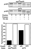
prAPI is protease accessible in the apg5ts mutant. Spheroplasts isolated from apg5ts pep4Δ and ypt7Δ cells were shifted to 38°C for 5 min, labeled for 5 min, and then chased for 60 min. A portion of the sample was removed and immunoprecipitated as a total (T) control. After lysis in PS200 with 5 mM MgCl2 and centrifugation at 5000 × g, supernatant (S) and pellet (P) fractions were collected and subjected to protease treatment, as indicated. The resulting fractions were subsequently immunoprecipitated with antiserum against API, CPY, and hexokinase (cytosolic marker). Hexokinase was recovered 78 and 81% in the supernatant fraction in apg5ts pep4Δ and ypt7Δ cells, respectively, indicating efficient lysis of the spheroplasts. The graph represents the percentage of prAPI and p2CPY recovered in the pellet fraction that is protease resistant (lane 4 divided by lane 3). TX-100, Triton X-100.
Figure 7
Figure 7
Electron microscopic analysis reveals that prAPI is associated with membrane sac structures in the apg5ts mutant. The apg5ts strain was grown to log phase in YPD and shifted to 38°C for 3 h. Samples were prepared for electron microscopy by rapid freezing and freeze-substitution fixation and stained with lead citrate for 1 min (a–c) or stained with lead citrate for 30 s and immunostained with anti-API antibodies (d), as described in MATERIALS AND METHODS. The arrows mark membranous structures around the Cvt complex. V, vacuole.
Figure 8
Figure 8
prAPI is membrane associated in the apg5Δ mutant. The apg5Δ strain was grown to log phase in YPD and shifted to SD(−N) medium for 3 h to induce autophagy. Samples were prepared for electron microscopy with the use of rapid freezing and freeze-substitution fixation and stained with lead citrate for 1 min (a–c) or stained with lead citrate for 30 s and immunostained with anti-API antibodies (d and e), as described in MATERIALS AND METHODS. The arrows mark membranous structures around the Cvt complex. V, vacuole; N, nucleus.
Figure 9
Figure 9
Two-pathway model for Cvt/Apg transport to the vacuole. (1) prAPI rapidly oligomerizes into a dodecameric conformation after translation. (2) The dodecamers assemble into Cvt complexes, which are subsequently sequestered and wrapped by double-membrane Cvt vesicles and autophagosomes in a process requiring Apg5p. (3) The vesicles are then targeted to the vacuole, where subsequent docking and fusion of the outer membrane results in the release of the inner vesicle into the lumen. (4) The inner membrane is degraded within the lumen, allowing vacuolar hydrolases access to the cargo. (5) The N-terminal propeptides of the prAPI molecules are proteolytically removed in a PEP4-dependent manner to generate mature API dodecamers.

Similar articles

Cited by

References

    1. Baba M, Osumi M, Scott SV, Klionsky DJ, Ohsumi Y. Two distinct pathways for targeting proteins from the cytosol to the vacuole/lysosome. J Cell Biol. 1997;139:1687–1695. - PMC - PubMed
    1. Baba M, Takeshige K, Baba N, Ohsumi Y. Ultrastructural analysis of the autophagic process: autophagosomes and their characterization. J Cell Biol. 1994;124:903–913. - PMC - PubMed
    1. Becherer KA, Rieder SE, Emr SD, Jones EW. Novel syntaxin homologue, Pep12p, required for the sorting of lumenal hydrolases to the lysosome-like vacuole in yeast. Mol Biol Cell. 1996;4:579–594. - PMC - PubMed
    1. Haas A, Scheglmann D, Lazar T, Gallwitz D, Wickner W. The GTPase Ypt7p of Saccharomyces cerevisiae is required on both partner vacuoles for the homotypic fusion step of vacuole inheritance. EMBO J. 1995;14:5258–5270. - PMC - PubMed
    1. Hammond EM, Brunet CL, Johnson GD, Parkhill J, Milner AE, Brady G, Gregory CD, Grand RJ. Homology between a human apoptosis specific protein and the product of APG5, a gene involved in autophagy in yeast. FEBS Lett. 1998;425:391–395. - PubMed

Publication types

MeSH terms