. 2000 Mar;105(5):577-87.
doi: 10.1172/JCI8316.

Hypoxia-inducible factor-1 mediates the biological effects of oxygen on human trophoblast differentiation through TGFbeta(3)

Affiliations

Hypoxia-inducible factor-1 mediates the biological effects of oxygen on human trophoblast differentiation through TGFbeta(3)

I Caniggia et al. J Clin Invest. 2000 Mar.

Abstract

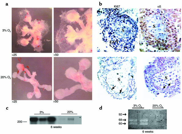
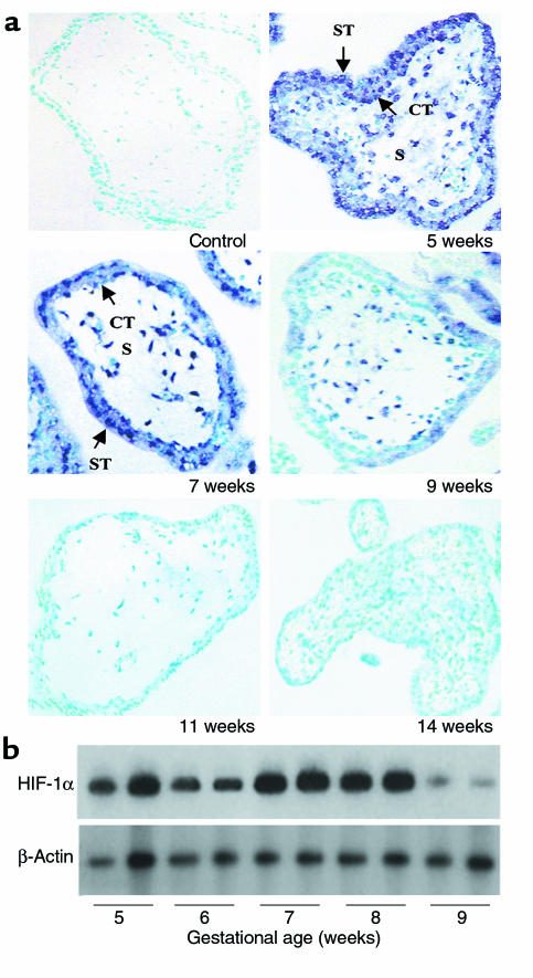
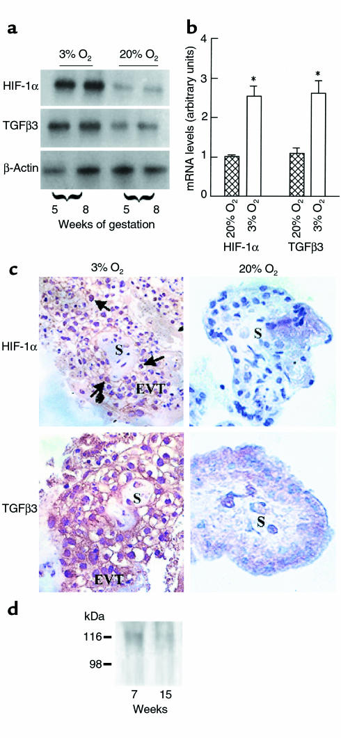
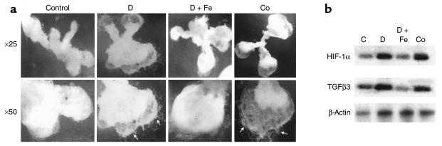
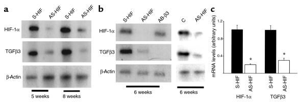
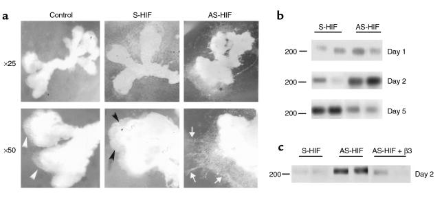
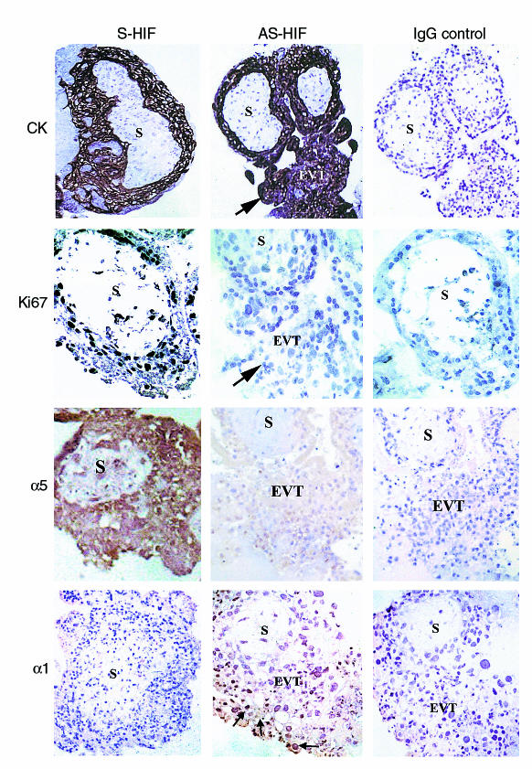
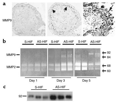
During early pregnancy, placentation occurs in a relatively hypoxic environment that is essential for appropriate embryonic development. Intervillous blood flow increases around 10 to 12 weeks of gestation and results in exposure of trophoblast cells to increased oxygen tension. Before this time, low oxygen appears to prevent trophoblast differentiation toward an invasive phenotype. Using human villous explants of 5-8 weeks' gestation, we found that low oxygen tension triggered trophoblast proliferation, fibronectin synthesis, alpha(5) integrin expression, and gelatinase A activity. These biochemical markers were barely detectable under oxic conditions. We therefore examined the placental expression of hypoxia-inducible factor-1 (HIF-1), a master regulator of oxygen homeostasis, and determined that expression of HIF-1alpha subunit during the first trimester of gestation parallels that of TGFbeta(3), an inhibitor of extravillous trophoblast differentiation. Expression of both molecules is high in early pregnancy and falls around 9 weeks of gestation, when placental pO(2) levels are believed to increase. Increasing oxygen tension induced a similar decrease in expression in cultured explants. Moreover, antisense inhibition of HIF-1alpha expression in hypoxic explants inhibited expression of TGFbeta(3), arrested cell proliferation, decreased alpha(5) expression and gelatinase A activity, and triggered biochemical markers of an invasive trophoblast phenotype such as alpha(1) integrin and gelatinase B expression. These data suggest that the oxygen-regulated early events of trophoblast differentiation are in part mediated by TGFbeta(3) through HIF-1 transcription factors.


Figures

Figure 1
Figure 1
Low oxygen tension induces trophoblast outgrowth and increases proliferation, fibronectin synthesis, and gelatinase activity in villous explant cultures. (a) Villous explants from 5 to 8 weeks of gestation were maintained in culture for 5 days under normal (20% O2) or low (3% O2) oxygen tension. Note that exposure of villous explants to 3% O2 dramatically increases budding and outgrowth of EVT from the distal end of the villous tips when compared with villous explants kept at 20% O2. ×25, ×50. (b) Section of explants, cultured in either 3% or 20% O2, were stained for Ki67, a marker of cellular proliferation and α5 integrin. Strong positive nickel-enhanced immunoreactivity for Ki67 was observed in EVT of the outgrowth (EVT) of explants cultured in 3% O2. Similar immunolocalization was observed for α5 integrin. A few immunopositive cells for both Ki67 and α5 integrin were noted in explants kept at 20% O2 (arrows). S, villous stroma. (c) Explants incubated for 4 days in either low or normal pO2 conditions were metabolically labeled with [35S]methionine for 18 hours. Fibronectin was isolated from conditioned medium using gelatin-Sepharose beads. Samples were subjected to SDS-PAGE, and the position of the marker (with Mr = 200 × 103) is indicated. (d) Samples of medium conditioned by explants, cultured in either 3 or 20% O2, were collected at day 5 and subjected to analysis by gelatin zymography. Arrows indicate positions of gelatinase activity (gelatinase A/MMP-2: 60 and 68 kDa; gelatinase B/MMP-9: 92 kDa).
Figure 2
Figure 2
Expression of HIF-1α in human placenta in the first trimester of gestation (a) Expression of HIF-1α mRNA was also assessed by in situ hybridization to placental sections at 5–14 weeks of gestation with digoxigenin-labeled sense and antisense HIF-1α riboprobes. Endogenous alkaline phosphatase was blocked by the addition of levamisole. Sections were counterstained with methyl green. Note that HIF-1α mRNA expression, revealed by blue staining, is high at 5–7 weeks in chorionic villi, decreases around 9 weeks, and is absent at 11–14 weeks. Controls using a sense HIF-1α riboprobe were negative. ×100. (b) Message expression of HIF-1α and TGFβ3 were assessed by low-cycle RT-PCR followed by Southern blot analysis using specific probes for HIF-1α and TGFβ3 and the control housekeeping gene β-actin. Note that mRNA expression of HIF-1α is high between 5 to 8 weeks of gestation and declines thereafter.
Figure 3
Figure 3
Expression of HIF-1α villous explants exposed to different oxygen tension (a) Exposure of villous explants to 20% O2 downregulates the mRNA expression for HIF-1α and TGFβ3. (b) Bands were quantified by densitometric analysis. Shown are the changes in HIF-1α and TGFβ3 mRNA after normalization to control cultures kept at 20% O2. All data are expressed as the mean ± SEM of 5 separate experiments carried out in triplicate. (c) Immunoperoxidase staining of HIF-1α and TGFβ3 was performed in sections from explants kept at either 3% or 20% O2.. Sections of explants kept at 3% O2 show positive HIF-1α and TGFβ3 immunoreactivity in EVT cells of the outgrowth (EVT), whereas no immunoreactivity for HIF-1α and low immunoreactivity for TGFβ3 are noted in sections of explants kept at 20% O2. S, villous stroma. (d) Western blot analysis for HIF-1α in human placental tissue. HIF-1α protein (116 kDa) was detected in placentas of 7 weeks’ gestation, but not 15 weeks’ gestation.
Figure 4
Figure 4
Desferoxamine and cobalt chloride mimic the low-oxygen effect on EVT outgrowth and HIF-1α and TGFβ3 expression. (a) Exposure of villous explants, cultured in 20% O2 to either desferoxamine (10 μM) or CoCl2 (100 μM) increased EVT outgrowth (arrows). Addition of FeCl2 (20 μM) to the desferoxamine-treated cultures prevented the outgrowth. (b) Exposure of villous explants to either desferoxamine (10 μM) or cobalt chloride (100 μM) increased mRNA expression for HIf-1α and TGFβ3. C, control medium; D, desferoxamine; Co, cobalt chloride; D+Fe, desferoxamine + iron chloride. ×25, ×50.
Figure 5
Figure 5
Antisense oligonucleotides to HIF-1α inhibit HIF-1α and TGFβ3 mRNA expression. Explants of 5–8 weeks’ gestation were treated for 5 days with 10 μM antisense oligonucleotides to HIF-1α (AS-HIF) and antisense oligonucleotides to TGFβ3 (AS-β3). Control experiments were run in parallel using either sense oligonucleotides to HIF-1α (S-HIF) and TGF-β3 (S-β3) or medium alone (C). Expression of HIF-1α and TGFβ3 mRNA was measured by low-cycle RT-PCR followed by Southern blot analysis using specific probes for HIF-1α, TGF-β3, and the control housekeeping gene β-actin. (a) Antisense HIF-1α treatment of villous explants inhibits both HIF-1α and TGFβ3 mRNA. (b) Antisense TGFβ3 treatment of villous explants inhibits TGFβ3 mRNA expression but not that of HIF-1α, whereas antisense HIF-1α exposure results in inhibition of both HIF-1α and TGFβ3 mRNA expression. (c) Bands were quantified by densitometric analysis. Shown are the changes in HIF-1α and TGFβ3 mRNA after normalization to control cultures using sense oligonucleotides. All data are expressed as the mean ± SEM of 3 separate experiments carried out in triplicate.
Figure 6
Figure 6
Antisense oligonucleotides to HIF-1α induce extravillous trophoblast invasion and fibronectin production in villous explant cultures. (a) Villous explants of 5–8 weeks’ gestation were maintained in culture for 5 days at 3% O2 in the presence of 10 μM antisense oligonucleotides to HIF-1α (AS-HIF). Control experiments were run in parallel using explants from the same placentas cultured with medium alone or medium containing sense oligonucleotides (S-HIF). Note that antisense HIF-1α–treatment dramatically increases EVT invasion into the surrounding Matrigel (arrows) when compared with control villous explants, which show the typical low pO2-induced outgrowth (arrowheads). ×25, ×50. (b) Antisense HIF-1α treatment increases fibronectin synthesis as early as 2 days after oligonucleotide exposure. Note that after 5 days of treatment the antisense-stimulatory effect on fibronectin synthesis is lost. (c) Shown is a representative experiment demonstrating that addition of recombinant TGFβ3 to the antisense HIF-1α–treated explants abolished the early antisense-stimulatory effect on fibronectin production.
Figure 7
Figure 7
Antisense oligonucleotides to HIF-1α inhibit proliferation and α5 integrin expression and induce α1 integrin expression in villous explant cultures. Immunoperoxidase staining of cytokeratin, Ki67, integrin α5, and α1 integrin was performed in placental sections from villous explants cultured under hypoxic condition with antisense and control sense oligonucleotides to HIF-1α. Sections of explants, cultured with antisense oligonucleotides, have no positive Ki67 immunoreactivity and markedly reduced staining for α5 integrin but are immunopositive for α1 integrin in EVT cells of the invading column (arrow, EVT). Sections of control cultures in the presence of sense oligonucleotides show positive immunoreactivity for Ki67 and α5 integrin but no positive α1 integrin staining in the EVT of the outgrowth induced by 3% O2 exposure. Independent of treatment, trophoblast cells stained positive for cytokeratin (CK), a marker for epithelial cells. Control immunostaining experiments were performed using control IgG. S, villous stroma.
Figure 8
Figure 8
Antisense oligonucleotides to HIF-1α trigger gelatinase expression and activity in villous explants cultured at 3% O2. Explants of 5–8 weeks’ gestation were treated with antisense (AS-HIF) or control sense (S-HIF) oligonucleotides to HIF-1α (a) Section of explants treated for 5 days with antisense HIF-1α show positive immunoreactivity for gelatinase B/MMP9 in the invading EVT (arrow, EVT) when compared with control sense–treated explants. Samples of conditioned medium were collected at days 1, 3, and 5 and subjected to analysis by gelatin zymography (b) or at day 5 for Western blotting with gelatinase B/MMP9 antisera (c). Arrows indicate positions of gelatinase activity (gelatinase A/MMP-2: 60 and 68 kDa; gelatinase B/MMP-9: 84 and 92 kDa).

Comment in

Similar articles

Cited by

References

    1. Cross JC, Werb Z, Fisher SJ. Implantation and the placenta: key pieces of the development puzzle. Science. 1994;266:1508–1518. - PubMed
    1. Strickland S, Richards WG. Invasion of trophoblasts. Cell. 1992;71:355–357. - PubMed
    1. Aplin JD. Implantation, trophoblast differentiation and haemochorial placentation: mechanistic evidence in vivo and in vitro. J Cell Sci. 1991;99:681–692. - PubMed
    1. Rodesch F, Simon P, Donner C, Jauniaux E. Oxygen measurements in endometrial and trophoblastic tissues during early pregnancy. Obstet Gynecol. 1992;80:283–285. - PubMed
    1. Genbacev O, Joslin R, Damsky CH, Polliotti BM, Fisher SJ. Hypoxia alters early gestation human cytotrophoblast differentiation/invasion in vitro and models the placental defects that occur in preeclampsia. J Clin Invest. 1996;97:540–550. - PMC - PubMed

Publication types

MeSH terms