. 2000 Apr;74(7):3353-65.
doi: 10.1128/jvi.74.7.3353-3365.2000.

Vaccinia virus envelope H3L protein binds to cell surface heparan sulfate and is important for intracellular mature virion morphogenesis and virus infection in vitro and in vivo

Affiliations

Vaccinia virus envelope H3L protein binds to cell surface heparan sulfate and is important for intracellular mature virion morphogenesis and virus infection in vitro and in vivo

C L Lin et al. J Virol. 2000 Apr.

Abstract

An immunodominant antigen, p35, is expressed on the envelope of intracellular mature virions (IMV) of vaccinia virus. p35 is encoded by the viral late gene H3L, but its role in the virus life cycle is not known. This report demonstrates that soluble H3L protein binds to heparan sulfate on the cell surface and competes with the binding of vaccinia virus, indicating a role for H3L protein in IMV adsorption to mammalian cells. A mutant virus defective in expression of H3L (H3L(-)) was constructed; the mutant virus has a small plaque phenotype and 10-fold lower IMV and extracellular enveloped virion titers than the wild-type virus. Virion morphogenesis is severely blocked and intermediate viral structures such as viral factories and crescents accumulate in cells infected with the H3L(-) mutant virus. IMV from the H3L(-) mutant virus are somewhat altered and less infectious than wild-type virions. However, cells infected by the mutant virus form multinucleated syncytia after low pH treatment, suggesting that H3L protein is not required for cell fusion. Mice inoculated intranasally with wild-type virus show high mortality and severe weight loss, whereas mice infected with H3L(-) mutant virus survive and recover faster, indicating that inactivation of the H3L gene attenuates virus virulence in vivo. In summary, these data indicate that H3L protein mediates vaccinia virus adsorption to cell surface heparan sulfate and is important for vaccinia virus infection in vitro and in vivo. In addition, H3L protein plays a role in virion assembly.


Figures
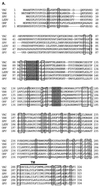
FIG. 1
FIG. 1
(A) Alignment of amino acid sequences deduced from vaccinia virus H3L gene and its homologues in poxvirus family. VAC, vaccinia virus H3L gene (SP/P07240); VAR, variola virus (IND3, isolated in India in 1967) immunodominant envelope protein P35 gene (accession no. sp/P33059) (53); SPV, sheep poxvirus P32 antigen gene (accession no. gb/AF124517) (20); LSDV, lumpy skin disease virus P32 antigen gene (accession no. gb/AF124516) (20); ORF, orf virus (strain NZ2) immunodominant envelope antigen F1L gene (accession no. gi/AF097215) (21); and SFV, shope fibroma virus S071L gene (D. H. Evans, personal communication). The boxed areas indicate identical amino acids, and the shaded areas are potential GAG-binding sites, as described previously (4). TM, transmembrane region. (B) Neutralization of vaccinia virus infection by antisera against H3L protein. BSC40 cells were infected with vaccinia virus in the absence or the presence of various dilutions of antisera, as indicated, and an agar overlay was added for plaque determination. The number of plaques obtained in the absence of sera, approximately 150 PFU per 60-mm-diameter dish, was used as the 100% value. (C) Purified soluble H3L protein (sH3L) stained by Coomassie blue after SDS–12% PAGE. Protein marker masses, in kilodaltons, are shown at the left side of the gel. (D) sH3L protein blocked viral early gene expression. BSC40 cells were infected with vMJ360 expressing lacZ at an MOI of 10 PFU per cell in the presence of various amounts of sH3L (0, 1, 10, or 50 μg) and harvested at 2 h p.i. for β-gal assays as described previously (12). Cells infected with vaccinia virus without sH3L protein were used as a control. (E) sH3L protein blocked vaccinia virus adsorption to cells. BSC40 cells were infected with vaccinia virus at an MOI of 10 PFU per cell at 4°C for 30 min. After washing, these cells were immediately harvested and cell-associated virions were determined by plaque assay on BSC40 cells (22). Controls were the same as described for panel D.
FIG. 1
FIG. 1
(A) Alignment of amino acid sequences deduced from vaccinia virus H3L gene and its homologues in poxvirus family. VAC, vaccinia virus H3L gene (SP/P07240); VAR, variola virus (IND3, isolated in India in 1967) immunodominant envelope protein P35 gene (accession no. sp/P33059) (53); SPV, sheep poxvirus P32 antigen gene (accession no. gb/AF124517) (20); LSDV, lumpy skin disease virus P32 antigen gene (accession no. gb/AF124516) (20); ORF, orf virus (strain NZ2) immunodominant envelope antigen F1L gene (accession no. gi/AF097215) (21); and SFV, shope fibroma virus S071L gene (D. H. Evans, personal communication). The boxed areas indicate identical amino acids, and the shaded areas are potential GAG-binding sites, as described previously (4). TM, transmembrane region. (B) Neutralization of vaccinia virus infection by antisera against H3L protein. BSC40 cells were infected with vaccinia virus in the absence or the presence of various dilutions of antisera, as indicated, and an agar overlay was added for plaque determination. The number of plaques obtained in the absence of sera, approximately 150 PFU per 60-mm-diameter dish, was used as the 100% value. (C) Purified soluble H3L protein (sH3L) stained by Coomassie blue after SDS–12% PAGE. Protein marker masses, in kilodaltons, are shown at the left side of the gel. (D) sH3L protein blocked viral early gene expression. BSC40 cells were infected with vMJ360 expressing lacZ at an MOI of 10 PFU per cell in the presence of various amounts of sH3L (0, 1, 10, or 50 μg) and harvested at 2 h p.i. for β-gal assays as described previously (12). Cells infected with vaccinia virus without sH3L protein were used as a control. (E) sH3L protein blocked vaccinia virus adsorption to cells. BSC40 cells were infected with vaccinia virus at an MOI of 10 PFU per cell at 4°C for 30 min. After washing, these cells were immediately harvested and cell-associated virions were determined by plaque assay on BSC40 cells (22). Controls were the same as described for panel D.
FIG. 2
FIG. 2
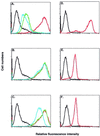
sH3L protein bound to cell surface HS. BSC40 (A to C), L (D), gro2C (E), or sog9 (F) cells were incubated with PBS (black line) or biotinylated H3L (red line) and analyzed with a FACS as described previously (22). Alternatively, as shown in panels A to C, biotinylated H3L protein was mixed with 10 μg/ml (results shown by green line) or with 100 μg/ml (results shown by blue line) of soluble GAGs and analyzed with a FACS. The soluble GAGs used in competitions were HS (A), CS (B), or DS (C).
FIG. 3
FIG. 3
(A) H3L mutant virus formed smaller plaques on BSC40 cells. BSC40 cells were infected with WT (left) or H3L mutant virus (right) for plaque assay with neutral red as described above, and photographed at 3 days p.i. (B) Expression of H3L gene was inactivated in H3L mutant virus. Purified virions from WT (lane 2) and H3L mutant (lane 4) virus as well as infected cell lysates from WT (lane 3) and H3L mutant (lane 5) virus were loaded onto a SDS–12% PAGE gel and transferred for Western blot analysis with antiserum B (1:1,000) against H3L protein. Mock lysates (lane 1) were used as a negative control. (C) H3L mutant virus was resistant to antiserum C. BSC40 cells were infected with WT or H3L mutant virus in the presence of various dilutions of an antiserum against H3L protein (serum C) or a MAb against L1R protein (2D5), as indicated at the bottom of the figures, followed by plaque determination (39). The number of plaques obtained in the absence of sera, around 150 PFU per 60-mm-diameter dish, was used as the 100% value.
FIG. 4
FIG. 4
One-step growth curve analysis of WT vaccinia virus (vv) and H3L mutant virus. BSC40 cells were infected with WT and H3L mutant viruses at an MOI of 5 PFU per cell and harvested at various times for plaque assays as described previously (22). Expt., experiment.
FIG. 5
FIG. 5
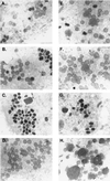
Electron micrographs of vaccinia virion morphogenesis. BSC40 cells were infected with WT (A to D) and H3L mutant viruses (E to H), fixed at 12 h (A and E), 24 h (B and F), or 48 h (C, D, G, and H) p.i. and analyzed with a Zeiss 902 transmission electron microscope. Magnification, ×13,000.
FIG. 6
FIG. 6
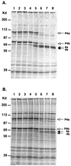
Processing of P4a and P4b precursor core proteins in H3L mutant virus. BSC40 cells were infected with WT (A) or H3L mutant virus (B) at an MOI of 10 PFU per cell, and the cultures were pulsed with 35S-methionine (50 μCi/ml) for 30 min for viral late protein synthesis (lane 1) and chased with normal growth medium for various times, as follows: 15 min (lane 2), 30 min (lane 3), 1 h (lane 4), 2 h (lane 5), 4 h (lane 6), 12 h (lane 7), and 24 h (lane 8). At each time one set of samples was harvested, lysed in SDS-containing buffer, separated on a SDS–10% PAGE gel, and analyzed by autoradiography. The white arrows mark the positions of two precursor core proteins, P4a and P4b, and the black arrows indicate the processed forms of core proteins 4a and 4b as described previously (60).
FIG. 7
FIG. 7
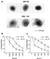
Structures of H3L mutant IMV and the neutralization sensitivity to antibodies against A27L and D8L proteins. (A) Electron microscope images of purified WT and H3L mutant virions after negative staining as described in Materials and Methods. (B and C) BSC40 cells were infected with WT or H3L mutant vaccinia virus (vv) in the presence of various dilutions of antisera against A27L (B) or D8L (C) proteins. After infection, cells were washed and overlaid with 1% agar, and plaque numbers were determined. The plaque numbers obtained in the absence of antiserum were used as the 100% values.
FIG. 8
FIG. 8
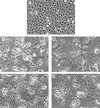
H3L protein is not required for fusion of virus-infected cells. BSC40 cells were either mock infected (A) or infected with WT (B and D) or H3L mutant virus (C and E) at an MOI of 5 PFU per cell. At 24 h p.i., cells were briefly treated with PBS at a pH of 7.2 (B and C) or with PBS at a pH of 4.8 (D and E), incubated for another 3 h, and photographed with a Nikon inverted microscope.
FIG. 9
FIG. 9
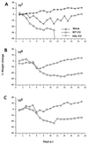
Virulence of H3L mutant virus is attenuated in vivo. BALB/c mice were either mock infected or intranasally infected with WT or H3L mutant virus doses of 107 PFU (A), 106 PFU (B), and 105 PFU (C) at day 0 as indicated in each graph. Body weight was measured on a daily basis. There were five mice per group. All (100%) of the mice infected with 107 PFU of the WT virus 50% of mice infected with 106 PFU of the WT virus died between 10 to 14 days p.i.

Similar articles

Cited by

References

    1. Appleyard G, Hapel A J, Boulter E A. An antigenic difference between intracellular and extracellular rabbitpox virus. J Gen Virol. 1971;13:9–17. - PubMed
    1. Banfield B W, Leduc Y, Esford L, Schubert K, Tufaro F. Sequential isolation of proteoglycan synthesis mutants by using herpes simplex virus as a selective agent: evidence for a proteoglycan-independent virus entry pathway. J Virol. 1995;69:3290–3298. - PMC - PubMed
    1. Banfield B W, Leduc Y, Esford L, Visalli R J, Brandt C R, Tufaro F. Evidence for an interaction of herpes simplex virus with chondroitin sulfate proteoglycans during infection. Virology. 1995;208:531–539. - PubMed
    1. Cardin A D, Weintraub H J R. Molecular modeling of protein-glycosaminoglycan interactions. Arteriosclerosis. 1989;9:21–32. - PubMed
    1. Chakrabarti S, Brechling K, Moss B. Vaccinia virus expression vector: coexpression of β-galactosidase provides visual screening of recombinant virus plaques. Mol Cell Biol. 1985;5:3403–3409. - PMC - PubMed

Publication types

MeSH terms