Pathogenesis of primary R5 human immunodeficiency virus type 1 clones in SCID-hu mice
- PMID: 10708437
- PMCID: PMC111821
- DOI: 10.1128/jvi.74.7.3205-3216.2000
Pathogenesis of primary R5 human immunodeficiency virus type 1 clones in SCID-hu mice
Abstract
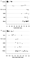
We studied the replication and cytopathicity in SCID-hu mice of R5 human immunodeficiency virus type 1 (HIV-1) biological clones from early and late stages of infection of three patients who never developed MT-2 cell syncytium-inducing (SI; R5X4 or X4) viruses. Several of the late-stage non-MT-2 cell syncytium-inducing (NSI; R5) viruses from these patients depleted human CD4(+) thymocytes from SCID-hu mice. Earlier clones from the same patients did not deplete CD4(+) thymocytes from SCID-hu mice as well as later clones. We studied three R5 HIV-1 clones from patient ACH142 in greater detail. Two of these clones were obtained prior to the onset of AIDS; the third was obtained following the AIDS diagnosis. In GHOST cell infection assays, all three ACH142 R5 HIV-1 clones could infect GHOST cells expressing CCR5 but not GHOST cells expressing any of nine other HIV coreceptors tested. Furthermore, these patient clones efficiently infected stimulated peripheral blood mononuclear cells from a normal donor but not those from a homozygous CCR5Delta32 individual. Statistical analyses of data obtained from infection of SCID-hu mice with patient ACH142 R5 clones revealed that only the AIDS-associated clone significantly depleted CD4(+) thymocytes from SCID-hu mice. This clone also replicated to higher levels in SCID-hu mice than the two earlier clones, and a significant correlation between viral replication and CD4(+) thymocyte depletion was observed. Our results indicate that an intrinsic property of AIDS-associated R5 patient clones causes their increased replication and cytopathic effects in SCID-hu mice and likely contributes to the development of AIDS in patients who harbor only R5 quasispecies of HIV-1.
Figures


Similar articles
-
HIV-1 replication and pathogenesis in the human thymus.Curr HIV Res. 2003 Jul;1(3):275-85. doi: 10.2174/1570162033485258. Curr HIV Res. 2003. PMID: 15046252 Free PMC article. Review.
-
Human immunodeficiency virus type 1 pathogenesis in SCID-hu mice correlates with syncytium-inducing phenotype and viral replication.J Virol. 2000 Apr;74(7):3196-204. doi: 10.1128/jvi.74.7.3196-3204.2000. J Virol. 2000. PMID: 10708436 Free PMC article.
-
Human immunodeficiency virus type 1 strains R5 and X4 induce different pathogenic effects in hu-PBL-SCID mice, depending on the state of activation/differentiation of human target cells at the time of primary infection.J Virol. 1999 Aug;73(8):6453-9. doi: 10.1128/JVI.73.8.6453-6459.1999. J Virol. 1999. PMID: 10400739 Free PMC article.
-
R5 human immunodeficiency virus type 1 infection of fetal thymic organ culture induces cytokine and CCR5 expression.J Virol. 2005 Jan;79(1):458-71. doi: 10.1128/JVI.79.1.458-471.2005. J Virol. 2005. PMID: 15596839 Free PMC article.
-
HIV-1-associated pathology in hemato-lymphoid organs and the experimental evaluation in the SCID-hu mouse.Int J Hematol. 1996 Jun;63(4):253-64. doi: 10.1016/0925-5710(96)00452-5. Int J Hematol. 1996. PMID: 8762809 Review.
Cited by
-
Cytopathicity of human immunodeficiency virus type 1 primary isolates depends on coreceptor usage and not patient disease status.J Virol. 2001 Sep;75(18):8842-7. doi: 10.1128/jvi.75.18.8842-8847.2001. J Virol. 2001. PMID: 11507229 Free PMC article.
-
Host and Viral Factors in HIV-Mediated Bystander Apoptosis.Viruses. 2017 Aug 22;9(8):237. doi: 10.3390/v9080237. Viruses. 2017. PMID: 28829402 Free PMC article. Review.
-
HIV-1 induced bystander apoptosis.Viruses. 2012 Nov 9;4(11):3020-43. doi: 10.3390/v4113020. Viruses. 2012. PMID: 23202514 Free PMC article. Review.
-
HIV-1 replication and pathogenesis in the human thymus.Curr HIV Res. 2003 Jul;1(3):275-85. doi: 10.2174/1570162033485258. Curr HIV Res. 2003. PMID: 15046252 Free PMC article. Review.
-
Determination of coreceptor usage of human immunodeficiency virus type 1 from patient plasma samples by using a recombinant phenotypic assay.J Virol. 2001 Jan;75(1):251-9. doi: 10.1128/JVI.75.1.251-259.2001. J Virol. 2001. PMID: 11119595 Free PMC article.
References
-
- Aldrovandi G M, Feuer G, Gao L, Jamieson B, Kristeva M, Chen I S, Zack J A. The SCID-hu mouse as a model for HIV-1 infection. Nature. 1993;363:732–736. - PubMed
-
- Alkhatib G, Berger E A, Murphy P M, Pease J E. Determinants of HIV-1 coreceptor function on CC chemokine receptor 3. Importance of both extracellular and transmembrane/cytoplasmic regions. J Biol Chem. 1997;272:20420–20426. - PubMed
-
- Alkhatib G, Combadiere C, Broder C C, Feng Y, Kennedy P E, Murphy P M, Berger E A. CC CKR5: a RANTES, MIP-1alpha, MIP-1beta receptor as a fusion cofactor for macrophage-tropic HIV-1. Science. 1996;272:1955–1958. - PubMed
-
- Asjo B, Morfeldt-Manson L, Albert J, Biberfeld G, Karlsson A, Lidman K, Fenyo E M. Replicative capacity of human immunodeficiency virus from patients with varying severity of HIV infection. Lancet. 1986;ii:660–662. - PubMed
Publication types
MeSH terms
Substances
Grants and funding
LinkOut - more resources
Full Text Sources
Molecular Biology Databases
Research Materials

