. 2000 Feb 15;14(4):403-13.

The carboxyl terminus of vertebrate poly(A) polymerase interacts with U2AF 65 to couple 3'-end processing and splicing

Affiliations

The carboxyl terminus of vertebrate poly(A) polymerase interacts with U2AF 65 to couple 3'-end processing and splicing

S Vagner et al. Genes Dev. .

Abstract

Although it has been established that the processing factors involved in pre-mRNA splicing and 3'-end formation can influence each other positively, the molecular basis of this coupling interaction was not known. Stimulation of pre-mRNA splicing by an adjacent cis-linked cleavage and polyadenylation site in HeLa cell nuclear extract is shown to occur at an early step in splicing, the binding of U2AF 65 to the pyrimidine tract of the intron 3' splice site. The carboxyl terminus of poly(A) polymerase (PAP) previously has been implicated indirectly in the coupling process. We demonstrate that a fusion protein containing the 20 carboxy-terminal amino acids of PAP, when tethered downstream of an intron, increases splicing efficiency and, like the entire 3'-end formation machinery, stimulates U2AF 65 binding to the intron. The carboxy-terminal domain of PAP makes a direct and specific interaction with residues 17-47 of U2AF 65, implicating this interaction in the coupling of splicing and 3'-end formation.


Figures
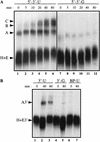
Figure 1
Figure 1
A cleavage/polyadenylation site stimulates 3′ splice site recognition. Shown is time course of splicing complex assembly. RNAs 593 nucleotides long containing the adenovirus I major late intron and a wild-type or mutant adenovirus L3 cleavage/polyadenylation site (5′ → 3′-U and 5′ → 3′-G) (A) or RNAs 211 nucleotides long containing the 3′ splice site of the adenovirus I major late intron and the wild-type or mutant adenovirus L3 cleavage/polyadenylation site (3′-U and 3′-G) or a branchpoint mutation and a wild-type cleavage/polyadenylation site (BP-U) (B) were incubated with HeLa nuclear extract for the times indicated. Heparin was then added, and an aliquot of each reaction was loaded on a native gel. The bands corresponding to the H, E, A, B, C, E3′ and A3′ complexes are indicated.
Figure 2
Figure 2
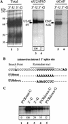
A cleavage/polyadenylation site stimulates U2AF 65 binding to an upstream 3′ splice site. (A) RNAs 211 nucleotides long containing the 3′ splice site of the adenovirus I major late intron and the wild-type or mutant adenovirus L3 cleavage and polyadenylation site (3′-U and 3′-G) were incubated in HeLa nuclear extract and cross-linked by UV irradiation. Cross-linked polypeptides were either fractionated directly by SDS-PAGE (lanes 1,2, total) or immunoprecipitated before fractionation with the MC3 monoclonal antibody directed against U2AF 65 (Gama-Carvalho et al. 1997) (lanes 3,4, αU2AF 65) or the 64-kD subunit of CstF (MacDonald et al. 1994) (lanes 5,6, αCstF). The results of six independent U2AF 65 immunoprecipitation experiments were averaged, and the result is presented in lanes 3 and 4. The value for the 3′-U substrate was set to 100. (B) Details of the sequence of the wild-type adenovirus I major late 3′ splice site and the two mutants used. (PYRmut) The pyrimidine tract has been almost completely mutated to a purine tract; (PYRdown) the pyrimidine tract has been changed to that of the rat preprotachykinin exon 4, 3′ splice site. (C) RNAs 211 nucleotides long containing the 3′ splice site and the wild-type or mutant adenovirus L3 cleavage/polyadenylation site were cross-linked by UV irradiation. Cross-linked polypeptides were immunoprecipitated with monoclonal antibodies directed against U2AF 65 and separated by SDS-PAGE. Three separate experiments were quantified, and the average values are given below. The 3′-U substrate was set to a value of 100.
Figure 3
Figure 3
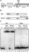
The carboxy-terminal 20 amino acids of PAP, when tethered to an adenovirus 1 major late intron, stimulate in vitro splicing. (A) The splicing substrates used contained the adenovirus 1 major late intron followed by a 237-nucleotide-long 3′ exon containing a wild-type IRE (TGCTTCAACAGTGCTTGGAC) or a mutant IRE with a single C deletion (TGCTTCAAAGTGCTTGGAC) (Hentze et al. 1987). (B) The recombinant fusion proteins produced in E. coli are the IRP fused at its amino terminus to a histidine tag and the IRP fused at its amino terminus to a histidine tag and at its carboxyl terminus to the 20 carboxy-terminal residues of PAP. (C) Gel mobility shift analysis of interaction between the two recombinant fusion proteins described in B and the wild-type IRE or mutant IRE (IREm) containing RNA. Lanes 1 and 8 show the input RNAs. Fifty nanograms (lanes 2,5,9,12), 100 ng (lanes 3,6,10,13), or 200 ng (lanes 4,7,11,14) of one of the two recombinant proteins was added to the 32P-labeled RNA substrates as indicated. (D) Effect of the addition of the recombinant proteins described in B on the splicing activity of the 32P-labeled wild-type IRE-containing splicing substrate (lanes 1–8) or the 32P-labeled mutant IRE-containing splicing substrate (lanes 9–12). (Lanes 1,9) Input RNAs; (lanes 2,10) splicing in nuclear extract (NE). Increasing amounts of the two recombinant proteins were added to the reaction: 50 ng (lanes 3,6); 100 ng (lanes 4,7); 200 ng (lanes 5,8,11,12). The positions of the precursor RNAs and the products of the reactions are indicated at left.
Figure 3
Figure 3
The carboxy-terminal 20 amino acids of PAP, when tethered to an adenovirus 1 major late intron, stimulate in vitro splicing. (A) The splicing substrates used contained the adenovirus 1 major late intron followed by a 237-nucleotide-long 3′ exon containing a wild-type IRE (TGCTTCAACAGTGCTTGGAC) or a mutant IRE with a single C deletion (TGCTTCAAAGTGCTTGGAC) (Hentze et al. 1987). (B) The recombinant fusion proteins produced in E. coli are the IRP fused at its amino terminus to a histidine tag and the IRP fused at its amino terminus to a histidine tag and at its carboxyl terminus to the 20 carboxy-terminal residues of PAP. (C) Gel mobility shift analysis of interaction between the two recombinant fusion proteins described in B and the wild-type IRE or mutant IRE (IREm) containing RNA. Lanes 1 and 8 show the input RNAs. Fifty nanograms (lanes 2,5,9,12), 100 ng (lanes 3,6,10,13), or 200 ng (lanes 4,7,11,14) of one of the two recombinant proteins was added to the 32P-labeled RNA substrates as indicated. (D) Effect of the addition of the recombinant proteins described in B on the splicing activity of the 32P-labeled wild-type IRE-containing splicing substrate (lanes 1–8) or the 32P-labeled mutant IRE-containing splicing substrate (lanes 9–12). (Lanes 1,9) Input RNAs; (lanes 2,10) splicing in nuclear extract (NE). Increasing amounts of the two recombinant proteins were added to the reaction: 50 ng (lanes 3,6); 100 ng (lanes 4,7); 200 ng (lanes 5,8,11,12). The positions of the precursor RNAs and the products of the reactions are indicated at left.
Figure 4
Figure 4
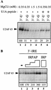
Both the AAUAAA sequence and the RNA-bound PAP carboxyl terminus stimulate U2AF 65 binding to the 3′ splice site. (A) RNAs 211 nucleotides long containing the 3′ splice site of the adenovirus I major late intron and the wild-type or mutant adenovirus L3 cleavage/polyadenylation site (3′-U and 3′-G) were incubated in HeLa nuclear extract at 0.35 mm MgCl2 (lanes 2,5,6) or 1.5 mm MgCl2 (lanes 3,4) or in the presence of 0.5 μg of a U1A peptide that uncouples splicing from 3′-end formation (Gunderson et al. 1997). The U1A peptide consists of the sequence ERDRKREKRKPKS, located between residues 103 and 115 of U1A. After UV irradiation, cross-linked polypeptides were immunoprecipitated with the MC3 monoclonal antibody directed against U2AF 65. (B) A 237-nucleotide -long RNA containing the adenovirus I major late intron 3′ splice site and the IRE element was incubated in HeLa nuclear extract supplemented with 50 (lane 2), 100 (lane 3), or 200 (lane 4) ng of the IRPAP recombinant protein or 200 ng of the IRP recombinant protein (lane 5). UV cross-linked proteins were immunoprecipitated with MC3 as in A. Three separate experiments were quantified, and the average values are given below. The 3′-U substrate was set to a value of 100 in A and the 3′ IRE substrate in B.
Figure 5
Figure 5
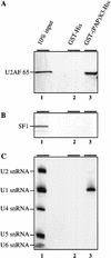
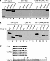
The 20 carboxy-terminal residues of PAP interact specifically with U2AF 65 in HeLa nuclear extracts. GST pull-down experiments with a recombinant GST–His fusion protein or a GST–His fusion protein containing three copies of the last carboxy-terminal 20 residues of PAP (GST–(PAP)X3–His; Gunderson et al. 1997). Five micrograms of the E. coli-produced GST–His fusion proteins were bound to glutathione–agarose beads and incubated with 200 μg of HeLa nuclear extract in splicing conditions. Bound proteins were eluted, loaded onto SDS-polyacrylamide gels, and vizualized by Western blot. (A) U2AF 65 binding; (B) SF1/mBBP binding; (C) bound RNAs were eluted, purified and loaded on a denaturing polyacrylamide gel before Northern blot analysis with U1, U2, U4, U5, and U6 snRNA probes. (Lane 1) Ten percent of the binding reaction inputs are loaded. Pull-down with the GST–His or the GST–(PAP)X3–His fusion proteins are in lanes 2 and 3. U2AF 65, SF1, and the various snRNAs are indicated at left.
Figure 6
Figure 6
The 20 carboxy-terminal residues of PAP interact specifically with residues 17–47 of U2AF 65. Five micrograms of E. coli-produced GST–His fusion proteins were bound to glutathione–agarose beads and used in binding assays with [35S] methionine labeled U1A, SF1-H, SF1–Bo, and U2AF 65 proteins (A) or various [35S]methionine-labeled U2AF 65 deletion mutants (B). (C) Comparison of the U1A and U1 70K motifs that bind to PAP (Gunderson et al. 1998) with the U2AF 65 sequence between residues 17 and 47.
Figure 7
Figure 7
Depletion of U1 snRNP does not block the AAUAAA-mediated stimulation of U2AF 65. (A) Antisense affinity depletion of U1 and U2 snRNP from HeLa nuclear extract (Barabino et al. 1990). RNAs recovered from mock-depleted extract (lane 1), U1 snRNP-depleted extract (lane 2), or U2 snRNP-depleted extract (lane 3) were analyzed by Northern hybridization with U1 and U2 snRNA-specific probes. U1 and U2 snRNAs are indicated. (B) UV cross-linking/immunoprecipitation, as in Fig. 2, was performed with the depleted extracts, as indicated at top. The 3′-U RNA substrate was used.

Similar articles

Cited by

References

    1. Abovich N, Rosbash M. Cross-intron bridging interactions in the yeast commitment complex are conserved in mammals. Cell. 1997;89:403–412. - PubMed
    1. Arias JA, Peterson SR, Dynan WS. Promoter-dependent phosphorylation of RNA polymerase II by a template-bound kinase. Association with transcriptional initiation. J Biol Chem. 1991;266:8055–8061. - PubMed
    1. Arning S, Grüter P, Bilbe G, Krämer A. Mammalian splicing factor SF1 is encoded by variant cDNAs and binds to RNA. RNA. 1996;2:794–810. - PMC - PubMed
    1. Barabino SML, Blencowe BJ, Ryder U, Sproat BS, Lamond AI. Targeted snRNP depletion reveals an additional role for mammalian U1 snRNP in spliceosome assembly. Cell. 1990;63:293–302. - PubMed
    1. Bauren G, Belikov S, Wieslander L. Transcriptional termination in the Balbiani ring 1 gene is closely coupled to 3′-end formation and excision of the 3′-terminal intron. Genes & Dev. 1998;12:2759–2769. - PMC - PubMed

Publication types

MeSH terms