. 2000 Feb 21;191(4):669-82.
doi: 10.1084/jem.191.4.669.

Fcgamma receptor-mediated phagocytosis in macrophages lacking the Src family tyrosine kinases Hck, Fgr, and Lyn

Affiliations

Fcgamma receptor-mediated phagocytosis in macrophages lacking the Src family tyrosine kinases Hck, Fgr, and Lyn

C J Fitzer-Attas et al. J Exp Med. .

Abstract

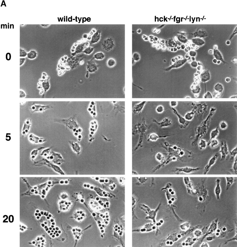
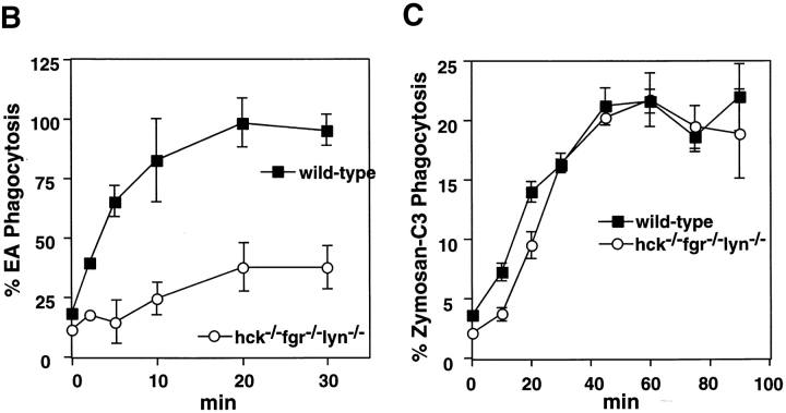
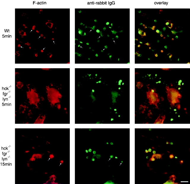
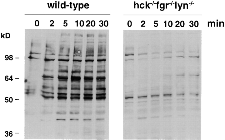
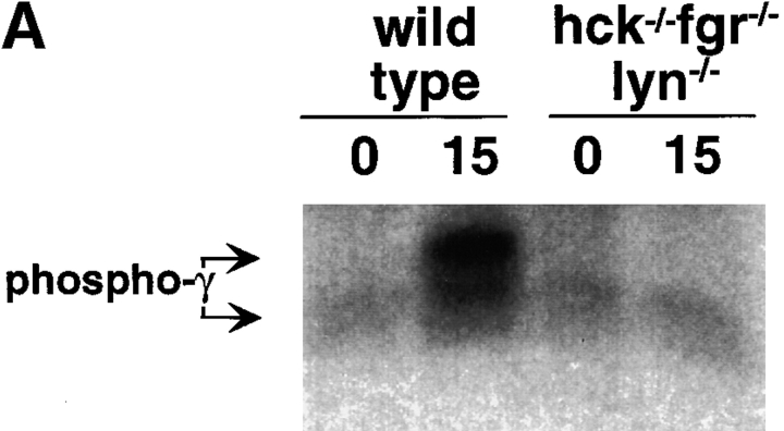
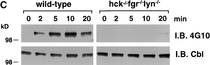
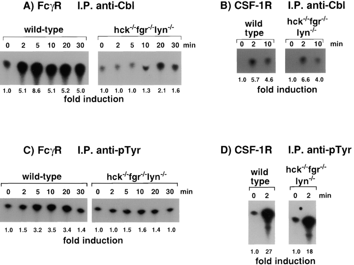
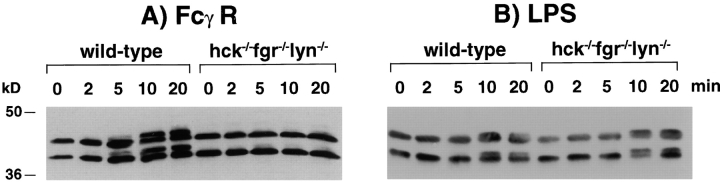
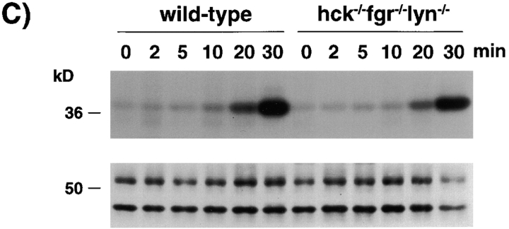
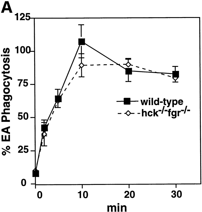
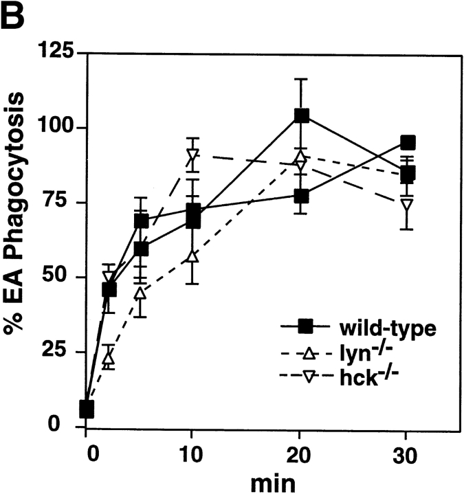
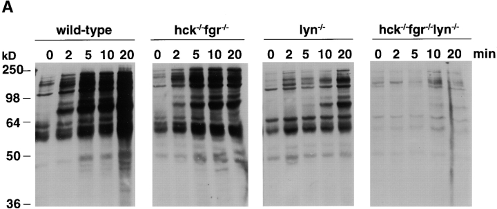
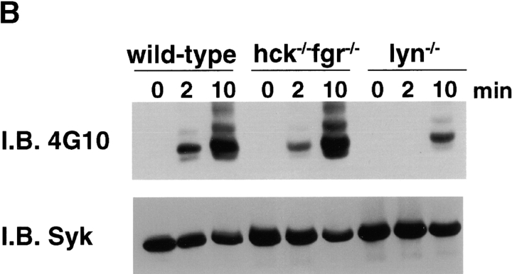
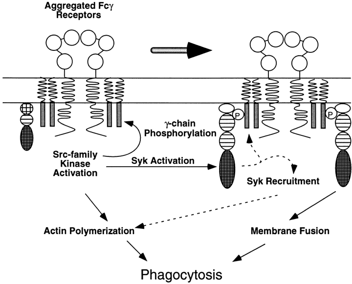
Macrophage Fcgamma receptors (FcgammaRs) mediate the uptake and destruction of antibody-coated viruses, bacteria, and parasites. We examined FcgammaR signaling and phagocytic function in bone marrow-derived macrophages from mutant mice lacking the major Src family kinases expressed in these cells, Hck, Fgr, and Lyn. Many FcgammaR-induced functional responses and signaling events were diminished or delayed in these macrophages, including immunoglobulin (Ig)G-coated erythrocyte phagocytosis, respiratory burst, actin cup formation, and activation of Syk, phosphatidylinositol 3-kinase, and extracellular signal-regulated kinases 1 and 2. Significant reduction of IgG-dependent phagocytosis was not seen in hck(-)(/)-fgr(-)(/)- or lyn(-)(/)- cells, although the single mutant lyn(-)(/)- macrophages did manifest signaling defects. Thus, Src family kinases clearly have roles in two events leading to FcgammaR-mediated phagocytosis, one involving initiation of actin polymerization and the second involving activation of Syk and subsequent internalization. Since FcgammaR-mediated phagocytosis did occur at modest levels in a delayed fashion in triple mutant macrophages, these Src family kinases are not absolutely required for uptake of IgG-opsonized particles.


Figures

Figure 1
Figure 1
Reduced phagocytosis in hck /fgr /lyn /− macrophages. (A) EAs were bound to bone marrow–derived macrophages on ice (0 min) before initiation of phagocytosis at 37°C for the indicated time periods. Uningested EAs were removed by hypotonic lysis, and cells were fixed for photomicroscopy. Bar, 10 μm. (B) Phagocytosis assays were performed as in A with 51Cr-EAs. After hypotonic lysis, the amount of 51Cr-EAs ingested was determined by quantitation of radioactivity in total cell lysates. Data shown are representative of 26 (wild-type) or 22 (mutant) independent experiments on cells pooled from 2–5 mice. Error bars represent the SEM of quadruplicate samples. (C) Phagocytosis of C3-opsonized FITC-labeled zymosan particles. C3-zymosan phagocytosis was initiated by binding of particles to macrophages at 4°C, unbound particles were removed by washing, and 37°C medium was added. Phagocytosis was stopped by addition of 2% paraformaldehyde, and the fluorescence from nonengulfed particles was quenched by addition of trypan blue. Data shown are representative of six independent experiments on cells pooled from two to five mice. Error bars represent the SEM of six determinations per time point. (D) Equivalent expression of FcγRs on wild-type and triple mutant cells as demonstrated by flow cytometry after staining with mAb 2.4G2 (rat anti-FcγRII and III, solid line) or control rat IgG2b (broken line).
Figure 1
Figure 1
Reduced phagocytosis in hck /fgr /lyn /− macrophages. (A) EAs were bound to bone marrow–derived macrophages on ice (0 min) before initiation of phagocytosis at 37°C for the indicated time periods. Uningested EAs were removed by hypotonic lysis, and cells were fixed for photomicroscopy. Bar, 10 μm. (B) Phagocytosis assays were performed as in A with 51Cr-EAs. After hypotonic lysis, the amount of 51Cr-EAs ingested was determined by quantitation of radioactivity in total cell lysates. Data shown are representative of 26 (wild-type) or 22 (mutant) independent experiments on cells pooled from 2–5 mice. Error bars represent the SEM of quadruplicate samples. (C) Phagocytosis of C3-opsonized FITC-labeled zymosan particles. C3-zymosan phagocytosis was initiated by binding of particles to macrophages at 4°C, unbound particles were removed by washing, and 37°C medium was added. Phagocytosis was stopped by addition of 2% paraformaldehyde, and the fluorescence from nonengulfed particles was quenched by addition of trypan blue. Data shown are representative of six independent experiments on cells pooled from two to five mice. Error bars represent the SEM of six determinations per time point. (D) Equivalent expression of FcγRs on wild-type and triple mutant cells as demonstrated by flow cytometry after staining with mAb 2.4G2 (rat anti-FcγRII and III, solid line) or control rat IgG2b (broken line).
Figure 2
Figure 2
Normal immune complex endocytosis but impaired FcγR-mediated activation of respiratory burst in hck /fgr /lyn /− macrophages. (A) Suspension macrophages (resting or primed by 12-h incubation in LPS/IFN-γ) were incubated with 120 μg/ml of Fc OxyBURST® immune complexes, and the development of fluorescence resulting from oxidative burst was monitored by flow cytometry. (B) Endocytosis of heat-aggregated rabbit IgG was determined by exposing cells to PE-conjugated immune complexes for varying periods of time, then removing the surface-bound complexes by acid treatment. The amount of remaining fluorescence representing internalized immune complexes was determined by flow cytometry. The data shown are for a 20-min incubation of cells with immune complexes at 37°C (solid lines) or 4°C (broken lines). Equivalent uptake of complexes was seen also in wild-type and mutant cells at earlier and later time points (not shown).
Figure 3
Figure 3
Reduced actin cup formation in hck /fgr /lyn /− macrophages. Phagocytosis of EAs in wild-type (top panels, Wt) and triple mutant (middle and bottom panels) macrophages was initiated as described in the legend to Fig. 1. After 5 min (top and middle panels) or 15 min (bottom panels), unbound EAs were removed by washing and macrophages were fixed in 3% paraformaldehyde. Cells were stained with rhodamine-conjugated phalloidin to detect F-actin at sites of phagocytic cup formation (left), and with FITC-conjugated anti–rabbit IgG to detect the bound SRBCs (middle). An overlay of the F-actin and anti–rabbit IgG staining demonstrates colocalization of bound EAs with phagocytic cups (right). Cells were examined in all focal planes for the presence of actin cups. The middle panels show a focal plane that includes the nucleus of the triple mutant cells to localize cellular structures, since these cells had very low F-actin formation at all focal planes. Arrows indicate representative phagocytic cups colocalized with SRBCs. Bar, 10 μm.
Figure 4
Figure 4
Impaired FcγR-induced tyrosine phosphorylation of cellular proteins in hck /fgr /lyn /− macrophages. Cells were stimulated by incubation with anti-FcγR mAb 2.4G2 followed by anti–rat IgG cross-linking for the indicated times. Cells were lysed, and equivalent amounts of cellular protein were subjected to SDS-PAGE and immunoblotting with antiphosphotyrosine antibodies.
Figure 5
Figure 5
Impairment of proximal FcγR-induced phosphorylation events in hck /fgr /lyn /− macrophages. 32PO4-labeled (A) or unlabeled (B and C) macrophages were stimulated by incubation with anti-FcγR mAb 2.4G2 followed by anti–rat IgG cross-linking for the indicated times. Cells were lysed, and equivalent amounts of cellular protein were subjected to immunoprecipitation with anti-γ chain (A), anti-Syk (B), or anti-Cbl (C) antibodies. Each immunoprecipitation experiment was performed on lysates from an independent set of FcγR-stimulated macrophages. After SDS-PAGE, phosphoproteins were detected either by autoradiography (A) or by immunoblotting (I.B.) with 4G10 antiphosphotyrosine antibodies (B and C). Membranes from B and C were stripped and reprobed with anti-Syk and anti-Cbl antibodies, respectively.
Figure 5
Figure 5
Impairment of proximal FcγR-induced phosphorylation events in hck /fgr /lyn /− macrophages. 32PO4-labeled (A) or unlabeled (B and C) macrophages were stimulated by incubation with anti-FcγR mAb 2.4G2 followed by anti–rat IgG cross-linking for the indicated times. Cells were lysed, and equivalent amounts of cellular protein were subjected to immunoprecipitation with anti-γ chain (A), anti-Syk (B), or anti-Cbl (C) antibodies. Each immunoprecipitation experiment was performed on lysates from an independent set of FcγR-stimulated macrophages. After SDS-PAGE, phosphoproteins were detected either by autoradiography (A) or by immunoblotting (I.B.) with 4G10 antiphosphotyrosine antibodies (B and C). Membranes from B and C were stripped and reprobed with anti-Syk and anti-Cbl antibodies, respectively.
Figure 6
Figure 6
Impaired FcγR-, but not CSF-1R–induced signaling through the PI 3-kinase pathway in hck /fgr /lyn /− macrophages. Cells were stimulated through the FcγR as in the legend to Fig. 3 (A and C) or by incubation with LCM (B and D), and cell lysates were immunoprecipitated (I.P.) with anti-Cbl (A and B) or antiphosphotyrosine (C and D, anti-pTyr) antibodies. Washed immunoprecipitates were then subjected to a lipid kinase assay using phosphatidylinositol as a substrate. The spots shown represent γ-32P–labeled phosphatidylinositol 3-phosphate after TLC chromatography and autoradiography. Fold induction, shown below each lane, was quantitated using a PhosphorImager® and ImageQuant® software (Molecular Probes).
Figure 7
Figure 7
MAP kinase and JNK activation in hck /fgr /lyn /− macrophages. Cells were stimulated by incubation with anti-FcγR mAb 2.4G2 followed by anti–rat IgG cross-linking (A and C) or by incubation with 1 μg/ml LPS (B) for the indicated times. After cell lysis, equivalent amounts of protein were subjected to SDS-PAGE and immunoblotting with anti-Erk1/2 antibodies (A and B). For C, equivalent amounts of cellular lysates were subjected to immunoprecipitation with polyclonal anti-JNK antibodies followed by an in vitro kinase assay using a c-Jun GST fusion protein as a substrate. After SDS-PAGE and transfer to nitrocellulose, γ-32P–labeled substrate was detected by autoradiography (top). The membrane was reprobed with an anti-JNK1 mAb to verify similar amounts of immunoprecipitated protein (bottom).
Figure 7
Figure 7
MAP kinase and JNK activation in hck /fgr /lyn /− macrophages. Cells were stimulated by incubation with anti-FcγR mAb 2.4G2 followed by anti–rat IgG cross-linking (A and C) or by incubation with 1 μg/ml LPS (B) for the indicated times. After cell lysis, equivalent amounts of protein were subjected to SDS-PAGE and immunoblotting with anti-Erk1/2 antibodies (A and B). For C, equivalent amounts of cellular lysates were subjected to immunoprecipitation with polyclonal anti-JNK antibodies followed by an in vitro kinase assay using a c-Jun GST fusion protein as a substrate. After SDS-PAGE and transfer to nitrocellulose, γ-32P–labeled substrate was detected by autoradiography (top). The membrane was reprobed with an anti-JNK1 mAb to verify similar amounts of immunoprecipitated protein (bottom).
Figure 8
Figure 8
Normal phagocytosis in single and double mutant macrophages. Phagocytosis assays using 51Cr-EAs were performed as in the legend to Fig. 1 with bone marrow–derived macrophages from hck /fgr /− double mutant (A) or hck /− and lyn /− single mutant mice (B). Data shown are representative of four independent experiments on cells pooled from two to four mice per experiment. Two separate wild-type macrophage cultures are shown in B to illustrate normal variation between genetically identical samples.
Figure 8
Figure 8
Normal phagocytosis in single and double mutant macrophages. Phagocytosis assays using 51Cr-EAs were performed as in the legend to Fig. 1 with bone marrow–derived macrophages from hck /fgr /− double mutant (A) or hck /− and lyn /− single mutant mice (B). Data shown are representative of four independent experiments on cells pooled from two to four mice per experiment. Two separate wild-type macrophage cultures are shown in B to illustrate normal variation between genetically identical samples.
Figure 9
Figure 9
Dominant role of Lyn in FcγR-mediated signaling events. Cells were stimulated by incubation with anti-FcγR mAb 2.4G2 followed by anti–rat IgG cross-linking for the indicated times. After cell lysis, equivalent amounts of cellular protein were subjected to SDS-PAGE and immunoblotting (I.B.) with antiphosphotyrosine antibodies (A) or to immunoprecipitation with anti-Syk antibodies followed by antiphosphotyrosine immunoblotting (B). The membrane in B was stripped and reprobed with anti-Syk antibodies to verify equivalent amounts of immunoprecipitated protein.
Figure 9
Figure 9
Dominant role of Lyn in FcγR-mediated signaling events. Cells were stimulated by incubation with anti-FcγR mAb 2.4G2 followed by anti–rat IgG cross-linking for the indicated times. After cell lysis, equivalent amounts of cellular protein were subjected to SDS-PAGE and immunoblotting (I.B.) with antiphosphotyrosine antibodies (A) or to immunoprecipitation with anti-Syk antibodies followed by antiphosphotyrosine immunoblotting (B). The membrane in B was stripped and reprobed with anti-Syk antibodies to verify equivalent amounts of immunoprecipitated protein.
Figure 10
Figure 10
Model for the role of Src family kinases in FcγR-mediated phagocytosis. Src family kinases are responsible for phosphorylation of the FcR γ chain, recruitment of Syk to the receptor, and activation of Syk, which then mediates signaling leading to internalization of the bound particle. The results presented here, however, demonstrate an additional function of Src family kinases: to promote actin polymerization and phagocytic cup formation, as evidenced by the significant delay in these events in hck /fgr /lyn /− macrophages and their normal kinetics in Syk-deficient macrophages. Other signaling events possibly involving Syk (indicated by the broken arrows) include ITAM phosphorylation, stimulation of slow actin cup formation, and recruitment and/or autoactivation. These additional signaling reactions may account for residual phagocytosis in the absence of the Src family kinases.

Similar articles

Cited by

References

    1. Daeron M. Fc receptor biology. Annu. Rev. Immunol. 1997;15:203–234. - PubMed
    1. Aderem A., Underhill D.M. Mechanisms of phagocytosis in macrophages. Annu. Rev. Immunol. 1999;17:593–623. - PubMed
    1. Clynes R., Takechi Y., Moroi Y., Houghton A., Ravetch J.V. Fc receptors are required in passive and active immunity to melanoma. Proc. Natl. Acad. Sci. USA. 1998;95:652–656. - PMC - PubMed
    1. Deo Y.M., Graziano R.F., Repp R., van de Winkel J.G. Clinical significance of IgG Fc receptors and FcγR-directed immunotherapies. Immunol. Today. 1997;18:127–135. - PubMed
    1. Strzelecka A., Kwiatkowska K., Sobota A. Tyrosine phosphorylation and Fcγ receptor-mediated phagocytosis. FEBS Lett. 1997;400:11–14. - PubMed

Publication types

MeSH terms