. 2000 Feb 15;19(4):662-71.
doi: 10.1093/emboj/19.4.662.

Regulation of E2F1 activity by acetylation

Affiliations

Regulation of E2F1 activity by acetylation

M A Martínez-Balbás et al. EMBO J. .

Abstract

During the G(1) phase of the cell cycle, an E2F-RB complex represses transcription, via the recruitment of histone deacetylase activity. Phosphorylation of RB at the G(1)/S boundary generates a pool of 'free' E2F, which then stimulates transcription of S-phase genes. Given that E2F1 activity is stimulated by p300/CBP acetylase and repressed by an RB-associated deacetylase, we asked if E2F1 was subject to modification by acetylation. We show that the p300/CBP-associated factor P/CAF, and to a lesser extent p300/CBP itself, can acetylate E2F1 in vitro and that intracellular E2F1 is acetylated. The acetylation sites lie adjacent to the E2F1 DNA-binding domain and involve lysine residues highly conserved in E2F1, 2 and 3. Acetylation by P/CAF has three functional consequences on E2F1 activity: increased DNA-binding ability, activation potential and protein half-life. These results suggest that acetylation stimulates the functions of the non-RB bound 'free' form of E2F1. Consistent with this, we find that the RB-associated histone deacetylase can deacetylate E2F1. These results identify acetylation as a novel regulatory modification that stimulates E2F1's activation functions.


Figures
None
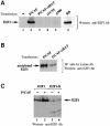
Fig. 1. Acetylation of E2F1 by P/CAF, GCN5, CBP and p300 acetylases. (A) In vitro acetylation of E2F1. Purified GST–E2F1 (amino acids 89–437) was incubated with [14C]acetyl-CoA and recombinant GST–P/CAF (amino acids 352–832, lanes 1 and 5), His-GCN5 (amino acids 1–427, lanes 2 and 6), GST–CBP (amino acids 1098–1877, lanes 3 and 7) and His-p300 (amino acids 1071–1715, lanes 4 and 8). Reaction products were separated by SDS–PAGE and gels were autoradiographed. Lanes 5–8 are a longer exposure (8 days) of lanes 1–4 (6 h) to visualize acetylation of E2F1 by GCN5, CBP and p300. (B) Control acetylation reactions with GST–E2F1, GST and BSA were performed as described in (A) and show that E2F1 (lane 2) in the presence of [14C]acetyl-CoA, but in the absence of P/CAF (lane 1) is not acetylated, and GST (lane 3) or BSA (lane 4), are not targets for P/CAF. (C) In vivo acetylation of E2F1. Extracts from U2OS cells were prepared and E2F1 was immunoprecipitated using an anti-E2F1 (C20) antibody or an anti-HA antibody as a control. Immunoprecipitates were analysed by SDS–PAGE and Western blot analysis using an antibody raised against acetylated histone H4.
None
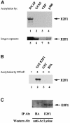
Fig. 2. E2F1 interacts with P/CAF. (A) U2OS cells were transfected with 8 μg of pCX-P/CAF (Flag-tagged), and afterwards whole-cell extracts were prepared and incubated with anti-Flag antibody and, as controls, either with anti-HA antibody or without any antibody. By incubation with protein A–Sepharose, immunoprecipitates were collected. The precipitates were then analysed by SDS–PAGE and Western blotting with the anti-E2F1 (C20) antibody for the presence of co-immunoprecipitated E2F1. (B) U2OS cells were transfected and extracted, as described in (A). In this experiment, immunoprecipitation of E2F1 was performed with an anti-E2F1 antibody (C20). The control reactions were the same as in (A). An anti-Flag Western blot was used for detection of co-immunoprecipitated P/CAF. (C) Schematic representation of P/CAF fragments used to identify the E2F interaction region in P/CAF. E2F1 binding to different P/CAF fragments is indicated in the scheme. (D) The P/CAF fragments were expressed as GST fusion proteins, purified and used in GST pull-down assays with in vitro translated, 35S-labelled E2F1 protein. Equivalent P/CAF protein amounts of the different mutants were used. Interaction between E2F1 and P/CAF was analysed by SDS–PAGE of the pull-down reactions and autoradiography (lanes 3–9). The input (lane 1) contains 20% of the E2F1 IVT. GST protein (lane 2) was used as a control.
None
Fig. 3. The E2F1 acetylation sites are located directly adjacent to the DNA-binding domain. (A) Identification and evolutionary conservation of the acetylation sites in E2F1. Schematic representation of E2F1, its functional domains, the three lysine clusters that were mutated to arginines, and polypeptide sequence alignment of E2F1 (amino acids 114–185) with E2F2, E2F3, E2F4, E2F5 and EMA. The arrows indicate the mutated lysine residues, the conservation of which is highlighted by boxes. The corresponding secondary structure of the E2F4 DNA-binding domain (Zheng et al., 1999) with its four α–helices and two β–sheets is shown underneath the sequence alignment. The acetylated lysines are indicated by asterisks. (B) The E2F1 fragments indicated (lanes 1–4) were expressed as GST fusion proteins, purified and 2 μg of each were assayed for acetylation by GST–P/CAF as described in Figure 1A. (C) Wild-type (lane 1) and mutated E2F1 proteins (lanes 2–4), which were mutated at the indicated lysines to arginines, were expressed, purified and 2 μg of each were assayed for acetylation by P/CAF. (D) To test in vivo acetylation of the E2F1–R mutant [E2F1–K(117,120,125)R, (C) lane 4], P/CAF (8 μg of pCX-P/CAF) was co-transfected with either E2F1 (3 μg of pcDNA3-E2F1) or E2F1–R (6 μg of pcDNA3-E2F1–R) into 293T cells. Acetylation of E2F1 and the mutant was investigated by immunoprecipitation of equivalent amounts of E2F1 and E2F1–R protein with an anti-acetylated lysine antibody covalently coupled to protein A–Sepharose and subsequent E2F1 Western blot analysis (with KH95 antibody) of the precipitates.
None
Fig. 4. DNA-binding activity of E2F1 is augmented by acetylation. (A) Acetylation of E2F1-DBD increases its DNA binding. The His-E2F1-DBD (amino acids 92–195) was bacterially expressed and purified. DNA binding of unmodified [in the presence of GST–P/CAF (amino acids 352–632), but the absence of AcCoA (lanes 5–7)] versus in vitro acetylated E2F1-DBD [in the presence of GST–P/CAF and AcCoA (lanes 2–4)] was tested by EMSA by incubation of increasing amounts of these proteins with 32P-labelled E2F-binding site (50 fmol per reaction). Reaction products were resolved by electrophoresis and visualized by autoradiography. (B) E2F1–DP1 binds DNA more efficiently following acetylation of E2F1. His-E2F1 full-length protein was bacterially expressed, purified and in vitro acetylated [in the presence of P/CAF and AcCoA (lanes 7 and 8)] or not acetylated [in the presence of either P/CAF, but not AcCoA (lanes 3 and 4) or P/CAF-ΔHAT and AcCoA (lanes 5 and 6)]. DNA binding was investigated by incubation of these proteins with GST–DP1 and 32P-labelled E2F-binding site (50 fmol per reaction). Complexes were separated by electrophoresis and autoradiographed. The DNA-binding ability of either E2F1 or DP1 alone is shown in lanes 1 and 2, respectively. (C) Increased DNA binding of acetylated E2F1 is due to acetylation. The E2F1–R mutant, which contains lysines 117, 120 and 125 mutated to arginines, and which cannot be acetylated (Figure 3C and D), was expressed in bacteria as a GST fusion protein, purified and tested for its DNA-binding ability after incubation with either P/CAF (lanes 4–6) or P/CAF-ΔHAT (lanes 1–3) as described in (A) and (B).
None
Fig. 5. Acetylation of E2F1 stimulates its transcriptional activity. (A) U2OS cells were transfected with 0.5 μg of the CAT reporter gene plasmid and 1 μg of the indicated expression plasmid for E2F1 and DP1, 5 μg of pCX-P/CAF and P/CAF-ΔHAT, respectively, or equivalent empty vector. Whole-cell extracts were used in CAT assays and the results were quantified on a PhosphoImager. The basal promoter activity of the CAT reporter (TK promoter with 3× E2F sites) in the presence of empty expression vector was normalized to 1.0, and the activities of the remaining transfection reactions were expressed relative to this, as fold activation of the basal promoter. The graph shows the average of three independent experiments. (B) The effects of P/CAF on the transactivation potential of E2F1–R is shown, as assayed under the same conditions as described in (A).
None
Fig. 6. Acetylation by P/CAF increases E2F1 protein level and prolonges its half-life. (A) 293T cells were either not transfected (lane 1) or transfected with 5 μg of the indicated eukaryotic expression plasmids for P/CAF (lane 2), P/CAF-ΔHAT (lane 3), GCN5 (lane 4) and p300 (lane 5), and 3 μg of RB (lane 6). At 24 h after the medium change, the cells were lysed in SDS-containing sample buffer and analysed by SDS–PAGE and anti-E2F1 Western blot (C20) for endogenous E2F1 expression levels. (B) To investigate if the increase in E2F1 protein level is accompanied by in vivo acetylation of endogenous E2F1, P/CAF- and P/CAF ΔHAT-transfected cells [as in (A)] were analysed by immunoprecipitation with anti-acetylated lysine antibody (covalently coupled) and Western blot of the precipitates with anti-E2F (KH95) antibody. Equivalent amounts of E2F1 protein (of both transfection) were used for the immunoprecipitations. (C) 293T cells were transfected with E2F1 (lanes 1 and 2) or E2F1–R (lanes 3 and 4) in the presence (lanes 2 and 4) or absence (lanes 1 and 3) of P/CAF overexpression. SDS–PAGE and Western blot analysis with the anti-E2F1 (C20) antibody revealed the effect of acetylation by P/CAF on the exogenous E2F1 protein level. (D) To determine the protein half-life of E2F1 in the presence or absence of P/CAF, 293T cells were transfected with 2 μg of pcDNA3-E2F1 in the absence or presence of 8 μg of pCX-P/CAF. At 24 h after the medium change, cells were pulse-labelled for 2 h with a [35S]methionine–cysteine mix. Chase was performed in medium supplemented with 10-fold excess of cold methionine and cysteine for the time periods indicated. Cells were lysed and immuno– precipitations were performed with the anti-E2F (C20) antibody. Immunoprecipitates were separated by SDS–PAGE, blotted and autoradio– graphed. The intensity of the 35S-labelled proteins was measured densitometrically and calculated in comparison with the amount of E2F protein present at time point zero, which was set at 100%. (E) The effects of P/CAF on the half-life of E2F1–R were assayed under the same conditions as described in (D). Therefore, 6 μg of pcDNA3-E2F1–R were transfected with or without P/CAF into 293T cells.
None
Fig. 7. RB-associated HDAC deacetylates E2F1. (A) The RB-associated deacetylase activity was purified from HeLa nuclear extract using GST–RB fusion protein bound to glutathione–Sepharose beads. As a control, only glutathione–Sepharose beads and GST protein were used. The recovered endogenous deacetylase complex was then added to 1 μg of recombinant acetylated His-E2F1 full-length fusion protein [previously in vitro acetylated by GST–P/CAF (amino acids 352–632) in the presence of [3H]AcCoA]. The deacetylase assay was performed for 1 h at 37°C as described in Materials and methods. The histone deacetylase activity is given as d.p.m. of [3H]acetate released from the full-length acetylated E2F1. (B) RB facilitates the deacetylation of E2F1. Active HDAC1 deacetylase was expressed from baculovirus, purified to homogeneity and assayed for its ability to deacetylate 1 μg of in vitro acetylated His-E2F1 full-length protein (lane 2) alone or in the presence of a saturating amount of either GST–RB fusion protein (lane 3) or a GST–RB pocket mutant (GST–RBΔ22), which cannot bind E2F (lane 4). (C) RB tethers the deacetylase to E2F1. A 1 μg aliquot of in vitro acetylated His-E2F1-DBD was subjected to deacetylation by purified recombinant, baculovirus-expressed HDAC1 in the absence of RB (lane 2) or in the presence of a saturating amount of either GST–RB recombinant protein (lane 3) or GST–RB pocket mutant (GST–RBΔ22). The histone deacetylase activity is given as d.p.m. of [3H]acetate released from the acetylated His–E2F1–DBD.

Similar articles

Cited by

References

    1. Allen K.E., de la Luna, S., Kerkhoven, R.M., Bernards, R. and La Thangue, N.B. (1997) Distinct mechanisms of nuclear accumulation regulate the functional consequence of E2F transcription factors. J. Cell Sci., 110, 2819–2831. - PubMed
    1. Bagchi S., Weinmann, R. and Raychaudhuri, P. (1991) The retinoblastoma protein copurifies with E2F-I, an E1A-regulated inhibitor of the transcription factor E2F. Cell, 65, 1063–1072. - PubMed
    1. Bannister A.J. and Kouzarides, T. (1996) The CBP co-activator is a histone acetyltransferase. Nature, 384, 641–643. - PubMed
    1. Boyes J., Byfield, P., Nakatani, Y. and Ogryzko, V. (1998) Regulation of activity of the transcription factor GATA-1 by acetylation. Nature, 396, 594–598. - PubMed
    1. Brehm A., Miska, E.A., McCance, D.J., Reid, J.L., Bannister, A.J. and Kouzarides, T. (1998) Retinoblastoma protein recruits deacetylase to repress transcription. Nature, 391, 597–601. - PubMed

Publication types

MeSH terms