. 2000 Feb 1;19(3):434-44.
doi: 10.1093/emboj/19.3.434.

Serum-stimulated, rapamycin-sensitive phosphorylation sites in the eukaryotic translation initiation factor 4GI

Affiliations

Serum-stimulated, rapamycin-sensitive phosphorylation sites in the eukaryotic translation initiation factor 4GI

B Raught et al. EMBO J. .

Abstract

The eukaryotic translation initiation factor 4G (eIF4G) proteins play a critical role in the recruitment of the translational machinery to mRNA. The eIF4Gs are phosphoproteins. However, the location of the phosphorylation sites, how phosphorylation of these proteins is modulated and the identity of the intracellular signaling pathways regulating eIF4G phosphorylation have not been established. In this report, two-dimensional phosphopeptide mapping demonstrates that the phosphorylation state of specific eIF4GI residues is altered by serum and mitogens. Phosphopeptides resolved by this method were mapped to the C-terminal one-third of the protein. Mass spectrometry and mutational analyses identified the serum-stimulated phosphorylation sites in this region as serines 1108, 1148 and 1192. Phosphoinositide-3-kinase (PI3K) inhibitors and rapamycin, an inhibitor of the kinase FRAP/mTOR (FKBP12-rapamycin-associated protein/mammalian target of rapamycin), prevent the serum-induced phosphorylation of these residues. Finally, the phosphorylation state of N-terminally truncated eIF4GI proteins acquires resistance to kinase inhibitor treatment. These data suggest that the kinases phosphorylating serines 1108, 1148 and 1192 are not directly downstream of PI3K and FRAP/mTOR, but that the accessibility of the C-terminus to kinases is modulated by this pathway(s).


Figures

None
Fig. 1. Phosphorylation of specific sites in eIF4GI is modulated by serum. 32P–labeled eIF4GI immunoprecipitated from 293 cells starved of serum for 36 h (–Serum or –S), or starved of serum for 36 h then stimulated with serum for 30 min (+Serum or +S), was subjected to (A) SDS–8% PAGE, then (B and C) to two-dimensional tryptic peptide mapping. The directions of chromatography (vertical) and electrophoresis (horizontal) as well as the loading origin (arrow) are indicated. Major phosphopeptides are numbered.
None
Fig. 2. Localization of eIF4GI phosphorylation sites. (A) Tryptic map of endogenous eIF4GI. (BE) HA-tagged eIF4GI fusion protein fragments were expressed in 293T cells. Cells were metabolically labeled with 32P, and HA-tagged proteins immunoprecipitated, gel purified, and subjected to two-dimensional tryptic mapping. (F) Protein fragments tested in this study. The various protein binding regions of eIF4GI are indicated.
None
Fig. 3. The phospho-region of eIF4GI is poorly conserved within the human eIF4G family. (A) An alignment through the newly defined eIF4GI phospho-region of the members of the human eIF4G family. Locations of defined protein binding sites in eIF4G proteins are indicated, as well as percentage identity to the corresponding region in eIF4GI. Below the diagram is an alignment of the amino acid sequences of eIF4GI, eIF4GII and p97 in this region. Identical residues are boxed in black, similar residues are boxed in gray. (B) Fragments encompassing the phosphorylated region of eIF4GI (aa 1035–1206) and the corresponding regions of eIF4GII (aa 1057–1225) and p97 (aa 395–547) were expressed in 293T cells (5, 10 and 20 μg of DNA transfected) as GST fusion proteins. Cells were metabolically labeled with 32P. Proteins isolated with glutathione-coupled Sepharose beads were washed, gel-purifed, and transferred to nitrocellulose. Upper panel, Western blot using anti-GST antiserum. Lower panel, direct autoradiogram of the same nitrocellulose membrane.
None
Fig. 4. Identification of serum-stimulated phosphorylation sites by mass spectrometry. (A) Tandem mass spectrum resulting from the analysis of excised phosphopeptide 1 from the map in Figure 1C. This spectrum contained sequence information for a single phosphopeptide. Two separate ion series were recorded simultaneously; b- and y–ion series represent sequencing inward from the N- and C–termini, respectively. The computer program Sequest (Eng et al., 1994) was utilized to match this tandem mass spectrum to the sequence shown, with the serine residue being phosphorylated. (B) Tandem mass spectrum resulting from the analysis of excised phosphopeptide 3. The position of the phosphorylated serine residue is indicated.
None
Fig. 5. Identification of serum-inducible phosphorylation sites by mutational analysis. Mutations of serine residues identified by mass spectrometry as phosphorylation sites were introduced into the eIF4GI phospho-region (aa 1035–1206)–GST fusion proteins. Proteins were expressed in 293T cells, 32P–labeled, isolated and mapped. Tryptic maps of the GST–eIF4GI (aa 1035–1206) wild-type protein (A), and point mutants Ser1108Ala (B), Ser1148Ala (C) and Ser1192Ala (D). A novel phosphopeptide that does not co-migrate with any endogenous eIF4GI peptide is indicated with an asterisk. (E) Complete mutational analysis summary. An alignment of the eIF4GI phospho-region with the corresponding regions of eIF4GII and p97. Identical residues are boxed in black, similar residues are boxed in gray. eIF4GI residues mutated to alanine are indicated by an ‘x’ or arrow. Locations of the serum-stimulated, phosphorylated residues, as well as the phosphopeptides identified in this analysis, are also indicated.
None
Fig. 6. Serum-induced phosphorylation of eIF4GI is sensitive to inhibitors of PI3K and FRAP/mTOR. Two-dimensional tryptic peptide mapping of endogenous eIF4GI isolated from 293 cells (A) starved of serum, (B) then treated with serum for 30 min, (C) pre-treated with LY294002 for 20 min, then treated with serum for 30 min, or (D) pre-treated with rapamycin for 20 min, then treated with serum for 30 min. The map in (A) is the same as that shown in Figure 1B.
None
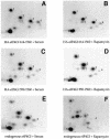
Fig. 7. N–terminal sequences confer kinase inhibitor sensitivity to the C–terminal phospho-region. HA-tagged eIF4GI fragments were expressed in 293T cells. Cells were metabolically labeled and incubated in 10% serum (A, C and E) or 10% serum + 25 ng/ml rapamycin (B, D and F) for 1 h. Following lysis, immunoprecipitation was conducted sequentially with anti-HA and anti-eIF4GI antisera. Isolated proteins were gel-purified and mapped. The HA–eIF4GI 550–1560 fragment interacts with eIF4E, while HA–eIF4GI 614–1560 does not.

Similar articles

Cited by

References

    1. Abraham R.T. (1998) Mammalian target of rapamycin—immunosuppressive drugs uncover a novel pathway of cytokine receptor signaling. Curr. Opin. Immunol., 10, 330–336. - PubMed
    1. Brown E.J. and Schreiber, S.L. (1996) A signaling pathway to translational control. Cell, 86, 517–520. - PubMed
    1. Browning K.S., Maia, D.M., Lax, S.R. and Ravel, J.M. (1987) Identification of a new protein synthesis initiation factor from wheat germ. J. Biol. Chem., 262, 538–541. - PubMed
    1. Chen C.A. and Okayama, H. (1988) Calcium phosphate-mediated gene transfer: a highly efficient transfection system for stably transforming cells with plasmid DNA. Biotechniques, 6, 632–638. - PubMed
    1. Craig A.W.B., Haghighat, A., Yu, A.T.K. and Sonenberg, N. (1998) Interaction of polyadenylate-binding protein with the eIF4G homologue PAIP enhances translation. Nature, 392, 520–523. - PubMed

Publication types

MeSH terms

LinkOut - more resources