. 2000 Jan 15;14(2):163-76.

Cell surface-localized matrix metalloproteinase-9 proteolytically activates TGF-beta and promotes tumor invasion and angiogenesis

Affiliations

Cell surface-localized matrix metalloproteinase-9 proteolytically activates TGF-beta and promotes tumor invasion and angiogenesis

Q Yu et al. Genes Dev. .

Abstract

We have uncovered a novel functional relationship between the hyaluronan receptor CD44, the matrix metalloproteinase-9 (MMP-9) and the multifunctional cytokine TGF-beta in the control of tumor-associated tissue remodeling. CD44 provides a cell surface docking receptor for proteolytically active MMP-9 and we show here that localization of MMP-9 to cell surface is required for its ability to promote tumor invasion and angiogenesis. Our observations also indicate that MMP-9, as well as MMP-2, proteolytically cleaves latent TGF-beta, providing a novel and potentially important mechanism for TGF-beta activation. In addition, we show that MMP-9 localization to the surface of normal keratinocytes is CD44 dependent and can activate latent TGF-beta. These observations suggest that coordinated CD44, MMP-9, and TGF-beta function may provide a physiological mechanism of tissue remodeling that can be adopted by malignant cells to promote tumor growth and invasion.


Figures

Figure 1
Figure 1
CD44 promotes tumor growth and angiogenesis. (A, B) Gross appearance of solid tumors derived from TA3wt (A) and TA3sCD44 cells (B). Bar, 7.5mm. (C) A total of 2 × 106 viable cells were injected subcutaneously into syngeneic A/Jax mice. The tumor specimens were collected 3 weeks after injection, and tumor weight was assessed. In each experiment, six mice were injected with each transfectant. Weight of tumors derived from TA3 transfectants is expressed relative to that of TA3wt 1 ±s.d. (D, E) Immunohistochemistry with anti-vWF antibody (Dako) on tissue sections of solid tumors derived from subcutaneously injected TA3wt (D) or TA3sCD44 cells (E). The average number of the capillary blood vessels, as indicated by anti-vWF antibody staining, per 10× power microscopic field from 10 randomly chosen fields is shown (F). Bar, 160 μm.
Figure 2
Figure 2
Characterization of the TA3 transfectants and G8 myoblast invasion assays. (A) Schematic representation of v5-tagged MMP-9 and the CD44–MMP-9 fusion protein. Amino acid sequences that form the fusion are indicated. (L) Leader peptide; (pro) prodomain; (Zn) zinc-binding domain (active site); (hemopex) hemopexin domain; (tm) transmembrane domain; (ic) intracellular domain; (v5) v5 peptide tag. (B) (a) Western blot of TA3 transfectant cell lysates and corresponding concentrated serum-free conditioned media (b), with anti-CD44 mAb IM7.8. Endogenous CD44 expression is comparable in all transfectants (a). Soluble CD44 expression is comparable in TA3sCD44, TA3sCD44mmp-9/CD44fp, TA3sCD44mmp-9v5 cells (b, lanes 3–8), whereas TA3wt cells do not express soluble CD44 (lanes 1,2). (c,d) Gelatin zymograms of (c) serum-free media and (d) crude membrane preparations of the transfected TA3 cells. Cell lysates (a), conditioned media (b,c), and crude membrane preparations (d) were from the following: (lanes 1, 2) TA3wt cells; (lanes 3,4) TA3sCD44 cells; (lanes 5,6) TA3sCD44mmp-9/CD44fp cells; (lanes 7,8) TA3sCD44MMP-9v5 cells. Molecular mass markers are indicated. (C) G8 myoblast monolayer invasion assay: Expression of the MMP-9–CD44 fusion protein by TA3sCD44mmp-9/CD44fp cells (a, arrowhead) rescues the ability of TA3sCD44 (b, arrow) to invade the G8 monolayer to an extent that is comparable with that of TA3wt cells (c, arrowhead), whereas overexpression of soluble MMP-9v5 has little effect (d, arrow). Bar, 80 μm.
Figure 2
Figure 2
Characterization of the TA3 transfectants and G8 myoblast invasion assays. (A) Schematic representation of v5-tagged MMP-9 and the CD44–MMP-9 fusion protein. Amino acid sequences that form the fusion are indicated. (L) Leader peptide; (pro) prodomain; (Zn) zinc-binding domain (active site); (hemopex) hemopexin domain; (tm) transmembrane domain; (ic) intracellular domain; (v5) v5 peptide tag. (B) (a) Western blot of TA3 transfectant cell lysates and corresponding concentrated serum-free conditioned media (b), with anti-CD44 mAb IM7.8. Endogenous CD44 expression is comparable in all transfectants (a). Soluble CD44 expression is comparable in TA3sCD44, TA3sCD44mmp-9/CD44fp, TA3sCD44mmp-9v5 cells (b, lanes 3–8), whereas TA3wt cells do not express soluble CD44 (lanes 1,2). (c,d) Gelatin zymograms of (c) serum-free media and (d) crude membrane preparations of the transfected TA3 cells. Cell lysates (a), conditioned media (b,c), and crude membrane preparations (d) were from the following: (lanes 1, 2) TA3wt cells; (lanes 3,4) TA3sCD44 cells; (lanes 5,6) TA3sCD44mmp-9/CD44fp cells; (lanes 7,8) TA3sCD44MMP-9v5 cells. Molecular mass markers are indicated. (C) G8 myoblast monolayer invasion assay: Expression of the MMP-9–CD44 fusion protein by TA3sCD44mmp-9/CD44fp cells (a, arrowhead) rescues the ability of TA3sCD44 (b, arrow) to invade the G8 monolayer to an extent that is comparable with that of TA3wt cells (c, arrowhead), whereas overexpression of soluble MMP-9v5 has little effect (d, arrow). Bar, 80 μm.
Figure 3
Figure 3
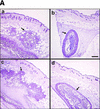
Expression of MMP-9–CD44 fusion proteins restores the invasiveness, growth, and angiogenesis of the TA3sCD44 cells in vivo. (A) Hematoxylin-stained sections of the representative tumors derived from the subcutaneously injected TA3wt (a), TA3sCD44 (b), TA3sCD44MMP-9/CD44fp (c), and TA3sCD44MMP-9v5 (d) cells. A total of 2 × 106 viable cells were injected, and the tumor specimens were collected 1 week after the injection. (B), In a separate set of animals, tumor weight was assessed 3 weeks after the injection. In each experiment, six mice were injected with each transfectant. Mean weight ±s.d. of tumors derived from the TA3 transfectants are shown. (C) Tumor angiogenesis was assessed and expressed as the average number of capillary blood vessels, as revealed by anti-vWF antibody, per 10× power microscopic field in the solid tumor sections derived from TA3 transfectants. The number ±s.d. represents the average blood vessel count per microscopic field from total counts in 10 randomly selected fields. Bar, 300 μm.
Figure 3
Figure 3
Expression of MMP-9–CD44 fusion proteins restores the invasiveness, growth, and angiogenesis of the TA3sCD44 cells in vivo. (A) Hematoxylin-stained sections of the representative tumors derived from the subcutaneously injected TA3wt (a), TA3sCD44 (b), TA3sCD44MMP-9/CD44fp (c), and TA3sCD44MMP-9v5 (d) cells. A total of 2 × 106 viable cells were injected, and the tumor specimens were collected 1 week after the injection. (B), In a separate set of animals, tumor weight was assessed 3 weeks after the injection. In each experiment, six mice were injected with each transfectant. Mean weight ±s.d. of tumors derived from the TA3 transfectants are shown. (C) Tumor angiogenesis was assessed and expressed as the average number of capillary blood vessels, as revealed by anti-vWF antibody, per 10× power microscopic field in the solid tumor sections derived from TA3 transfectants. The number ±s.d. represents the average blood vessel count per microscopic field from total counts in 10 randomly selected fields. Bar, 300 μm.
Figure 4
Figure 4
Endothelial cell tubule formation assay. BME cells (3 × 105/ml) were seeded onto type I collagen gels in medium supplemented with 10% calf serum. On the following day, the medium was aspirated and replaced with conditioned serum-free medium derived from cocultures of G8 myoblast monolayers and TA3wt (A) TA3sCD44 (B), TA3sCD44/MMP-9-CD44fp (C), or TA3sCD44/MMP-9v5 (D) cells. A total of 30 μg/ml of pan specific anti-TGF-β (E) or anti-bFGF (F) antibody were added to the TA3wt/G8-conditioned coculture medium prior to use in the assay. Tubules are indicated by arrows. Bar, 140 μm.
Figure 5
Figure 5
CD44-anchored MMP-9 activates TGF-β. (A) The ability of TA3 transfectant-G8 myoblast coculture media to stimulate TMLC luciferase activity is shown. Conditioned coculture media tested are indicated. (B) Pan-specific TGF-β antibody (30 μg/ml) or specific antibodies against TGF-β1 (100 ng/ml), TGF-β2(100ng/ml), or TGF-β3(100 ng/ml) were used to determine the activity of TGF-β isoforms in the coculture media. One unit of luciferase activity corresponds to the activity produced by 5 pg of purified human TGF-β1 (R & D).
Figure 5
Figure 5
CD44-anchored MMP-9 activates TGF-β. (A) The ability of TA3 transfectant-G8 myoblast coculture media to stimulate TMLC luciferase activity is shown. Conditioned coculture media tested are indicated. (B) Pan-specific TGF-β antibody (30 μg/ml) or specific antibodies against TGF-β1 (100 ng/ml), TGF-β2(100ng/ml), or TGF-β3(100 ng/ml) were used to determine the activity of TGF-β isoforms in the coculture media. One unit of luciferase activity corresponds to the activity produced by 5 pg of purified human TGF-β1 (R & D).
Figure 6
Figure 6
TGF-β is activated by purified MMP-9. (A) TMLC luciferase activity induced by concentrated COS cell supernatants containing the indicated latent TGF-β isoforms following incubation with the indicated purified AMPA-activated MMPs. Comparable amounts of latent TGF-β1, TGF-β2, and TGF-β3 were present in the supernatants as assessed by both Western blot analysis and TMLC-luciferase induction following heat treatment (80°C for 5min) (B) TMLC luciferase activity induced by affinity-purified TGF-β2 following incubation with the indicated purified activated MMPs. Activity is expressed in relative light units (RLU), in which 800 RLU corresponds to the luciferase activity generated by 1 pg of purified human TGF-β1 (R & D). (C) TGF-β is proteolytically cleaved by MMP-9 and MMP-2. Purified v5-tagged TGF-β2 (lane 1) was incubated with protein-A Sepharose-bound, AMPA-activated MMP-9 (lane 2), MMP-2 (lane 3), and MMP-3 (lane 4) for 90 min at 37°C. Following incubation, the supernatants were separated from the beads, subjected to SDS/12% PAGE, transferred to Hybond-C membranes, and blotted with anti-v5 antibody. (D) MMP-2/CD44 fusion protein expression promotes TGF-β activation in TA3sCD44 cells. TMLC luciferase assays were performed with serum-free coculture media from TA3sCD44 cells transiently transfected with the indicated cDNAs.
Figure 6
Figure 6
TGF-β is activated by purified MMP-9. (A) TMLC luciferase activity induced by concentrated COS cell supernatants containing the indicated latent TGF-β isoforms following incubation with the indicated purified AMPA-activated MMPs. Comparable amounts of latent TGF-β1, TGF-β2, and TGF-β3 were present in the supernatants as assessed by both Western blot analysis and TMLC-luciferase induction following heat treatment (80°C for 5min) (B) TMLC luciferase activity induced by affinity-purified TGF-β2 following incubation with the indicated purified activated MMPs. Activity is expressed in relative light units (RLU), in which 800 RLU corresponds to the luciferase activity generated by 1 pg of purified human TGF-β1 (R & D). (C) TGF-β is proteolytically cleaved by MMP-9 and MMP-2. Purified v5-tagged TGF-β2 (lane 1) was incubated with protein-A Sepharose-bound, AMPA-activated MMP-9 (lane 2), MMP-2 (lane 3), and MMP-3 (lane 4) for 90 min at 37°C. Following incubation, the supernatants were separated from the beads, subjected to SDS/12% PAGE, transferred to Hybond-C membranes, and blotted with anti-v5 antibody. (D) MMP-2/CD44 fusion protein expression promotes TGF-β activation in TA3sCD44 cells. TMLC luciferase assays were performed with serum-free coculture media from TA3sCD44 cells transiently transfected with the indicated cDNAs.
Figure 7
Figure 7
TGF-β activation by CD44-anchored MMP-9 is detected in tumor extracts and keratinocyte cultures. (A) Seven and 10-day solid tumors derived from TA3wt, TA3sCD44, TA3sCD44/MMP-9-CD44fp, and TA3sCD44/MMP-9v5 cells were extracted in 50 mm Tris-HCl, (pH7.5), 75 mm NaCl, 10 mm EDTA, containing AEBSF (1 μg/ml), aprotinin (0.05 units), leupeptin (1 μg/ml), pepstatin A (2 μg/ml) and E64 (1 μg/ml), and 100 μg of the extracted proteins were added to TMLCs, and resulting luciferase activity was determined. (B) Gelatin zymogram analysis of serum-free conditioned media (a) and crude cell membrane extracts (b) of wild-type (lane 1 in a and b) and CD44-null (lane 2 in a and b) keratinocytes. (C) TMLC luciferase assay with serum-free conditioned media from wild-type and CD44-null keratinocytes. (D) TMLC luciferase assay with serum-free conditioned media from CD44 null keratinocyte cultures transiently transfected with MMP-9/CD44fp, MMP-9v5, and sCD44 cDNAs. Luciferase activity is expressed in relative light units (RLU) in which 800 RLU correspond to the activity generated by 1 pg of purified human TGF-β1 (R & D).
Figure 7
Figure 7
TGF-β activation by CD44-anchored MMP-9 is detected in tumor extracts and keratinocyte cultures. (A) Seven and 10-day solid tumors derived from TA3wt, TA3sCD44, TA3sCD44/MMP-9-CD44fp, and TA3sCD44/MMP-9v5 cells were extracted in 50 mm Tris-HCl, (pH7.5), 75 mm NaCl, 10 mm EDTA, containing AEBSF (1 μg/ml), aprotinin (0.05 units), leupeptin (1 μg/ml), pepstatin A (2 μg/ml) and E64 (1 μg/ml), and 100 μg of the extracted proteins were added to TMLCs, and resulting luciferase activity was determined. (B) Gelatin zymogram analysis of serum-free conditioned media (a) and crude cell membrane extracts (b) of wild-type (lane 1 in a and b) and CD44-null (lane 2 in a and b) keratinocytes. (C) TMLC luciferase assay with serum-free conditioned media from wild-type and CD44-null keratinocytes. (D) TMLC luciferase assay with serum-free conditioned media from CD44 null keratinocyte cultures transiently transfected with MMP-9/CD44fp, MMP-9v5, and sCD44 cDNAs. Luciferase activity is expressed in relative light units (RLU) in which 800 RLU correspond to the activity generated by 1 pg of purified human TGF-β1 (R & D).

Similar articles

Cited by

References

    1. Aruffo A, Stamenkovic I, Melnick M, Underhill CB, Seed B. CD44 is the principal cell surface receptor for hyaluronate. Cell. 1990;61:1303–1313. - PubMed
    1. Bartolazzi A, Peach R, Aruffo A, Stamenkovic I. Interaction between CD44 and hyaluronan is directly implicated in the regulation of tumor development. J Exp Med. 1994;180:53–66. - PMC - PubMed
    1. Border WA, Ruoslahti E. Transforming growth factor-β in disease: The dark-side of tissue repair. J Clin Invest. 1992;90:1–7. - PMC - PubMed
    1. Bourguignon LYW, Gunja-Smith Z, Iida N, Zhu HB, Young LJT, Muller WJ, Ardiff RD. CD44v3, 8–10 is involved in cytoskeleton-mediated tumor cell migration and matrix metalloproteinase (MMP-9) association in metastatic breast cancer cells. J Cell Physiol. 1998;176:206–215. - PubMed
    1. Brooks PC, Stromblad S, Sanders LC, von Schalscha TL, Aimes RT, Stetler-Stevenson WG, Quigley JP, Cheresh DA. Localization of matrix metalloproteinase MMP-2 to the surface of invasive cells by interaction with integrin αvβ3. Cell. 1996;85:683–693. - PubMed

Publication types

MeSH terms

LinkOut - more resources