. 2000 Jan 10;148(1):203-16.
doi: 10.1083/jcb.148.1.203.

Monocytes induce reversible focal changes in vascular endothelial cadherin complex during transendothelial migration under flow

Affiliations

Monocytes induce reversible focal changes in vascular endothelial cadherin complex during transendothelial migration under flow

J R Allport et al. J Cell Biol. .

Abstract

The vascular endothelial cell cadherin complex (VE-cadherin, alpha-, beta-, and gamma-catenin, and p120/p100) localizes to adherens junctions surrounding vascular endothelial cells and may play a critical role in the transendothelial migration of circulating blood leukocytes. Previously, we have reported that neutrophil adhesion to human umbilical vein endothelial cell (HUVEC) monolayers, under static conditions, results in a dramatic loss of the VE-cadherin complex. Subsequent studies by us and others (Moll, T., E. Dejana, and D. Vestweber. 1998. J. Cell Biol. 140:403-407) suggested that this phenomenon might reflect degradation by neutrophil proteases released during specimen preparation. We postulated that some form of disruption of the VE-cadherin complex might, nonetheless, be a physiological process during leukocyte transmigration. In the present study, the findings demonstrate a specific, localized effect of migrating leukocytes on the VE-cadherin complex in cytokine-activated HUVEC monolayers. Monocytes and in vitro differentiated U937 cells induce focal loss in the staining of VE-cadherin, alpha-catenin, beta-catenin, and plakoglobin during transendothelial migration under physiological flow conditions. These events are inhibited by antibodies that prevent transendothelial migration and are reversed following transmigration. Together, these data suggest that an endothelial-dependent step of transient and focal disruption of the VE-cadherin complex occurs during leukocyte transmigration.


Figures
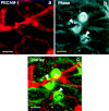
Figure 1
Figure 1
Live-time assessment of monocytes transmigration across activated HUVEC monolayers at lateral junctions. Activated HUVEC monolayers, preincubated with 5 μg/ml anti–PECAM-1 antibody, P1.1, and anti-mouse IgG Texas red were washed and then perfused with PBMC (5 × 10⁵/ml) for 15 min. Live-time immunofluorescence images and corresponding phase-contrast micrographs were taken at intervals and subsequently overlaid in register. PECAM-1 staining is shown in red (a) and the corresponding phase image is shown in b. Actively migrating monocytes are indicated by arrows and a newly arrested monocyte is indicated by the asterisk. An overlay of the two images is indicated in c. Bars, 10 μm.
Figure 2
Figure 2
Adhesion and transmigration of U937L-Dif cells induces focal disruption of VE-cadherin staining under flow. U937L or U937L-Dif cells (106/ml) were perfused across 4 h TNF-α activated HUVEC monolayers for 10 min at 1.8 dynes/cm2. Coverslips were stained for VE-cadherin as described. In the presence of U937L cells (a and b) there was no detectable disruption of VE-cadherin staining (b), even where cells were aligned directly over a lateral junction (arrowheads). In contrast, in the presence of U937L-Dif cells (c and d), there was loss of VE-cadherin staining (d, arrowheads) in areas of leukocyte adhesion/transmigration. Bars, 12 μm.
Figure 3
Figure 3
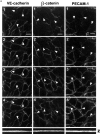
Transendothelial migration of monocytes under flow in vitro induces focal changes in the VE-cadherin complex. Monocytes (0.5 × 10⁶/ml) or buffer alone were perfused across activated HUVEC monolayers for 5 min at 1.0 dynes/cm2. The monolayers were recovered from the chamber and were immediately fixed in 2% paraformaldehyde (RT, 10 min) and stained for adherens junction proteins as described in Materials and Methods. Serial Z-sections were taken on the laser scanning confocal microscope where the CY3 and FITC channels were acquired sequentially using excitation wavelengths of 568 and 488 nm, respectively, to prevent bleed-through, and subsequently merged (a and b). Each series of panels (1–4) depicts sections from a Z-series from the apical (1) to the basolateral (4) surface. The Z-series was also projected in the x-z direction and shown at the bottom of each corresponding Z-series (Z). Selected images from b were further processed using NIH Image as described in Materials and Methods to generate c. a, Adherens junctional protein staining (VE-cadherin, left; β-catenin, middle; PECAM-1, right) is shown in the absence of monocytes. There were no detectable gaps in the VE-cadherin staining. Any apparent gaps in the staining pattern (arrowheads) were eliminated when neighboring sections were examined. b, In the presence of monocytes, gaps in the VE-cadherin staining pattern were seen where a monocyte was actively transmigrating the monolayer (arrow). There was no loss of staining where a monocyte was merely adherent to the apical surface of the monolayer (asterisk). Similarly, actively transmigrating monocytes induced loss of β-catenin staining (middle panels, arrows). In contrast, no detectable loss of PECAM-1 staining was observed in the presence of actively migrating monocytes. c, Selected regions (A, B, and C, bound by white lines) were projected in the x-z direction and collapsed to generate single images of VE-cadherin staining, β-catenin staining, and PECAM-1 staining in the presence of monocytes. Bars, 10 μm.
Figure 3
Figure 3
Transendothelial migration of monocytes under flow in vitro induces focal changes in the VE-cadherin complex. Monocytes (0.5 × 10⁶/ml) or buffer alone were perfused across activated HUVEC monolayers for 5 min at 1.0 dynes/cm2. The monolayers were recovered from the chamber and were immediately fixed in 2% paraformaldehyde (RT, 10 min) and stained for adherens junction proteins as described in Materials and Methods. Serial Z-sections were taken on the laser scanning confocal microscope where the CY3 and FITC channels were acquired sequentially using excitation wavelengths of 568 and 488 nm, respectively, to prevent bleed-through, and subsequently merged (a and b). Each series of panels (1–4) depicts sections from a Z-series from the apical (1) to the basolateral (4) surface. The Z-series was also projected in the x-z direction and shown at the bottom of each corresponding Z-series (Z). Selected images from b were further processed using NIH Image as described in Materials and Methods to generate c. a, Adherens junctional protein staining (VE-cadherin, left; β-catenin, middle; PECAM-1, right) is shown in the absence of monocytes. There were no detectable gaps in the VE-cadherin staining. Any apparent gaps in the staining pattern (arrowheads) were eliminated when neighboring sections were examined. b, In the presence of monocytes, gaps in the VE-cadherin staining pattern were seen where a monocyte was actively transmigrating the monolayer (arrow). There was no loss of staining where a monocyte was merely adherent to the apical surface of the monolayer (asterisk). Similarly, actively transmigrating monocytes induced loss of β-catenin staining (middle panels, arrows). In contrast, no detectable loss of PECAM-1 staining was observed in the presence of actively migrating monocytes. c, Selected regions (A, B, and C, bound by white lines) were projected in the x-z direction and collapsed to generate single images of VE-cadherin staining, β-catenin staining, and PECAM-1 staining in the presence of monocytes. Bars, 10 μm.
Figure 4
Figure 4
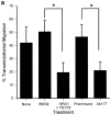
Inhibition of monocyte transmigration prevents monocyte-dependent changes in VE-cadherin staining. PBMC (106/ml) were incubated in the presence of either binding control (W6/32 mAb, 10 μg/ml) or the combination of α4-integrin (mAb HP2.1) and β2-integrin (TS1/18 mAb, each at 10 μg/ml) for 15 min at room temperature. In additional experiments, PBMC and the HUVEC monolayer were incubated in the presence of either Ab 177 or preimmune IgG (20 μg/ml) for 30 min before use in flow assays. a, The total interacting cells (rolling, adherent, and transmigrated) were comparable in each case, 602 ± 157 cells/mm2 in the presence of HP2.1 and TS1/18; 573 ± 53 cells/mm2 in the presence of Ab177; and 575.8 ± 170 cells/mm2 in the presence of preimmune IgG. Data are the mean ± SD, n = 4 (four coverslips analyzed per experiment). b, The number of monocytes associated with changes in VE-cadherin were quantified as described in Materials and Methods. Three fields were taken from each coverslip (two coverslips analyzed per experiment). Data are expressed as the mean ± SD, n = 4. The range of total leukocytes per field was 13–65 cells, with most fields falling between 25 and 47 cells.
Figure 4
Figure 4
Inhibition of monocyte transmigration prevents monocyte-dependent changes in VE-cadherin staining. PBMC (106/ml) were incubated in the presence of either binding control (W6/32 mAb, 10 μg/ml) or the combination of α4-integrin (mAb HP2.1) and β2-integrin (TS1/18 mAb, each at 10 μg/ml) for 15 min at room temperature. In additional experiments, PBMC and the HUVEC monolayer were incubated in the presence of either Ab 177 or preimmune IgG (20 μg/ml) for 30 min before use in flow assays. a, The total interacting cells (rolling, adherent, and transmigrated) were comparable in each case, 602 ± 157 cells/mm2 in the presence of HP2.1 and TS1/18; 573 ± 53 cells/mm2 in the presence of Ab177; and 575.8 ± 170 cells/mm2 in the presence of preimmune IgG. Data are the mean ± SD, n = 4 (four coverslips analyzed per experiment). b, The number of monocytes associated with changes in VE-cadherin were quantified as described in Materials and Methods. Three fields were taken from each coverslip (two coverslips analyzed per experiment). Data are expressed as the mean ± SD, n = 4. The range of total leukocytes per field was 13–65 cells, with most fields falling between 25 and 47 cells.
Figure 5
Figure 5
Monocyte-dependent loss of VE-cadherin is reversible over time. PBMC or isolated CD4+ lymphocytes were perfused across activated HUVEC as indicated. Leukocyte–endothelial interactions were recorded by live-time videomicroscopy and analyzed from at least six fields at 3, 7, and 12 min of perfusion. Total interacting cells were 436 ± 44, 770 ± 336, and 1,155 ± 353 cells/mm2, respectively. Fixed monolayers were stained for VE-cadherin and the number of monocytes or lymphocytes associated with loss of VE-cadherin were determined as described. The range of total leukocytes per field was 23–68 cells. Data are expressed as mean ± SD, n = 3 (three coverslips analyzed per experiment, three fields per coverslip). *Indicates significance when compared with the 3-min time-point. Transmigration data are shown as filled symbols (•, PBMC; ▪, CD4+ lymphocytes), VE-cadherin staining data are shown as open symbols (○, monocytes; □, CD4+ lymphocytes).

Similar articles

Cited by

References

    1. Ali J., Liao F., Martens E., Muller W.A. Vascular endothelial cadherin (VE-cadherin)cloning and role in endothelial cell–cell adhesion. Microcirculation. 1997;4:267–277. - PubMed
    1. Allport J.R., Ding H., Collins T., Gerritsen M.E., Luscinskas F.W. Endothelial-dependent mechanisms regulate leukocyte transmigrationa process involving the proteasome and disruption of the vascular endothelial–cadherin complex at endothelial cell-to-cell junctions. J. Exp. Med. 1997;186:517–527. - PMC - PubMed
    1. Berman M.E., Muller W.A. Ligation of platelet/endothelial cell adhesion molecule-1 (PECAM-1/CD31) on monocytes and neutrophils increases binding capacity of leukocyte CR3 (CD11b/CD18) J. Immunol. 1995;154:299–307. - PubMed
    1. Breviario F., Caveda L., Corada M., Martin-Padura I., Navarro P., Golay J., Introna M., Gulino D., Lampugnani M.G., Dejana E. Functional properties of human vascular endothelial cadherin (7B4/cadherin-5), an endothelium-specific cadherin. Arterioscler. Thromb. Vasc. Biol. 1995;15:1229–1239. - PubMed
    1. Burns A.R., Walker D.C., Brown E.S., Thurmon L.T., Bowden R.A., Keese C.R., Simon S.I., Entman M.L., Smith C.W. Neutrophil transendothelial migration is independent of tight junctions and occurs preferentially at tricellular corners. J. Immunol. 1997;159:2893–2903. - PubMed

Publication types