. 2000 Jan;74(2):627-43.
doi: 10.1128/jvi.74.2.627-643.2000.

A recombinant human immunodeficiency virus type 1 envelope glycoprotein complex stabilized by an intermolecular disulfide bond between the gp120 and gp41 subunits is an antigenic mimic of the trimeric virion-associated structure

Affiliations

A recombinant human immunodeficiency virus type 1 envelope glycoprotein complex stabilized by an intermolecular disulfide bond between the gp120 and gp41 subunits is an antigenic mimic of the trimeric virion-associated structure

J M Binley et al. J Virol. 2000 Jan.

Abstract

The few antibodies that can potently neutralize human immunodeficiency virus type 1 (HIV-1) recognize the limited number of envelope glycoprotein epitopes exposed on infectious virions. These native envelope glycoprotein complexes comprise three gp120 subunits noncovalently and weakly associated with three gp41 moieties. The individual subunits induce neutralizing antibodies inefficiently but raise many nonneutralizing antibodies. Consequently, recombinant envelope glycoproteins do not elicit strong antiviral antibody responses, particularly against primary HIV-1 isolates. To try to develop recombinant proteins that are better antigenic mimics of the native envelope glycoprotein complex, we have introduced a disulfide bond between the C-terminal region of gp120 and the immunodominant segment of the gp41 ectodomain. The resulting gp140 protein is processed efficiently, producing a properly folded envelope glycoprotein complex. The association of gp120 with gp41 is now stabilized by the supplementary intermolecular disulfide bond, which forms with approximately 50% efficiency. The gp140 protein has antigenic properties which resemble those of the virion-associated complex. This type of gp140 protein may be worth evaluating for immunogenicity as a component of a multivalent HIV-1 vaccine.


Figures

FIG. 1
FIG. 1
Different forms of the HIV-1 envelope glycoproteins. (A) Monomeric gp120. (B) Full-length recombinant gp160 (in practice, this protein may form higher-order aggregates in solution because of associations between various hydrophobic domains). (C) Proteolytically unprocessed gp140 trimer with the peptide bond maintained between gp120 and gp41 (gp140UNC or gp140NON). (D) The SOS gp140 protein, a proteolytically processed gp140 stabilized by an intermolecular disulfide bond. (E) Native, virion-associated gp120-gp41 trimer. The topologies of these proteins are inferred from previous reports cited in the text and from studies described in this paper. The shading of the gp140UNC protein (C) indicates the major antibody-accessible regions that are poorly or not exposed on the SOS gp140 protein or on the native gp120-gp41 trimer. The trimeric state of the SOS gp140 protein (D) has not yet been confirmed experimentally.
FIG. 2
FIG. 2
Cotransfection of furin increases the efficiency of cleavage of the peptide bond between gp120 and gp41. 293T cells were transfected with DNA expressing the HIV-1 JR-FL gp140WT or gp140UNC (gp120-gp41 cleavage site mutant) proteins, in the presence or absence of a cotransfected furin-expressing plasmid. The 35S-labeled envelope glycoproteins secreted from the cells were immunoprecipitated with the anti-gp120 MAb 2G12, boiled with SDS, and analyzed by SDS-PAGE. Lanes: 1, gp140WT (gp140/gp120 doublet); 2, gp140WT plus furin (gp120 only); 3, gp140UNC (gp140 only); 4, gp140UNC plus furin (gp140 only). The approximate molecular masses, in kilodaltons, of the major species are recorded on the left, as are higher-molecular-mass aggregates. Only one-fifth of the immunoprecipitated proteins from the transfections shown in lanes 1 and 3 were loaded onto the gel, to ensure that the amounts of envelope glycoproteins analyzed in each lane were approximately comparable.
FIG. 3
FIG. 3
Positions of cysteine substitutions in JR-FL gp140. The various residues of the JR-FL gp140WT protein that have been mutated to cysteines in one or more mutants are indicated by arrows on the schematics of the gp120 and gp41ECTO subunits. The positions of the alanine-501 and threonine-605 residues that are both mutated to cysteine in the SOS gp140 protein are indicated by the larger arrows. (A) The depiction of JR-FL gp120, including the positioning of canonical sites for complex and high-mannose N-linked carbohydrates, is based on that of Leonard et al. (56), adjusted to reflect the sequence numbering of HIV-1 HxB2. (B) The cartoon of the JR-FL gp41-ectodomain is derived from reference , also adjusted to reflect the HxB2 sequence numbering. The open boxes at the C terminus of gp120 and the N terminus of gp41 indicate the regions that are mutated in the gp140UNC protein to eliminate the cleavage site between gp120 and gp41.
FIG. 4
FIG. 4
Immunoprecipitation analysis of selected double cysteine mutants of JR-FL gp140. The 35S-labeled envelope glycoproteins secreted from transfected 293T cells were immunoprecipitated with an anti-gp120 MAb, boiled with SDS, and analyzed by SDS-PAGE. The MAbs used were either 2G12 (odd-numbered lanes) or F91, which recognizes a CD4-binding site-related epitope (even-numbered lanes). The positions of the two cysteine substitutions in each protein (one in gp120, the other in gp41ECTO) are noted above the lanes. The gp140WT protein is shown in lane 15. All proteins were expressed in the presence of cotransfected furin, except for the gp140WT protein in lane 15, which serves as a reference standard for the position of 120-kDa (gp120) and 140-kDa (gp140NON) bands. Note that in this and all subsequent figures (except Fig. 10) that depict the outcome of RIPA experiments, the photographs have been cropped to show only the 120- and 140-kDa bands, since other regions of the gels were not informative.
FIG. 5
FIG. 5
The efficiency of intermolecular disulfide bond formation is dependent upon the positions of the cysteine substitutions. The 35S-labeled envelope glycoproteins secreted from 293T cells cotransfected with furin and the various gp140 mutants were immunoprecipitated with the anti-gp120 MAb 2G12, boiled with SDS, and analyzed by SDS-PAGE. For each mutant, the intensities of the 140- and 120-kDa bands were determined by densitometry and the ratio of gp140 to gp140 + gp120 was calculated and recorded. The extent of shading is proportional to the magnitude of the ratio. The positions of the amino acid substitutions in gp41 and the C1 and C5 domains of gp120 are recorded along the top and down the sides, respectively. N.D., not done.
FIG. 6
FIG. 6
Confirmation that an intermolecular gp120-gp41 bond forms in the SOS gp140 protein. 293T cells were transfected with plasmids expressing gp140 proteins and, when indicated, a furin-expressing plasmid. The secreted, 35S-labeled glycoproteins were immunoprecipitated with the anti-gp120 MAb 2G12, boiled in the presence of SDS or, when indicated, SDS plus DTT, and analyzed by SDS-PAGE. (A) Lanes: 1 and 4, SOS gp140 protein (double cysteine mutant A501C/T605C) plus furin; 2 and 5, gp140WT protein, no furin; 3 and 6, gp140UNC protein, no furin. In lanes 1 to 3 the immunoprecipitated proteins were boiled with SDS; in lanes 4 to 6 they were boiled with SDS plus DTT. (B) Lanes: 1 and 3, SOS gp140 protein plus furin; 2 and 4, SOS gp140 protein without furin. In lanes 1 and 2 the immunoprecipitated proteins were boiled with SDS; in lanes 3 and 4 they were boiled with SDS plus DTT. (C) Lanes: 1, SOS gp140 protein (double cysteine mutant A501C/T605C) plus furin; 2, single cysteine gp140 mutant A501C plus furin; 3, single cysteine gp140 mutant T605C plus furin. The immunoprecipitated proteins in each case were boiled with SDS. High-molecular-weight aggregates were also present in immunoprecipitates of the SOS gp140 protein (data not shown but see Fig. 10).
FIG. 7
FIG. 7
Analysis of cysteine mutants of JR-FL gp140. The 35S-labeled envelope glycoproteins secreted from gp140-transfected 293T cells in the presence of cotransfected furin were immunoprecipitated with the anti-gp120 MAb 2G12, boiled with SDS, and analyzed by SDS-PAGE. Lanes: 1 to 8, each of the different gp140 double cysteine mutants contained the T605C substitution in gp41, in combination with a second cysteine substitution at the indicated residue in the C5 region of gp120 (the SOS gp140 protein is in lane 3); 9 to 11, gp140 proteins containing the A501C/T605C double cysteine substitutions together with the indicated lysine to alanine substitutions at residue 500 (lane 9), residue 502 (lane 10) or both residues 500 and 502 (lane 11); 12 to 14, gp140 proteins containing quadruple cysteine substitutions; each protein contained the W45C, T605C, and P609C substitutions, plus K500C (lane 12), A501C (lane 13), or K502C (lane 14).
FIG. 8
FIG. 8
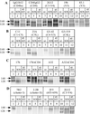
Comparison of the antigenic structures of the SOS gp140 protein, the gp140NON protein and gp120. The 35S-labeled envelope glycoproteins secreted from transfected 293T cells were immunoprecipitated with different anti-gp120 (A to C) or anti-gp41 (D) MAbs, boiled with SDS, and analyzed by SDS-PAGE. Lanes: 1, 4, 7, 10, and 13, gp140WT with no cotransfected furin, producing gp120 and the gp140NON protein; 2, 5, 8, 11, and 14, SOS gp140 protein plus cotransfected furin; 3, 6, 9, 12, and 15, gp140 protein containing the W45C/T605C double cysteine substitutions, plus co-transfected furin. Brief descriptions of the epitopes recognized by each MAb are noted above each lane; for more details, see the primary references listed in Materials and Methods. D, discontinuous epitope; L, linear epitope.
FIG. 9
FIG. 9
Comparison of the antigenic structures of the gp140NON and gp140UNC proteins. The 35S-labeled envelope glycoproteins secreted from transfected 293T cells were immunoprecipitated with different anti-gp120 MAbs, boiled with SDS, and analyzed by SDS-PAGE. Odd-numbered lanes contained gp140WT with no cotransfected furin, producing gp120 and the gp140NON protein. Even-numbered lanes contained gp140UNC protein with no cotransfected furin.
FIG. 10
FIG. 10
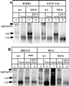
Preparation of disulfide bond-stabilized gp140 proteins from various HIV-1 isolates. 293T cells were transfected with plasmids expressing gp140 proteins from different isolates and, when indicated, a furin-expressing plasmid. The secreted, 35S-labeled glycoproteins were immunoprecipitated with the anti-gp120 MAb 2G12, boiled with SDS (and, when indicated, DTT), and analyzed by SDS-PAGE. The SOS gp140 protein from each isolate contained double cysteine substitutions at positions equivalent to alanine-501 and threonine-605 of the JR-FL gp140 protein. (A) HxB2 (lanes 1 to 4) and GUN-1wt (lanes 5 to 8). Lanes: 1 and 5, gp140WT with no cotransfected furin, producing gp120 and the gp140NON protein; 2 and 6, gp140WT plus furin, producing gp120; 3 and 7, SOS gp140 protein plus furin; 4 and 8, as lanes 3 and 7 except that the immunoprecipitates were boiled with both SDS and DTT prior to SDS-PAGE. (B) DH123 (lanes 1 to 4) and 89.6 (lanes 5 to 9). The layout of the lanes is as in panel A, except that for 89.6, lane 8 is the same as lane 7 but with only one-fifth of the immunoprecipitate loaded onto the gel and lane 9 is the same as lane 8 but with the immunoprecipitates boiled with both SDS and DTT prior to SDS-PAGE. The positions of the 120- and 140-kDa bands, and of higher-molecular-mass aggregates, are indicated on the left of each panel. Only one-fifth of the immunoprecipitated proteins from the gp140WT plus furin transfections (lanes 2 and 6) was loaded onto each gel, to approximately compensate for the increased envelope glycoprotein expression that was observed with the JR-FL gp140WT protein under these conditions.
FIG. 11
FIG. 11
Sucrose gradient analysis of the JR-FL SOS gp140 and gp140UNC proteins. Envelope glycoproteins secreted from transfected 293T cells were concentrated 100-fold and then fractionated by sucrose velocity gradient centrifugation. The gradient fractions (500 μl) were immunoprecipitated with MAb 2G12, boiled with SDS, and analyzed by SDS-PAGE to detect envelope glycoproteins and determine the sizes of their denatured components. (A) JR-FL SOS gp140 protein. (B) JR-FL gp140UNC protein. The last lane in each panel shows an unconcentrated supernatant containing the JR-FL gp140WT protein expressed in the absence of furin and then immunoprecipitated with 2G12 to provide a reference standard for the positions of gp120 and gp140 proteins on the gel. These bands are marked on the right of each panel, together with the position of high-molecular-weight aggregates.
FIG. 12
FIG. 12
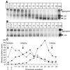
Gel filtration analysis of the JR-FL SOS gp140 and gp140UNC proteins. Envelope glycoproteins from transfected 293T cells were concentrated 100-fold and then fractionated by gel filtration chromatography. The fractions (250 μl) were immunoprecipitated with MAb 2G12, boiled with SDS, and analyzed by SDS-PAGE to detect envelope glycoproteins and determine the size of their constituent subunits. (A) JR-FL SOS gp140 protein. (B) JR-FL gp140UNC protein. The last lane in each panel shows an unconcentrated supernatant containing the protein under analysis and immunoprecipitated with 2G12 to provide a reference standard. These bands are marked on the right of each panel, together with the position of high-molecular-weight aggregates. (C) Densitometric analysis of the elution profile derived from the SOS gp140 protein (A and B). ▴, gp140UNC; ■, the 140-kDa component of SOS gp140; ●, the 120-kDa component of SOS gp140. The positions of molecular mass standards are indicated by arrows. These were thyroglobulin (669 kDa), ferritin (440 kDa), and aldolase (158 kDa).

Similar articles

Cited by

References

    1. Abacioglu Y H, Fouts T R, Laman J D, Claassen E, Pincus S H, Moore J P, Roby C A, Kamin-Lewis R, Lewis G K. Epitope mapping and topology of baculovirus expressed HIV-1 gp160 determined with a panel of murine monoclonal antibodies. AIDS Res Hum Retroviruses. 1994;10:371–381. - PubMed
    1. Allaway G P, Davis-Bruno K L, Beaudry G A, Garcia E B, Wong E L, Ryder A M, Hasel K W, Gauduin M-C, Koup R A, McDougal J S, Maddon P J. Expression and characterization of CD4lgG2, a novel heterotetramer which neutralizes primary HIV-1 isolates. AIDS Res Hum Retroviruses. 1995;11:533–540. - PubMed
    1. Baldinotti F, Matteucci D, Mazzetti P, Giannelli C, Bandecchi P, Tozzini F, Bendinelli M. Serum neutralization of feline immunodeficiency virus is markedly dependent on passage history of the virus and host system. J Virol. 1994;68:4572–4579. - PMC - PubMed
    1. Barnett S W, Rajasekar S, Legg H, Doe B, Fuller D H, Haynes J R, Walker C M, Steimer K S. Vaccination with HIV-1 gp120 DNA induces immune responses that are boosted by a recombinant gp120 protein subunit. Vaccine. 1997;15:869–873. - PubMed
    1. Belshe R B, Gorse G J, Mulligan M J, Evans T G, Keefer M C, Excler J-L, Duliege A M, Tartaglia J, Cox W I, McNamara J, Hwang K-L, Bradney A, Montefiori D C, Weinhold K J. Induction of immune responses to HIV-1 by canarypox virus (ALVAC) HIV-1 and gp120 SF-2 recombinant vaccines in uninfected volunteers. AIDS. 1998;12:2407–2415. - PubMed

Publication types

MeSH terms

LinkOut - more resources