. 2000 Jan 15;28(2):626-33.
doi: 10.1093/nar/28.2.626.

A novel snail-related transcription factor Smuc regulates basic helix-loop-helix transcription factor activities via specific E-box motifs

Affiliations

A novel snail-related transcription factor Smuc regulates basic helix-loop-helix transcription factor activities via specific E-box motifs

H Kataoka et al. Nucleic Acids Res. .

Abstract

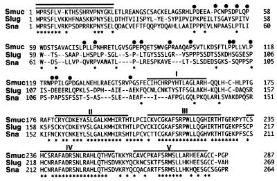
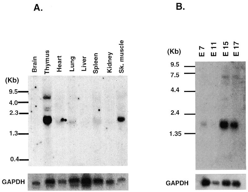
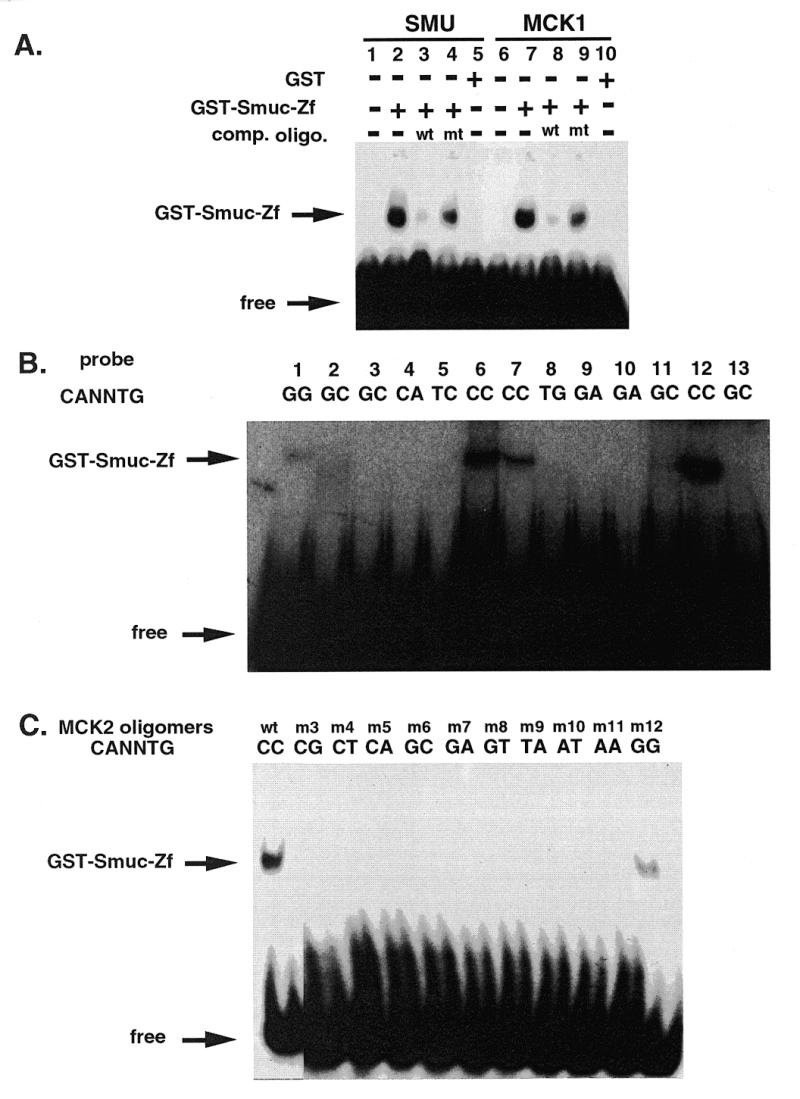
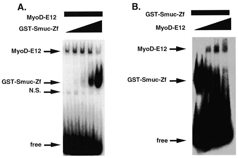
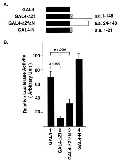
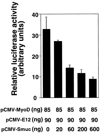
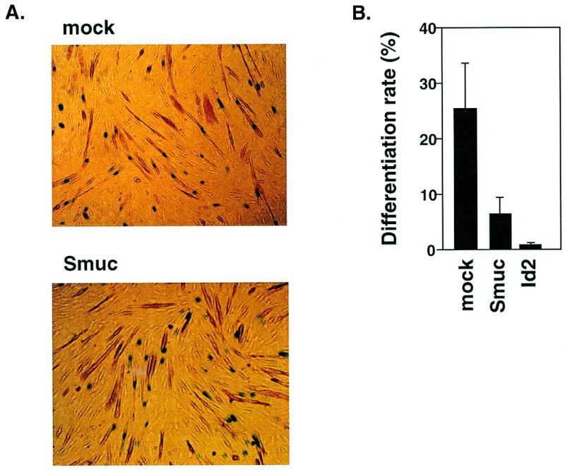
Snail family proteins are zinc finger transcriptional regulators first identified in Drosophila which play critical roles in cell fate determination. We identified a novel Snail -related gene from murine skeletalmusclecells designated Smuc. Northern blot analysis showed that Smuc was highly expressed in skeletal muscle and thymus. Smuc contains five putative DNA-binding zinc finger domains in its C-terminal half. In electrophoretic mobility shift assays, recombinant zinc finger domains of Smuc specifically bound to CAGGTG and CACCTG E-box motifs (CANNTG). Because basic helix-loop-helix transcription factors (bHLH) bind to the same E-box sequences, we examined whether Smuc competes with the myogenic bHLH factor MyoD for DNA binding. Smuc inhibited the binding of a MyoD-E12 complex to the CACCTG E-box sequence in a dose-dependent manner and suppressed the transcriptional activity of MyoD-E12. When heterologously targeted to the thymidine kinase promoter as fusion proteins with the GAL4 DNA-binding domain, the non-zinc finger domain of Smuc acted as a transcriptional repressor. Furthermore, overexpression of Smuc in myoblasts repressed transactivation of muscle differentiation marker Troponin T. Thus, Smuc might regulate bHLH transcription factors by zinc finger domains competing for E-box binding, and non-zinc finger repressor domains might also confer transcriptional repression to control differentiation processes.


Figures

Figure 1
Figure 1
Amino acid sequence of Smuc compared with those of mouse Sna (6,7) and mouse Slug (8). Identical residues among the three members are indicated by asterisks. The numbers of residues are indicated on both sides. The non-zinc finger domain of Smuc contains a region rich in proline residues, which are indicated by closed circles. Regions of amino acids corresponding to five zinc finger domains of Smuc and Slug and four of Sna are overlined. Conserved amino acid residues in the N-terminal region are indicated by double lines. The DNA sequence corresponding to the amino acids has been deposited in GenBank (accession no. AF133714).
Figure 2
Figure 2
Tissue distribution of Smuc mRNA. (A) Northern blot analysis of adult organs. Approximately 5 µg of poly(A)+ RNA derived from the indicated adult organs were hybridized with 32P-labeled Smuc probe as described in Materials and Methods. The size of Smuc was estimated to be 1.9 kb. Smuc was dominantly expressed in thymus and skeletal muscle with weak expression in lung, heart and spleen. (B) Expression of Smuc in developing mouse embryos. Each lane contains 2 µg of embryo poly(A)+ RNA. Smuc expression was detected at embryonic day 7 (E7) and up-regulated at later stages (E15 and E17). The presence of adequate amounts of RNA was confirmed by hybridization with GAPDH.
Figure 3
Figure 3
DNA binding properties of Smuc. (A) Binding of Smuc protein to CAGGTG E-box sites. EMSA was performed with GST–Smuc-Zf and double-stranded oligonucleotide probes containing the CAGGTG sequence. GST–Smuc-Zf was shown to bind to oligonucleotide probe SMU or MCK1, specifically to the CAGGTG sites in these two oligonucleotide probes. Competitive wild-type or mutant (mutant SMU or MCK1) oligonucleotide probes were added at 400 molar excess (lanes 3, 4, 8, and 9). (B) Preferential binding of Smuc zinc finger domains to CACCTG and CAGGTG E-box sequences. Various E-box and one N-box sequences were used as probes for binding of GST–Smuc-Zf. Oligonucleotide probes used in each lane were as follows: lane 1, kE2; lane 2, CGRP; lane 3, Troponin I; lane 4, N-box; lane 5, RIPE 3; lane 6, MLCA; lane 7, MCKR-1; lane 8, MCKL; lane 9, EF-1; lane 10, 8701; lane 11, MLCB; lane 12, MLCC; lane 13, CE2. The sequences of these oligonucleotides are described in Materials and Methods. GST–Smuc-Zf was shown to bind only to the E-box sequences containing CACCTG or CAGGTG (lane 1, 6, 7, and 12). One of four independent experimental results is shown. (C) Binding of GST–Smuc-Zf specifically to CACCTG and CAGGTG core E-box sequences. With other backbone sequences fixed, only the internal dinucleotides (CANNTG) of the MCK2 oligonucleotide probe were mutated and used in the EMSAs. GST–Smuc-Zf bound with high affinity only to the probes whose E-box sequences were CACCTG or CAGGTG.
Figure 4
Figure 4
Competition between Smuc and MyoD–E12 for the E-box DNA binding site. (A) Competition between GST–Smuc-Zf and bHLH proteins for a CACCTG site in the MCK2 oligonucleotide probe. The in vitro translated MyoD–E12 heterodimer (3 µl) was incubated with increasing amounts of GST–Smuc-Zf (0, 15, 50, 150 and 450 ng). Binding of MyoD–E12 heterodimer was decreased when increasing amounts of GST–Smuc-Zf were added. (B) Sixty nanograms of GST–Smuc-Zf was incubated with 32P-labeled MCK2 oligonucleotide with MyoD–E12 complex. GST–Smuc-Zf was displaced from the binding site by increasing amounts of MyoD–E12 complex (0, 2, 4 and 8 µl).
Figure 5
Figure 5
Non-zinc finger domain of Smuc as repressor domain. The non-zinc finger region of Smuc was expressed as a fusion protein with the GAL4 DBD and targeted heterologously to the tk promoter. An expression vector expressing only the GAL4 DBD was used as control (column 1). The reporter plasmid contained three GAL4 binding sites upstream of the luciferase gene. (A) Schematic illustrations of the GAL4 fusion constructs. Hatched and white boxes indicate the conserved N-terminal amino acids and the rest of the non-zinc finger domain, respectively. (B) The whole non-zinc finger domain (amino acids 1–148) repressed the transcriptional activity to 17% of the control level (column 2). Further deletion of the conserved N-terminal amino acids partially abolished the repression (column 3). However, the highly conserved 21 amino acids were insufficient to confer repression (column 4). Experiments were repeated four times in triplicate wells. One of the representative luciferase activities was statistically analyzed and is shown.
Figure 6
Figure 6
Function of Smuc as a bHLH regulator. Inhibition of the transcriptional activity of MyoD–E12 by Smuc was determined by luciferase reporter assays. C2C12 myoblast cells were transfected with expression vectors for MyoD and E12 (pCMV-MyoD, 85 ng/well; pCMV-E12, 90 ng/well). The reporter plasmid contains the luciferase gene under the control of 7× E-box sequences. Co-transfection of a Smuc expression vector suppressed the luciferase activity dose-dependently. Experiments were performed independently four times in triplicate wells. One of four independent results is shown.
Figure 7
Figure 7
Inhibition of myoblast differentiation by Smuc. (A) Smuc suppressed the expression of a muscle differentiation marker, Troponin T, in C2C12 myoblast cells in differentiation. C2C12 cells were transiently transfected with pCMV, p-CMV-Id2 or pCMV-Smuc, along with pNLS-LacZ, and then differentiated to muscles. Overexpression of Id2, a representative bHLH inhibitor, was considered as a positive control for negative regulation of the differentiation process. Nuclear staining of LacZ was used as a marker of incorporation of exogenous DNA. Percentages of Troponin T-positive cells (brown) among cells with LacZ nuclear staining (blue) [LacZ(+)] were measured in each well within several different microscopic fields. In total, 500 LacZ(+) cells were evaluated in each well. The percentages were defined as differentiation rates (percent Troponin T(+) + LacZ(+)/LacZ(+) = differentiation rate). (B) Differentiation rate of C2C12 cells was decreased with transfection of pCMV-Smuc. Bars indicate the percentages of differentiated Troponin T-positive cells among LacZ-positive cells. Experiments were performed three times in duplicate wells. The averages of these three independent results are indicated with SEM error bars.

Similar articles

Cited by

References

    1. Boulay J.L., Dennefeld,C. and Alberga,A. (1987) Nature, 330, 395–398. - PubMed
    1. Sefton M., Sanchez,S. and Nieto,M.A. (1998) Development, 125, 3111–3121. - PubMed
    1. Mauhin V., Lutz,Y., Dennefeld,C and Alberga,A. (1993) Nucleic Acids Res., 21, 3951–3957. - PMC - PubMed
    1. Fuse N., Hirose,S. and Hayashi,S. (1996) Development, 122, 1059–1067. - PubMed
    1. Braun T. and Arnold,H.H. (1991) Nucleic Acids Res., 19, 5645–5651. - PMC - PubMed

Publication types

MeSH terms

Associated data