Comparative Study
. 1999 Dec 1;13(23):3081-91.
doi: 10.1101/gad.13.23.3081.

The cAMP receptor protein CRP can function as an osmoregulator of transcription in Escherichia coli

Affiliations
Comparative Study

The cAMP receptor protein CRP can function as an osmoregulator of transcription in Escherichia coli

L Landis et al. Genes Dev. .

Erratum in

  • Genes Dev 2000 Feb 1;14(3):389

Abstract

Transcription of the P1 promoter of the Escherichia coli proP gene, which encodes a transporter of osmoprotectants, is strongly induced by a shift to hyperosmotic media. Unlike most other osmotically regulated promoters, the induction occurs for a brief period of time, corresponding to the replacement of intracellular K(+) glutamate with osmoprotecting compounds. This burst of proP transcription is correlated with the osmolarity-dependent binding of the cAMP receptor protein CRP to a site within the proP P1 promoter. We show that CRP-cAMP functions as an osmotically sensitive repressor of proP P1 transcription in vitro. Binding of CRP to the proP promoter in vivo is transiently destabilized after a hyperosmotic shift with kinetics that correspond to the derepression of transcription, whereas Fis and Lac repressor binding is not osmotically sensitive. Similar osmotic regulation of proP P1 transcription by the CRP* mutant implies that binding of cAMP is not responsible for the unusual osmotic sensitivity of CRP activity. Osmotic regulation of CRP activity is not limited to proP. Activation of the lac promoter by CRP is also transiently inhibited after an osmotic upshift, as is the binding of CRP to the galdelta4P1 promoter. These findings suggest that CRP functions in certain contexts to regulate gene expression in response to osmotic changes, in addition to its role in catabolite control.


Figures

Figure 1
Figure 1
The proP control region. (A) Schematic representation of the proP promoter region depicting the two promoters. P1 promoter activity is controlled by medium osmolarity and negatively regulated by CRP (Culham et al. 1993; Mellies et al. 1995, this paper). P2 promoter activity requires ς38 plus Fis binding at site I and is consequently activated in late exponential growth (Xu and Johnson 1995, 1997a). The weaker Fis-binding site II has a small negative effect on transcription from P1. (B) Sequence of the proP P1 promoter with the match to the CRP consensus noted. The G-to-C mutation strongly reduces CRP binding and leads to constitutive expression of the P1 promoter. The asterisks denote the positions of the guanines on the bottom strand that are protected from DMS reactivity by CRP–cAMP (Xu and Johnson 1997b).
Figure 2
Figure 2
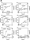
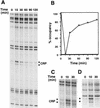
Osmotic induction of proP P1. (A) β-Galactosidase activities programmed by a proP–lacZ protein fusion in rich media during different levels of osmotic induction. RJ4373 was grown in LBN, and NaCl was added to a final concentration of 0.3–0.7 m as shown. The absence of Fis in RJ4373 insures that transcription initiates from the P1 promoter only. β-Galactosidase activities (▴) and cell growth (OD600, ●) were measured at the indicated times after addition of NaCl. β-Galactosidase activities are expressed as Miller units without normalization to cell densities (units per ml culture) to reflect synthesis rates. (B) Primer extension analysis of proP P1 RNA after addition of 0.5 m NaCl to CAG4000 growing in LBN. Times (min) after addition of NaCl (+) or LBN (−) are given at top. The sequence ladder was generated using the same primer used for the primer extension of the RNA. (C) Osmotic induction of proP P1 in minimal media. One-tenth volume of M9 plus glycerol containing 4 m or no NaCl was added to cultures of CAG4000 growing in M9 plus glycerol with or without 1 mm glycine betaine. RNA was extracted from cells collected at the indicated times after salt addition and subjected to primer extension as in B. The two panels represent images taken from one gel at the same exposure and thus are directly comparable.
Figure 2
Figure 2
Osmotic induction of proP P1. (A) β-Galactosidase activities programmed by a proP–lacZ protein fusion in rich media during different levels of osmotic induction. RJ4373 was grown in LBN, and NaCl was added to a final concentration of 0.3–0.7 m as shown. The absence of Fis in RJ4373 insures that transcription initiates from the P1 promoter only. β-Galactosidase activities (▴) and cell growth (OD600, ●) were measured at the indicated times after addition of NaCl. β-Galactosidase activities are expressed as Miller units without normalization to cell densities (units per ml culture) to reflect synthesis rates. (B) Primer extension analysis of proP P1 RNA after addition of 0.5 m NaCl to CAG4000 growing in LBN. Times (min) after addition of NaCl (+) or LBN (−) are given at top. The sequence ladder was generated using the same primer used for the primer extension of the RNA. (C) Osmotic induction of proP P1 in minimal media. One-tenth volume of M9 plus glycerol containing 4 m or no NaCl was added to cultures of CAG4000 growing in M9 plus glycerol with or without 1 mm glycine betaine. RNA was extracted from cells collected at the indicated times after salt addition and subjected to primer extension as in B. The two panels represent images taken from one gel at the same exposure and thus are directly comparable.
Figure 3
Figure 3
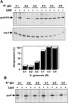
In vitro repression of transcription by CRP and Lac repressor. (A) In vitro single-round transcription reactions were performed using the proP substrate pRJ4069 in the presence (+) or absence (−) of CRP–cAMP and in the presence of 0.1–0.6 m K+ glutamate as denoted. The portions of the gel containing the P1 transcript and the vector rrn1 transcript are shown. The bar graph depicts the levels of the proP P1 transcript synthesized in the presence versus the absence of CRP–cAMP at each K+ glutamate concentration from the data shown above. (B) A similar set of reactions were performed using the substrate pKK223-3 containing the tac promoter in the presence (+) or absence (−) of Lac repressor.
Figure 4
Figure 4
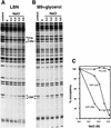
In vivo binding of CRP after addition of different concentrations of NaCl. (A) DMS footprinting reactions on cells growing in LBN and subjected to increasing concentrations of NaCl as denoted for each lane. (B) DMS footprinting reactions on cells growing in M9 plus glycerol and subjected to increasing concentrations of NaCl as in A. The cells were treated with DMS 15 min (A) or 30 min (B) after addition of NaCl. Media without NaCl was added to the cells labeled 0. The control lanes represent DMS reactions performed on plasmids after isolation and thus no cellular proteins are present. The locations of the guanines at −28 and −30 on the bottom strand that are protected from DMS modification in vitro by purified CRP or Fis protein at site I are designated with the asterisks. Fis-mediated protections in cells growing in M9 plus glycerol are weak because of the low Fis concentrations present under these growth conditions. (C) Graph depicting the percent occupancy of the CRP-binding site, as determined from the level of protection of the −28 guanine, in cells growing in LB and M9 plus glycerol. Occupancy of Fis site I is also shown from cells growing in LB after the different salt additions. The level of protection measured at 0 NaCl is defined as 100% occupancy relative to the control (0% occupancy).
Figure 5
Figure 5
Absence of osmotic sensitivity of Lac repressor binding and activity. (A) In vivo DMS footprinting reactions on cells growing in LBN and subjected to increasing concentrations of NaCl. This experiment was performed identically to Fig. 4A except that the cells contained pRZ4004, which carries the wild-type lac promoter region. (B) Lac repressor activity after an osmotic upshift. RJ3355 (lacI+PL8UV5 Z+Y+) was grown in LBN. β-Galactosidase (Miller units) was assayed 30 min after addition of no NaCl (control) or 0.5 m NaCl. For comparison, induction of β-galactosidase by the proP–lac fusion in RJ3265 under identical conditions is shown.
Figure 6
Figure 6
Kinetics of DNA binding by CRP after osmotic upshift. (A) In vivo DMS footprinting of cells growing in LBN immediately prior to time = 0 or at various times after NaCl addition as denoted. The locations of the guanines that are diagnostic for CRP binding are shown by the asterisks. (B) Graph depicting the occupancy of the CRP site at different times after osmotic upshift. The level of protection immediately preceding the osmotic upshift (time 0) was set at 100% occupancy and the amount of reactivity at 15 min was set at 0% occupancy. (C) Binding to the isolated proP CRP-binding site. In vivo DMS footprinting was performed on cells carrying pRJ1662 immediately prior to time 0, 10 min, or 30 min after addition of 0.6 m NaCl. (D) Binding to the CRP site in the galP1Δ4 promoter. Cells containing pRJ1663 were treated as in C.
Figure 7
Figure 7
Osmotic regulation of proP P1 by the cAMP-independent CRP* mutant. (A) Bar graph of β-galactosidase activities (Miller units) assayed immediately before or 30 min after addition of 0.5 mM NaCl to RJ3265 (cya+ crp+ proP-104–lacZ fis::str/spc) or RJ3372 (Δcya crp* proP-104–lacZ fis::str/spc) growing in LBN. (B) Primer extension assays of proP P1 mRNA. RNA was isolated from IT1131 (Δcya crp*) or IT1002 (Δcya crp+) growing in LBN (−) immediately before or 10 min after addition of 0.5 m NaCl (+). Time-course experiments showed that proP P1 mRNA in IT1131 decreased to basal levels 30 min after osmotic induction (data not shown).
Figure 7
Figure 7
Osmotic regulation of proP P1 by the cAMP-independent CRP* mutant. (A) Bar graph of β-galactosidase activities (Miller units) assayed immediately before or 30 min after addition of 0.5 mM NaCl to RJ3265 (cya+ crp+ proP-104–lacZ fis::str/spc) or RJ3372 (Δcya crp* proP-104–lacZ fis::str/spc) growing in LBN. (B) Primer extension assays of proP P1 mRNA. RNA was isolated from IT1131 (Δcya crp*) or IT1002 (Δcya crp+) growing in LBN (−) immediately before or 10 min after addition of 0.5 m NaCl (+). Time-course experiments showed that proP P1 mRNA in IT1131 decreased to basal levels 30 min after osmotic induction (data not shown).
Figure 8
Figure 8
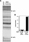
Osmotic sensitivity of CRP-mediated activation of the lac promoter. Primer extension analysis of transcription initiated at the lac promoter by the CRP-dependent wild-type lac promoter (RJ3354) or the CRP-independent lacL8UV5 promoter (RJ3355). Cells were grown in LBN + IPTG and 0.5 m NaCl was added to a portion of the culture. RNA was extracted 10 min and 30 min after salt addition and subjected to primer extension.

Similar articles

Cited by

References

    1. Bell A, Gaston K, Williams R, Chapman K, Kolb A, Buc H, Minchin S, Williams J, Busby S. Mutations that alter the ability of the Escherichia coli cyclic AMP receptor protein to activate transcription. Nucleic Acids Res. 1990;18:7243–7250. - PMC - PubMed
    1. Booth IR, Higgins CF. Enteric bacteria and osmotic stress: Intracellular potassium glutamate as a secondary signal of osmotic stress? FEMS Microbiol Rev. 1990;6:239–246. - PubMed
    1. Burgess RR, Jendrisak JJ. A procedure for the rapid, large-scale purification of Escherichia coli DNA-dependent RNA polymerase involving Polymin P precipitation and DNA-cellulose chromatography. Biochemistry. 1975;14:4634–4638. - PubMed
    1. Cairney J, Booth IR, Higgins CF. Salmonella typhimurium proP gene encodes a transport system for the osmoprotectant betaine. J Bacteriol. 1985;164:1218–1223. - PMC - PubMed
    1. Case CC, Roels SM, Gonzalez JE, Simons EL, Simons RW. Analysis of the promoters and transcripts involved in IS10 anti-sense RNA control. Gene. 1988;72:219–236. - PubMed

Publication types

MeSH terms

LinkOut - more resources