. 1999 Dec 6;190(11):1595-604.
doi: 10.1084/jem.190.11.1595.

Methylation-dependent gene silencing induced by interleukin 1beta via nitric oxide production

Affiliations

Methylation-dependent gene silencing induced by interleukin 1beta via nitric oxide production

A Hmadcha et al. J Exp Med. .

Abstract

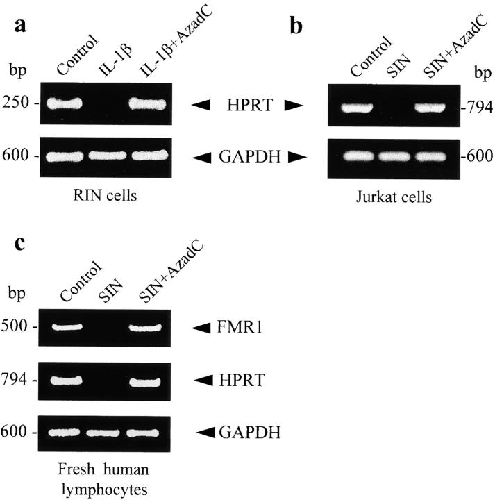
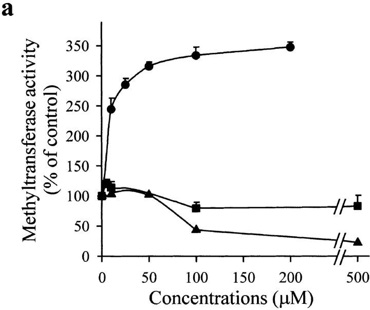
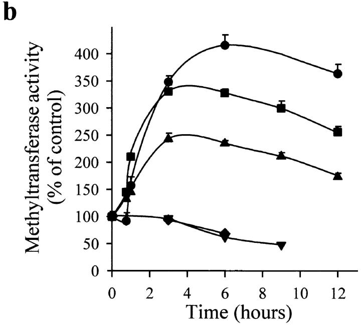
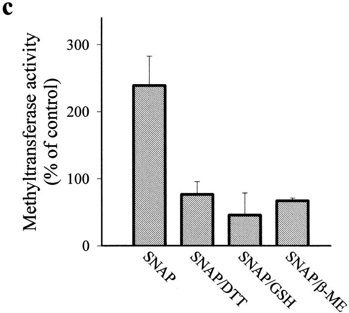
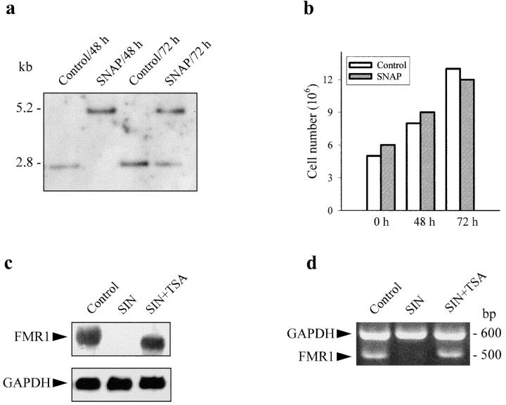
Interleukin (IL)-1beta is a pleiotropic cytokine implicated in a variety of activities, including damage of insulin-producing cells, brain injury, or neuromodulatory responses. Many of these effects are mediated by nitric oxide (NO) produced by the induction of NO synthase (iNOS) expression. We report here that IL-1beta provokes a marked repression of genes, such as fragile X mental retardation 1 (FMR1) and hypoxanthine phosphoribosyltransferase (HPRT), having a CpG island in their promoter region. This effect can be fully prevented by iNOS inhibitors and is dependent on DNA methylation. NO donors also cause FMR1 and HPRT gene silencing. NO-induced methylation of FMR1 CpG island can be reverted by demethylating agents which, in turn, produce the recovery of gene expression. The effects of IL-1beta and NO appear to be exerted through activation of DNA methyltransferase (DNA MeTase). Although exposure of the cells to NO does not increase DNA MeTase gene expression, the activity of the enzyme selectively increases when NO is applied directly on a nuclear protein extract. These findings reveal a previously unknown effect of IL-1beta and NO on gene expression, and demonstrate a novel pathway for gene silencing based on activation of DNA MeTase by NO and acute modification of CpG island methylation.


Figures

Figure 1
Figure 1
FMR1 expression in RIN cells treated with IL-1β and NO donors. In this and other figures, RT-PCR of KH domains and CGG repeats of FMR1 give bands of 500 and 242 bp, respectively; Na/K ATP-ase (250 bp) and GAPDH (600 bp) were used as control. (a) Time course of IL-1β (25 U/ml) induced FMR1 repression in RIN cells. (b) 16 h treatment with IL-1β (25 U/ml), SNP (100 μM), or IL-1β (25 U/ml) plus NMA (100 μM). (c) 16 h treatment with IL-1β (25 U/ml) or IL-1β plus a specific iNOS inhibitor (20 nM AMT, 100 nM EIT, or 10 μM L-NIL). (d) Different exposure times of RIN cells with ActD alone (5 μg/ml) or added simultaneously with SNP (100 μM). Data shown in this and the other figures are representative of two to six experiments.
Figure 2
Figure 2
NO-induced FMR1 silencing and methylation of FMR1 CpG island in Jurkat T cells. (a) RT-PCR of FMR1 KH domains after 16 h incubation with SNP (500 μM) or SIN (100 μM). GAPDH was used as control. (b) Southern blot of genomic DNA digested to completion with EcoRI-EagI. The 2.8-kb fragment results from cleavage by both enzymes, whereas the 5.2-kb fragment corresponds to digestion with EcoRI, indicating protection from restriction by methylation. Untreated cells (lane 1); cells incubated for 24 h with SNP (500 μM) or SIN (100 μM) (lanes 2 and 4); cells treated for 24 h simultaneously with SNP (500 μM) plus AzadC (2 μg/ml) or SIN (100 μM) plus AzadC (2 μg/ml) (lanes 3 and 5); cells incubated for 16 h with SIN (100 μM) (lane 6); and cells exposed for 16 h to SIN (100 μM), washed, and incubated for 24 h with AzadC (2 μg/ml) (lane 7). (c) Methylation pattern induced by 3, 4, 5, or 6 h incubation with SNAP (100 μM). (d) DNA MeTase activity in RIN and Jurkat T cells treated for 16 h with IL-1β (25 U/ml) and for 6 h with SIN (100 μM), respectively, expressed as cpm/μg protein (100 cpm/μg protein corresponds to a specific activity of 0.015 pmol/h/μg protein).
Figure 3
Figure 3
FMR1 expression in RIN and Jurkat T cells incubated with IL-1β or NO donors and the demethylating agent AzadC. (a) Northern blot of FMR1 and iNOS in RIN cells incubated for 24 h with IL-1β (25 U/ml) in the absence or presence of AzadC (2 μg/ml). (b) Northern blot of FMR1 in Jurkat T cells following the same protocol as in the legend to Fig. 2 b, except for the condition shown in lane 6. GAPDH expression was used as control.
Figure 4
Figure 4
Effect of NO on the expression of DNA MeTase in Jurkat T cells. (a) Northern blot of DNA MeTase in cells incubated for 16 h with 100 μM SNAP. FMR1 and GAPDH were used as control. (b) Western blot of DNA MeTase using a polyclonal antibody in cells incubated for 16 h with 100 μM SNAP.
Figure 6
Figure 6
HPRT expression in RIN and Jurkat T cells assessed by RT-PCR. (a) RIN cells unstimulated and treated for 24 h with IL-1β (25 U/ml) in the absence or presence of AzadC (2 μg/ml). (b) Similar protocol used with Jurkat T cells stimulated with 100 μM SIN. (c) RT-PCR of FMR1 and HPRT from fresh peripheral lymphocytes incubated for 24 h with SIN (100 μM) in the absence or presence of AzadC (2 μg/ml). RT-PCR of GAPDH was used as control.
Figure 5
Figure 5
Effect of NO on DNA MeTase activity in a nuclear protein extract of Jurkat T cells. (a) Dose-dependent effect of 3 h incubation with SNAP (•), sodium nitrite (▪), and peroxynitrite (▴) on the activity of DNA MeTase. (b) Time course of DNA MeTase activity in the presence of 50 μM SNAP (•), SNP (▪), SIN (▴), expired SIN (▾), or expired SNAP (♦). Values in plots a and b are expressed in percentage of control values by mean ± SEM of three or two independent experiments in duplicate. (c) Effect of reducing agents on DNA MeTase activity stimulated by 50 μM SNAP. The nuclear protein extract was incubated for 3 h without (control) or with 50 μM SNAP. Nuclear protein extracts were recovered, after washing, by centrifugation or by filtration in a Sephadex G-25 spin column. Samples were further incubated for 2 h with no addition (control and SNAP) or in the presence of 5 mM DTT (SNAP/DTT), 100 μM GSH (SNAP/GSH), or 100 μM β-ME (SNAP/β-ME). Values are expressed in percentage of control values by mean ± SEM of at least three independent experiments in duplicate. All the values of DNA MeTase activity in the presence of the reducing agents were significantly different (P < 0.001) from the value of enzymatic activity in the absence of the agents. The average DNA MeTase activity in basal conditions was 0.017 ± 0.005 (n = 15) pmol/h/μg protein. In each experiment, the basal DNA MeTase activity was set to 100%.
Figure 5
Figure 5
Effect of NO on DNA MeTase activity in a nuclear protein extract of Jurkat T cells. (a) Dose-dependent effect of 3 h incubation with SNAP (•), sodium nitrite (▪), and peroxynitrite (▴) on the activity of DNA MeTase. (b) Time course of DNA MeTase activity in the presence of 50 μM SNAP (•), SNP (▪), SIN (▴), expired SIN (▾), or expired SNAP (♦). Values in plots a and b are expressed in percentage of control values by mean ± SEM of three or two independent experiments in duplicate. (c) Effect of reducing agents on DNA MeTase activity stimulated by 50 μM SNAP. The nuclear protein extract was incubated for 3 h without (control) or with 50 μM SNAP. Nuclear protein extracts were recovered, after washing, by centrifugation or by filtration in a Sephadex G-25 spin column. Samples were further incubated for 2 h with no addition (control and SNAP) or in the presence of 5 mM DTT (SNAP/DTT), 100 μM GSH (SNAP/GSH), or 100 μM β-ME (SNAP/β-ME). Values are expressed in percentage of control values by mean ± SEM of at least three independent experiments in duplicate. All the values of DNA MeTase activity in the presence of the reducing agents were significantly different (P < 0.001) from the value of enzymatic activity in the absence of the agents. The average DNA MeTase activity in basal conditions was 0.017 ± 0.005 (n = 15) pmol/h/μg protein. In each experiment, the basal DNA MeTase activity was set to 100%.
Figure 7
Figure 7
Reversibility of CpG island methylation in dividing cells and reversion of FMR1 expression by a deacetylase inhibitor. (a) Jurkat T cells were incubated for 16 h with 100 μM SNAP. Methylation of the CpG island of FMR1 was measured by Southern blot at 48 and 72 h after washout of the stimulus. Control cells were treated in the same way but without exposure to SNAP. (b) Cell number was measured in the same experimental conditions as in a, with the MTT cell proliferation kit. (c) Northern blot of FMR1 in Jurkat T cells incubated for 24 h in the absence or presence of 2 μM TSA, an inhibitor of deacetylases. (d) RT-PCR of KH domain of FMR1 in the same experimental conditions as in c. In both cases, GAPDH was used as control.

Similar articles

Cited by

References

    1. Bird A.P. CpG islands and the function of DNA methylation. Nature. 1986;321:209–213. - PubMed
    1. Razin A., Cedar H. DNA methylation and gene expression. Microbiol. Rev. 1991;55:451–458. - PMC - PubMed
    1. Bird A. The essentials of DNA methylation. Cell. 1992;70:5–8. - PubMed
    1. Tate P.H., Bird A.P. Effects of DNA methylation on DNA-binding proteins and gene expression. Curr. Opin. Genet. Dev. 1993;3:226–231. - PubMed
    1. Eden S., Cedar H. Role of DNA methylation in the regulation of transcription. Curr. Opin. Genet. Dev. 1994;4:255–259. - PubMed

Publication types

MeSH terms

Substances