. 1999 Nov 29;147(5):1049-62.
doi: 10.1083/jcb.147.5.1049.

Programmed cell death of embryonic motoneurons triggered through the Fas death receptor

Affiliations

Programmed cell death of embryonic motoneurons triggered through the Fas death receptor

C Raoul et al. J Cell Biol. .

Abstract

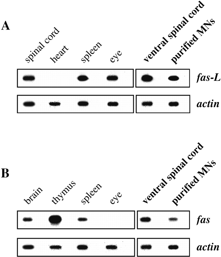
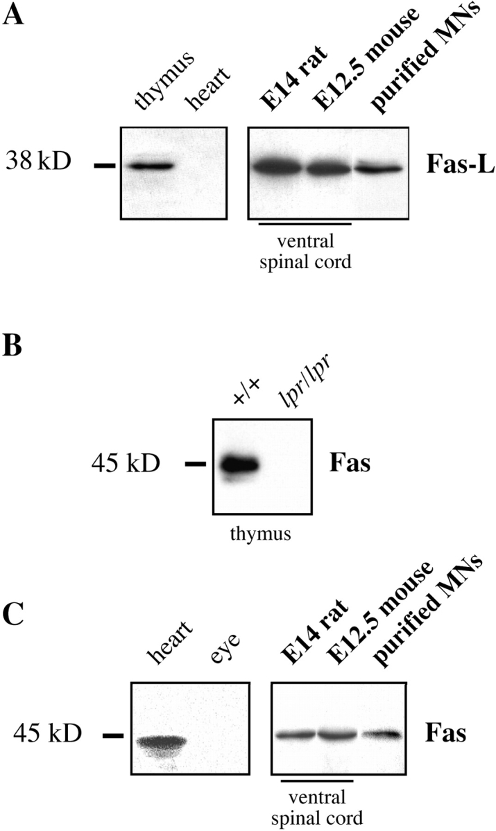
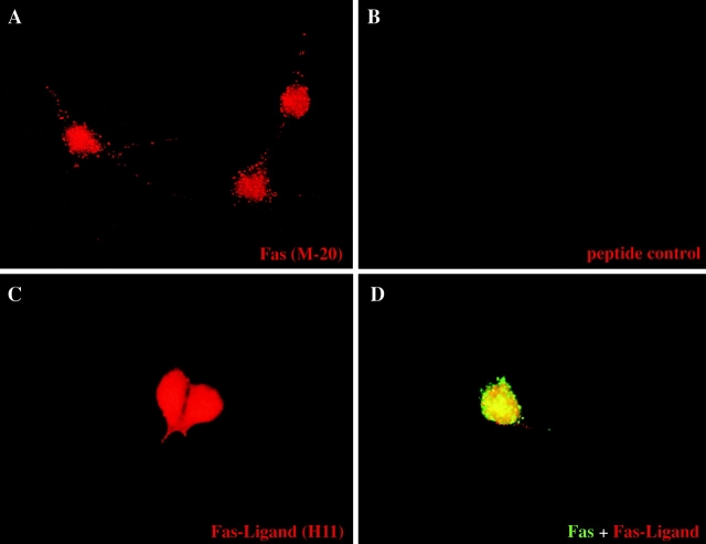
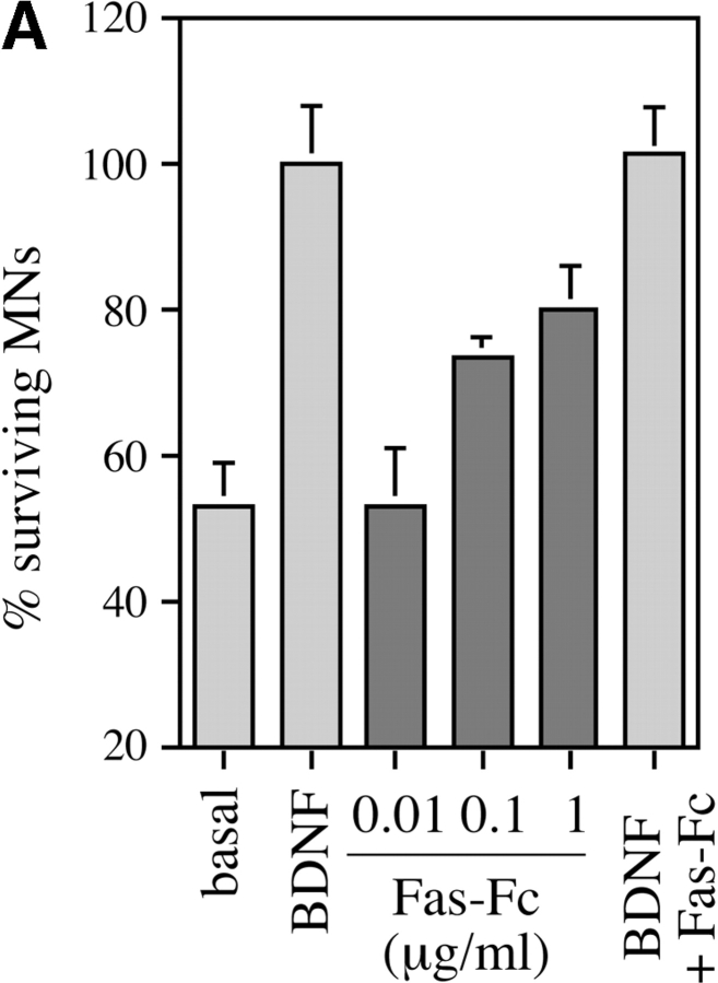
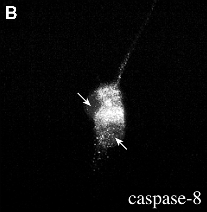
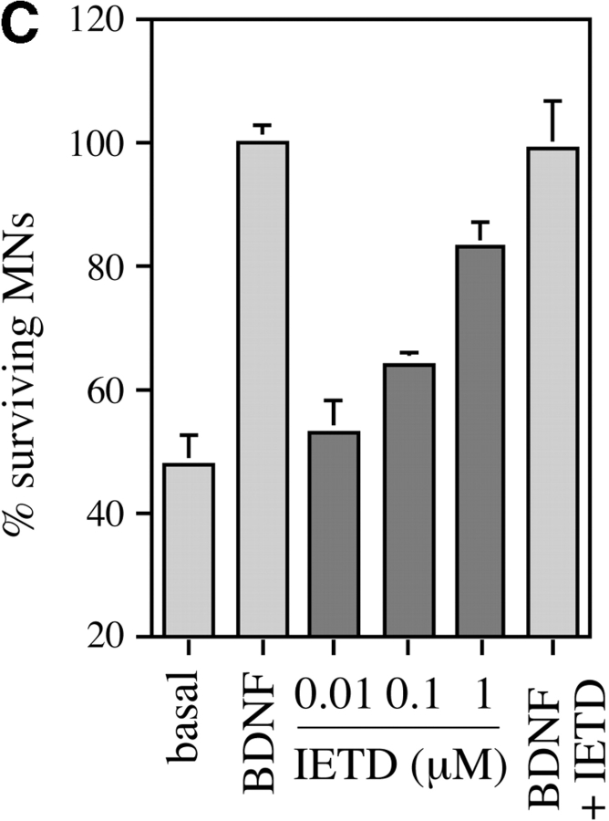
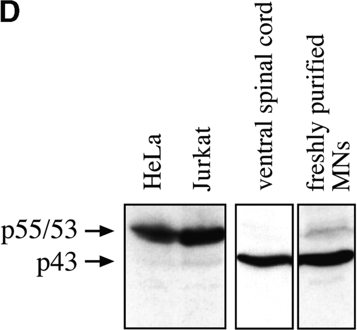
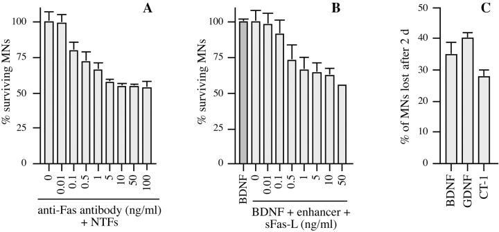
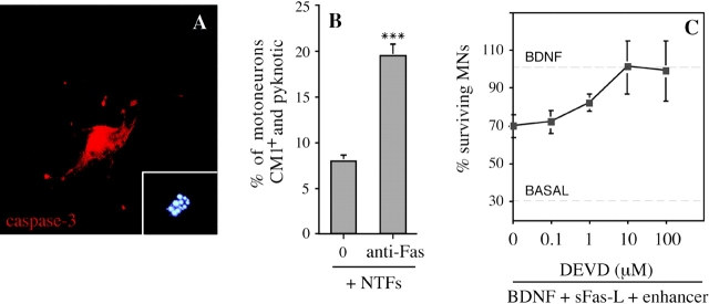
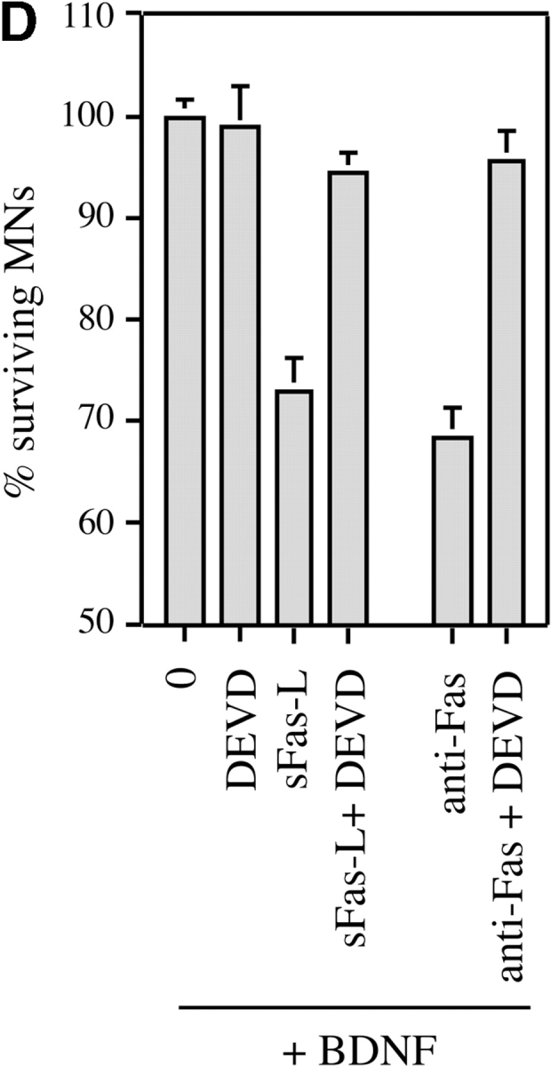
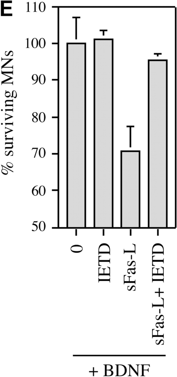
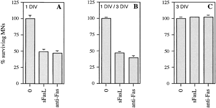
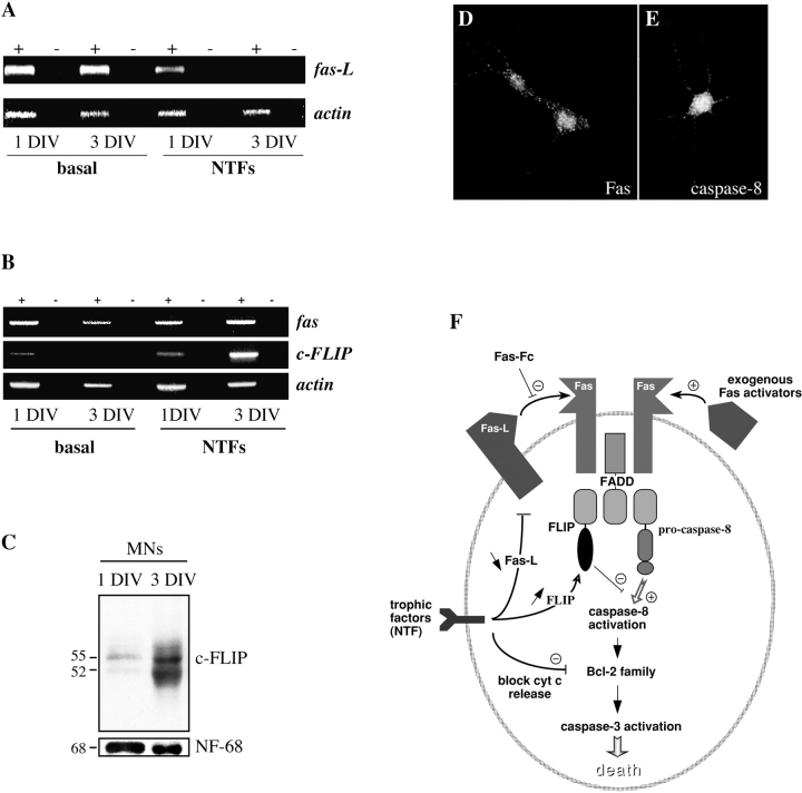
About 50% of spinal motoneurons undergo programmed cell death (PCD) after target contact, but little is known about how this process is initiated. Embryonic motoneurons coexpress the death receptor Fas and its ligand FasL at the stage at which PCD is about to begin. In the absence of trophic factors, many motoneurons die in culture within 2 d. Most (75%) of these were saved by Fas-Fc receptor body, which blocks interactions between Fas and FasL, or by the caspase-8 inhibitor tetrapeptide IETD. Therefore, activation of Fas by endogenous FasL underlies cell death induced by trophic deprivation. In the presence of neurotrophic factors, exogenous Fas activators such as soluble FasL or anti-Fas antibodies triggered PCD of 40-50% of purified motoneurons over the following 3-5 d; this treatment led to activation of caspase-3, and was blocked by IETD. Sensitivity to Fas activation is regulated: motoneurons cultured for 3 d with neurotrophic factors became completely resistant. Levels of Fas expressed by motoneurons varied little, but FasL was upregulated in the absence of neurotrophic factors. Motoneurons resistant to Fas activation expressed high levels of FLICE-inhibitory protein (FLIP), an endogenous inhibitor of caspase-8 activation. Our results suggest that Fas can act as a driving force for motoneuron PCD, and raise the possibility that active triggering of PCD may contribute to motoneuron loss during normal development and/or in pathological situations.


Figures

Figure 1
Figure 1
Embryonic motoneurons express Fas and FasL both in situ and after purification. Total RNA was prepared from the indicated rat tissues, and RT-PCR analysis followed by Southern blotting was performed using specific primers and internal probes for FasL (A) and Fas (B). RT-PCR for β-actin was used as an internal control to ensure that similar concentrations of mRNA were present in all samples. For each mRNA, the first panel corresponds to positive and negative control samples prepared from indicated tissues of adult rat; the results are in agreement with several reports in the literature. The second panel shows results from E14 rat embryos, using either freshly dissected ventral spinal cord or motoneurons purified by a metrizamide-immunoaffinity method in conditions that limit de novo synthesis of mRNA. Fas and FasL are present at significant levels in motoneurons. Alternate unlabeled lanes in all panels are control in which reverse transcriptase was omitted from the incubation.
Figure 2
Figure 2
Embryonic motoneurons express Fas and FasL polypeptides in vivo. Western blot analysis was performed using specific antibodies to FasL and Fas. (A) Antibodies to FasL specifically label a 38-kD band in P17 mouse thymus, but not in heart. In ventral spinal cord from rodent embryos dissected at the stage at which motoneuron PCD is about to begin, FasL is abundant. It is also clearly present in freshly purified motoneurons from E14 rat. (B) Controls for the specificity of immunoblot-purified anti-Fas antibodies (M-20) using thymus extracts from P17 mice of the C57BL/6 strain. Wild-type (+/+) and Fas mutant (lpr/lpr) samples are shown. (C) Using immunoblot-purified antibodies, Fas is abundant in adult rat heart but not in eye. Both ventral spinal cord and freshly purified motoneurons from embryos show clear expression of a single band corresponding to Fas.
Figure 3
Figure 3
Motoneurons in culture coexpress Fas and FasL. Immunolabeling of purified E12.5 mouse motoneurons cultured for 1 d in the presence of neurotrophic factors. (A) Cultured motoneurons labeled using blot-purified rabbit anti-Fas (M-20). Note punctate labeling of cell membranes and more diffuse labeling of nucleus. There was variation in intensity from cell to cell (not apparent in this field). (B) Control in which the same primary antibody was preincubated with the immunogenic peptide. Preincubation with an irrelevant peptide did not reduce staining (data not shown). (C) Immunostaining using a rat mAb against FasL (H11). Note the strong homogeneous staining. (D) Double immunolabeling for Fas (green) and FasL (red), showing the death receptor and its ligand present at the cell surface of the same motoneuron. Controls using an irrelevant rat mAb of the same class or purified rabbit antibodies gave no significant staining (data not shown).
Figure 4
Figure 4
Activation of Fas and caspase-8 by endogenous FasL is required for death of motoneurons in the absence of trophic support. (A) Freshly purified rat motoneurons were induced to die by culturing them in basal medium only for 24 h, or were kept alive by addition of an optimal concentration of BDNF (1 ng/ml). Increasing concentrations of Fas-Fc (0.01–1 μg/ml), which inhibits Fas activation by FasL, added to basal medium promoted their survival (expressed as a percentage of the number in BDNF) in a dose-dependent manner. Addition of 1 μg/ml of Fas-Fc to motoneurons cultured in the presence of BDNF (1 ng/ml) had no effect on their survival. (B) Immunolabeling of two cultured E12.5 mouse motoneurons using a specific antibody recognizing all forms of caspase-8. Double-labeling using DAPI (data not shown) allowed for localization of nuclei (arrows). All motoneurons presented clear granular labeling of the cytoplasm. (C) After seeding, rat motoneurons were incubated with increasing doses of the caspase-8 peptide inhibitor IETD-fmk (0.01–1 μM) without or with BDNF (1 ng/ml). The number of surviving cells was measured 24 h later and expressed relative to survival in BDNF. (D) Western blotting reveals the presence of caspase-8 in ventral spinal cord from mouse embryos, and in purified mouse motoneurons. The molecular mass of the major species (43 kD) is significantly lower than in nonneural cell lines (53–55 kD).
Figure 4
Figure 4
Activation of Fas and caspase-8 by endogenous FasL is required for death of motoneurons in the absence of trophic support. (A) Freshly purified rat motoneurons were induced to die by culturing them in basal medium only for 24 h, or were kept alive by addition of an optimal concentration of BDNF (1 ng/ml). Increasing concentrations of Fas-Fc (0.01–1 μg/ml), which inhibits Fas activation by FasL, added to basal medium promoted their survival (expressed as a percentage of the number in BDNF) in a dose-dependent manner. Addition of 1 μg/ml of Fas-Fc to motoneurons cultured in the presence of BDNF (1 ng/ml) had no effect on their survival. (B) Immunolabeling of two cultured E12.5 mouse motoneurons using a specific antibody recognizing all forms of caspase-8. Double-labeling using DAPI (data not shown) allowed for localization of nuclei (arrows). All motoneurons presented clear granular labeling of the cytoplasm. (C) After seeding, rat motoneurons were incubated with increasing doses of the caspase-8 peptide inhibitor IETD-fmk (0.01–1 μM) without or with BDNF (1 ng/ml). The number of surviving cells was measured 24 h later and expressed relative to survival in BDNF. (D) Western blotting reveals the presence of caspase-8 in ventral spinal cord from mouse embryos, and in purified mouse motoneurons. The molecular mass of the major species (43 kD) is significantly lower than in nonneural cell lines (53–55 kD).
Figure 5
Figure 5
Activation of Fas triggers PCD of motoneurons even in the presence of optimal trophic support. Different agents known to activate Fas receptor by clustering were tested for their ability to trigger motoneuron cell death. (A) Purified E12.5 mouse motoneurons were cultured in the presence of BDNF (1 ng/ml), CT-1 (10 ng/ml), and GDNF (0.1 ng/ml) (+NTFs) for 1 d. Subsequently, the indicated concentrations of anti-Fas antibody were added in the continued presence of NTFs, and motoneuron survival was counted 2 d later, at 3 DIV. Counts were expressed relative to the value for NTFs at 3 DIV. Fas antibody induced a dose-dependent loss of 45% of the motoneurons. (B) An analogous experiment using purified motoneurons from E14 rat, cultured in the continued presence of BDNF (1 ng/ml). After 1 DIV, tagged soluble FasL was added at the indicated concentrations in the presence of 1 μg/ml of enhancer antibody, which had no effect when tested alone. Survival was counted 2 d later. (C) Comparison of the fractions of E14 rat motoneurons lost after 2 d of incubation with 10 ng/ml sFasL, in the presence of indicated neurotrophic factors used at the same concentrations as for mouse motoneurons in A. The number of motoneurons lost was expressed as a percentage of the total number of motoneurons present in the same conditions but without sFasL. All histograms are representative of at least three independent experiments. Error bars represent the mean ± range of duplicate wells.
Figure 6
Figure 6
Motoneuron cell death triggered by Fas activation in the presence of trophic factors involves caspases-3 and -8. (A) Activation of caspase-3 in cultured mouse motoneurons treated with anti-Fas was visualized using the CM1 antibody, which specifically recognizes the activated form of caspase-3. The motoneuron illustrated was undergoing apoptosis, as visualized by the fragmented chromatin of the nucleus stained with DAPI (inset). Note the intense perinuclear staining for activated caspase-3. (B) Increase in the fraction of CM1-positive mouse motoneurons 30 h after treatment with anti-Fas antibodies in the presence of neurotrophic factors. After immunostaining, CM1-positive motoneurons were counted along two diameters of 14-mm coverslips (∼100 motoneurons counted for each). All CM1-positive cells showed pyknotic nuclei by DAPI staining. Values are means ± SEM of five coverslips, and are typical of two independent experiments (asterisks indicate P = 0.001 by t test). Total survival was not significantly different in the two conditions (P > 0.5; data not shown). (C) Death of E14 rat motoneurons triggered by sFasL (10 ng/ml) in the presence of BDNF (1 ng/ml) is inhibited in a dose-dependent fashion by the caspase-3 peptide inhibitor DEVD-fmk. (D) Death of mouse motoneurons triggered by sFasL (10 ng/ml) and enhancer (1 μg/ml), or anti-Fas antibodies (10 ng/ml), is blocked by DEVD-fmk (10 μM). Survival is expressed as the percentage of the number of motoneurons surviving in the presence of BDNF alone. (E) Death of rat motoneurons triggered by sFasL (10 ng/ml, in the presence of enhancer) is inhibited by the caspase-8 inhibitor IETD-fmk (1 μM). C, D, and E show combined mean values (± S.D.) from duplicate wells in two independent experiments.
Figure 6
Figure 6
Motoneuron cell death triggered by Fas activation in the presence of trophic factors involves caspases-3 and -8. (A) Activation of caspase-3 in cultured mouse motoneurons treated with anti-Fas was visualized using the CM1 antibody, which specifically recognizes the activated form of caspase-3. The motoneuron illustrated was undergoing apoptosis, as visualized by the fragmented chromatin of the nucleus stained with DAPI (inset). Note the intense perinuclear staining for activated caspase-3. (B) Increase in the fraction of CM1-positive mouse motoneurons 30 h after treatment with anti-Fas antibodies in the presence of neurotrophic factors. After immunostaining, CM1-positive motoneurons were counted along two diameters of 14-mm coverslips (∼100 motoneurons counted for each). All CM1-positive cells showed pyknotic nuclei by DAPI staining. Values are means ± SEM of five coverslips, and are typical of two independent experiments (asterisks indicate P = 0.001 by t test). Total survival was not significantly different in the two conditions (P > 0.5; data not shown). (C) Death of E14 rat motoneurons triggered by sFasL (10 ng/ml) in the presence of BDNF (1 ng/ml) is inhibited in a dose-dependent fashion by the caspase-3 peptide inhibitor DEVD-fmk. (D) Death of mouse motoneurons triggered by sFasL (10 ng/ml) and enhancer (1 μg/ml), or anti-Fas antibodies (10 ng/ml), is blocked by DEVD-fmk (10 μM). Survival is expressed as the percentage of the number of motoneurons surviving in the presence of BDNF alone. (E) Death of rat motoneurons triggered by sFasL (10 ng/ml, in the presence of enhancer) is inhibited by the caspase-8 inhibitor IETD-fmk (1 μM). C, D, and E show combined mean values (± S.D.) from duplicate wells in two independent experiments.
Figure 7
Figure 7
Resistance of motoneurons to the cell-killing effects of Fas activation is tightly regulated. Mouse motoneurons were cultured in the continued presence of BDNF, CNTF, and GDNF (NTFs) and then treated with the Fas activators sFasL (50 ng/ml) or anti-Fas antibodies (50 ng/ml). Motoneuron survival in all experiments was counted at 5 DIV, and expressed relative to the value in NTFs alone at that time. (A) Fas activators were added at 1 DIV only. (B) Fas activators were added at 1 and 3 DIV; no further motoneuron death was observed. (C) Fas activators were added at 3 DIV: all motoneurons became resistant to Fas activation. Histograms are representative of similar results obtained in three independent experiments. Error bars represent the mean ± range of duplicate values.
Figure 8
Figure 8
Regulation of FasL and FLIP by neurotrophic factors. (A and B) Levels of molecules involved in Fas signaling were followed using semiquantitative RT-PCR on motoneurons cultured for 1 and 3 DIV in the presence or absence of neurotrophic factors. (A) FasL mRNA is upregulated in motoneurons cultured in the absence of trophic factors. (B) Regulation of fas and FLIP mRNAs. mRNA samples were incubated with (+) or without (−) reverse transcriptase. Fas was expressed at relatively constant levels when normalized to actin, whereas FLIP was strongly upregulated in motoneurons cultured for 3 DIV in the presence of NTFs. (C) Strong upregulation of FLIP protein in the presence of neurotrophic factors. Western blots are shown of extracts of purified motoneurons cultured for 1 and 3 DIV in the presence of NTFs; two major forms of FLIP at 55 and 52 kD are observed. The loading control was neurofilament-68. (D) Immunolabeling of Fas-resistant mouse motoneurons cultured for 3 d in the presence of NTFs, using the blot-purified polyclonal antibodies against Fas characterized in Fig. 2. Fas continues to be expressed at the cell membrane. (E) Motoneurons treated for 3 d with NTFs also continue to express caspase-8. (F) Summary diagram showing the potential involvement of the Fas system in PCD of motoneurons. The main experimental strategies adopted in our study of the role of Fas are depicted. In addition, we have indicated the three levels at which neurotrophic factors may potentially act to block Fas-related motoneuron death.

Similar articles

Cited by

References

    1. Adachi M., Suematsu S., Kondo T., Ogasawara J., Tanaka T., Yoshida N., Nagata S. Targeted mutation in the Fas gene causes hyperplasia in peripheral lymphoid organs and liver. Nat. Genet. 1995;11:294–300. - PubMed
    1. Arce V., Garcès A., de Bovis B., Filippi P., Henderson C., Pettmann B., deLapeyrière O. Cardiotrophin-1 requires LIFRβ to promote survival of mouse motoneurons purified by a novel technique. J. Neurosci. Res. 1999;55:119–126. - PubMed
    1. Armstrong R.C., Aja T.J., Hoang K.D., Gaur S., Bai X., Alnemri E.S., Litwack G., Karanewsky D.S., Fritz L.C., Tomaselli K.J. Activation of the CED3/ICE-related protease CPP32 in cerebellar granule neurons undergoing apoptosis but not necrosis. J. Neurosci. 1997;17:553–562. - PMC - PubMed
    1. Ashkenazi A., Dixit V.M. Death receptorssignaling and modulation. Science. 1998;281:1305–1308. - PubMed
    1. Becher B., Barker P.A., Owens T., Antel J.P. CD95-CD95Lcan the brain learn from the immune system? Trends Neurosci. 21 1998. 114 117a - PubMed

Publication types

MeSH terms

Substances