. 1999 Dec;19(12):8469-78.
doi: 10.1128/MCB.19.12.8469.

BCL-2 is phosphorylated and inactivated by an ASK1/Jun N-terminal protein kinase pathway normally activated at G(2)/M

Affiliations

BCL-2 is phosphorylated and inactivated by an ASK1/Jun N-terminal protein kinase pathway normally activated at G(2)/M

K Yamamoto et al. Mol Cell Biol. 1999 Dec.

Abstract

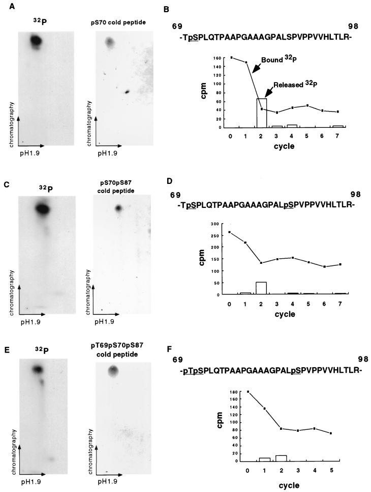
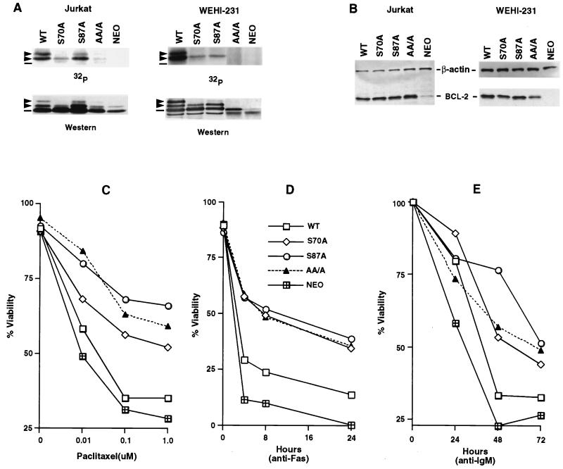
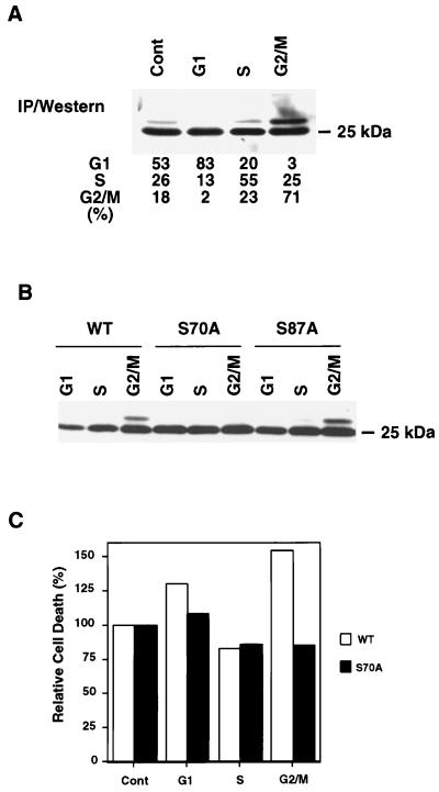
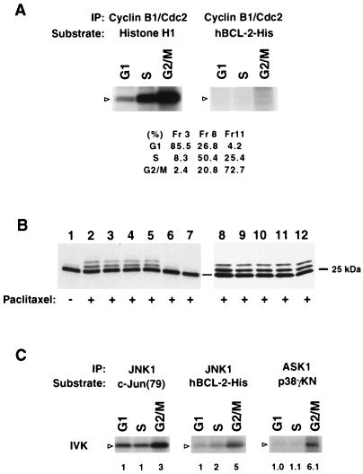
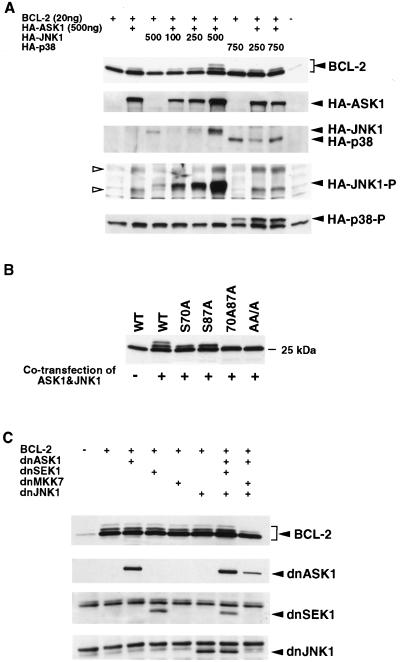
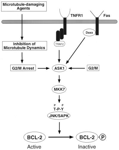
Multiple signal transduction pathways are capable of modifying BCL-2 family members to reset susceptibility to apoptosis. We used two-dimensional peptide mapping and sequencing to identify three residues (Ser70, Ser87, and Thr69) within the unstructured loop of BCL-2 that were phosphorylated in response to microtubule-damaging agents, which also arrest cells at G(2)/M. Changing these sites to alanine conferred more antiapoptotic activity on BCL-2 following physiologic death signals as well as paclitaxel, indicating that phosphorylation is inactivating. An examination of cycling cells enriched by elutriation for distinct phases of the cell cycle revealed that BCL-2 was phosphorylated at the G(2)/M phase of the cell cycle. G(2)/M-phase cells proved more susceptible to death signals, and phosphorylation of BCL-2 appeared to be responsible, as a Ser70Ala substitution restored resistance to apoptosis. We noted that ASK1 and JNK1 were normally activated at G(2)/M phase, and JNK was capable of phosphorylating BCL-2. Expression of a series of wild-type and dominant-negative kinases indicated an ASK1/Jun N-terminal protein kinase 1 (JNK1) pathway phosphorylated BCL-2 in vivo. Moreover, the combination of dominant negative ASK1, (dnASK1), dnMKK7, and dnJNK1 inhibited paclitaxel-induced BCL-2 phosphorylation. Thus, stress response kinases phosphorylate BCL-2 during cell cycle progression as a normal physiologic process to inactivate BCL-2 at G(2)/M.


Figures

FIG. 1
FIG. 1
BCL-2 is phosphorylated on serine and threonine in vivo. (A) The mobility shift of BCL-2 induced by paclitaxel is due to phosphorylation. BCL-2 was immunoprecipitated from 32P-orthophosphate-labeled Jurkat-BCL-2 cells treated with paclitaxel (+) or DMSO (−) by using anti-hBCL-2 Ab 6C8, separated by SDS-PAGE, and transferred to a nitrocellulose membrane. After autoradiography, the same membrane was subjected to Western analysis with anti-hBCL-2 Ab Bcl-2/100. The three 32P-labeled bands (arrows) corresponded to three mobility-shifted bands by immunoblotting. The position of nonphosphorylated BCL-2 is indicated by a dash. (B) λPPase treatment of immunoprecipitated BCL-2. Immunoprecipitated BCL-2 from Jurkat-BCL-2 cells treated with paclitaxel was incubated with λPPase at 30°C for 30 min with or without phosphatase inhibitors (50 mM NaF, 2 mM sodium orthovanadate, 5 mM EDTA, and 5 mM EGTA). The resultant samples were subjected to Western blot analysis. (C) Phosphoamino acid analysis of BCL-2. BCL-2 was immunoprecipitated from 32P-labeled Jurkat-BCL-2 cells treated with paclitaxel, separated by SDS-PAGE, and transferred to a polyvinylidene difluoride membrane. Each radioactive band on the membrane (Fig. 1A) was hydrolyzed with hydrochloric acid. The amino acid composition was determined by 2D electrophoresis on TLC plates. Encircled areas indicate the locations of phosphoserine (P-Ser), phosphothreonine (P-Thr), and phosphotyrosine (P-Tyr), visualized by ninhydrin staining; 1, 2, and 3 represent results from bands 1, 2, and 3, respectively.
FIG. 2
FIG. 2
Paclitaxel induces phosphorylation of Ser70, Ser87, and Thr69 in BCL-2 (A) 2D mapping and synthetic phosphopeptide comigration study of band 1. BCL-2 was immunoprecipitated from 32P-labeled Jurkat-BCL-2 cells treated with paclitaxel, size fractionated by SDS-PAGE, and transferred to a nitrocellulose membrane. The tryptic peptides of band 1 (Fig. 1A) were separated on a TLC plate by electrophoresis at pH 1.9 and chromatography. A synthetic pS70 phosphopeptide corresponding to band 1 migrated and was visualized on a TLC plate by ninhydrin staining. Moreover, the admixture of the pS70 peptide with the tryptic peptides revealed comigration on TLC plates (not shown). (B) Manual sequencing of tryptic phosphopeptide from band 1. The 32P-labeled peptide was eluted from the TLC plate, conjugated to a Sequelon-AA membrane, and subjected to manual Edman degradation. The radioactivity on the membrane (closed squares) or released into the liquid (open bars) was measured at the end of each cycle. (C and D) 2D mapping, synthetic phosphopeptide migration, and manual sequencing of band 2 as described above. (E and F) 2D mapping of synthetic phosphopeptide migration and manual sequencing of band 3 as described above. Tryptic peptides derived from bands 1, 2, and 3 were also eluted from the radioactive spots on TLC plates (A, C, and E) and subjected to phosphoamino acid analysis, confirming their composition.
FIG. 3
FIG. 3
Substitution of phosphorylation sites in BCL-2 further enhances antiapoptotic activity. (A) Jurkat clones and WEHI-231 clones stably expressing WT or alanine-substituted phosphorylation sites of BCL-2, consisting of Ser70 (S70A), Ser87 (S87A), or all three phosphorylation sites, including Thr69 plus the two serines (AA/A), or an empty vector (Neo) were generated. Cells were treated with 1 μM paclitaxel for Jurkat clones or 1 μM vincristine for WEHI-231 clones and in vivo labeled with 32P-orthophosphate. BCL-2 was immunoprecipitated, separated by SDS-PAGE, and transferred to a membrane. After autoradiography, the same membranes were used for immunoblotting. Arrowheads denote phosphorylated BCL-2, and dashes denote nonphosphorylated BCL-2. The residual-shifted band on Western analysis of Jurkat cells bearing AA/A or Neo represents the endogenous hBCL-2 of Jurkat cells. (B) BCL-2 expression levels in Jurkat and WEHI-231 clones, determined by Western blot analysis of lysates from equivalent cells of various Jurkat or WEHI-231 clones with anti-hBCL-2 Ab 6C8 and anti-β-actin Ab. (C to E) Cell viability assays of Jurkat clones (C and D) or WEHI-231 clones (E). Jurkat clones expressing comparable WT, S70A, S87A, AA/A BCL-2, or Neo control vector were stimulated with various doses of paclitaxel for 24 h (C) or anti-Fas antibody (100 ng/ml) (D), while WEHI-231 clones were treated with anti-IgM Ab (BET-2 supernatant at 1:100 dilution) (E). Viability was determined by PI exclusion using flow cytometry. The results represent the means of triplicate assays. Another independent set of clones expressing matched levels of WT or mutant BCL-2 showed comparable results.
FIG. 4
FIG. 4
Phosphorylation of BCL-2 during the cell cycle. (A) Phosphorylation of endogenous BCL-2 at G2/M phase. Jurkat-Neo cells were elutriated to enrich for G1, S, and G2/M fractions and compared to asynchronous cells (Cont). Cellular lysates were subjected to immunoprecipitation (IP) with anti-BCL-2 monoclonal Ab 6C8 and the subsequent Western blot was developed with anti-hBCL-2 Ab Bcl-2/100. The percentages of cells in G1, S, and G2/M were determined by PI staining using FACScan and CELLQUEST software. (B) Ser70 was phosphorylated in cycling cells. Jurkat clones expressing WT, S70A, or S87A BCL-2 were elutriated and subjected to Western analysis. (C) Cells with phosphorylated BCL-2 at G2/M demonstrated increased susceptibility to apoptosis. Jurkat clones expressing WT or S70A BCL-2 were elutriated, and typical G1-, S-, and G2/M-enriched fractions were compared to asynchronous cells (Cont). Cells were treated with 100 ng of anti-Fas Ab per ml, and cell viability was determined by PI exclusion 6 h later. Values represent the relative viability of each fraction when the viability of the asynchronous cells is set at 100%. Values are the means of duplicate assays.
FIG. 5
FIG. 5
G2/M-activated ASK1-JNK1 pathway and JNK1 phosphorylation of BCL-2 in vitro. (A) G2/M-activated cyclin B1-Cdc2 complex does not phosphorylate BCL-2. Elutriated G1 (fraction [Fr] 3), S (Fr 8), and G2/M (Fr 11) fractions of Jurkat cells were lysed and immunoprecipitated (IP) with anti-cyclin B1. The phosphorylation of substrate histone H1 was assessed. The activity of this kinase complex for recombinant hBCL-2-His was also assessed. Arrowheads denote the position of the substrate. Cell cycle status was assayed by PI staining using a FACScan and CELLQUEST software. (B) Genistein and staurosporine inhibit paclitaxel-induced BCL-2 phosphorylation. Jurkat cells expressing WT BCL-2 were pretreated with various kinase inhibitors including 50 μM PD98059 (lane 3), 10 μM SB203580 (lane 4), 10 μM SB202190 (lane 5), 10 μg of genistein per ml (lane 6), 0.1 μM staurosporine (lane 7), 100 μM Rp-cAMP (lane 9), 10 μM LY294002 (lane 10), 1 μM wortmannin (lane 11), 20 ng of rapamycin per ml (lane 12), or DMSO (lanes 1, 2, and 8) for 60 min and then treated with (+) or without (−) paclitaxel for 6 h. BCL-2 phosphorylation was examined by Western blot analysis. (C) In vitro kinase (IVK) assay. The ASK1-JNK1 pathway is activated at the G2/M stage in cycling cells, and JNK1 phosphorylates BCL-2 in vitro. Lysates from elutriated fractions of Jurkat cells were immunoprecipitated with anti-ASK1 (DAV) serum or anti-JNK1 antibody. The ASK1 complex was incubated with GST-MKK6 and then with GST-p38γKN as a substrate to measure kinase activity. JNK activity was determined with GST–c-Jun (79) as a substrate. Recombinant hBCL-2-His was also incubated with the JNK1 complex. The position of substrates is denoted by open arrowheads. The fold activation of kinase activity is indicated as detected by phosphorimage analysis with activity at G1 set at 1.0. Western analysis of immunoprecipitates confirmed an equivalent amount of the kinase protein in each fraction (not shown).
FIG. 6
FIG. 6
ASK1-JNK1 pathway phosphorylates BCL-2 in vivo. (A) Dose-dependent phosphorylation of BCL-2 by JNK1 in vivo. 293 cells were transfected with 20 ng of hBCL-2 or control vector together with indicated amounts of HA-ASK1, HA-JNK1, and/or HA-p38, as shown. The total amount of transfected DNA was kept constant by adding a compensating amount of empty vector pcDNA3. After 24 h, cells were lysed and the phosphorylation of BCL-2, expression level of each kinase, and activation of JNK and p38 were determined by Western analysis. Open arrowheads denote the activated endogenous JNKs (p46 and p54). (B) Substrate specificity of ASK1-JNK1 pathway in vivo. WT or phosphorylation site mutant BCL-2 expression vectors (20 ng) were cotransfected with ASK1 (500 ng) and JNK (750 ng) or pcDNA3 into 293 cells. Lysates generated 24 h after transfection were assessed for BCL-2 phosphorylation by Western analysis. (C) Inhibition of BCL-2 phosphorylation by dnASK1, dnMKK7, and dnJNK1. WT BCL-2 (20 ng) was cotransfected with dnASK1 (500 ng), dnSEK1 (500 ng), dnMKK7 (500 ng), and dnJNK1 (750 ng), as indicated, into 293 cells. The total amount of DNA transfected was normalized by adding pcDNA3. After 16 h, paclitaxel was added to a final concentration of 1 μM and cells were incubated an additional 8 h. BCL-2 phosphorylation and the expression levels of the dominant-negative (dn) kinases were determined by immunoblotting.
FIG. 7
FIG. 7
Schematic representation of the ASK1/MKK7/JNK1 pathway in BCL-2 phosphorylation. ASK1, a MAP3K, is activated by extracellular and intracellular stimuli to induce JNK pathway activation. JNK phosphorylates BCL-2, inactivating its antiapoptotic function. TNFR1, tumor necrosis factor receptor 1.

Similar articles

Cited by

References

    1. Bakhshi A, Jensen J P, Goldman P, Wright J J, McBride O W, Epstein A L, Korsmeyer S J. Cloning the chromosomal breakpoint of t(14;18) human lymphomas: clustering around JH on chromosome 14 and near a transcriptional unit on 18. Cell. 1985;41:899–906. - PubMed
    1. Blagosklonny M V, Giannakakou P, el-Deiry W S, Kingston D G, Higgs P I, Neckers L, Fojo T. Raf-1/bcl-2 phosphorylation: a step from microtubule damage to cell death. Cancer Res. 1997;57:130–135. - PubMed
    1. Blume-Jensen P, Janknecht R, Hunter T. The kit receptor promotes cell survival via activation of PI 3-kinase and subsequent Akt-mediated phosphorylation of Bad on Ser136. Curr Biol. 1998;8:779–782. - PubMed
    1. Boyle W J, van der Geer P, Hunter T. Phosphopeptide mapping and phosphoamino acid analysis by two-dimensional separation on thin-layer cellulose plates. Methods Enzymol. 1991;201:110–149. - PubMed
    1. Chang B S, Minn A J, Muchmore S W, Fesik S W, Thompson C B. Identification of a novel regulatory domain in Bcl-X(L) and Bcl-2. EMBO J. 1997;16:968–977. - PMC - PubMed

Publication types

MeSH terms