. 1999 Nov;10(11):3787-99.
doi: 10.1091/mbc.10.11.3787.

Mammalian transcription factor ATF6 is synthesized as a transmembrane protein and activated by proteolysis in response to endoplasmic reticulum stress

Affiliations
Free PMC article

Mammalian transcription factor ATF6 is synthesized as a transmembrane protein and activated by proteolysis in response to endoplasmic reticulum stress

K Haze et al. Mol Biol Cell. 1999 Nov.
Free PMC article

Abstract

The unfolded protein response (UPR) controls the levels of molecular chaperones and enzymes involved in protein folding in the endoplasmic reticulum (ER). We recently isolated ATF6 as a candidate for mammalian UPR-specific transcription factor. We report here that ATF6 constitutively expressed as a 90-kDa protein (p90ATF6) is directly converted to a 50-kDa protein (p50ATF6) in ER-stressed cells. Furthermore, we showed that the most important consequence of this conversion was altered subcellular localization; p90ATF6 is embedded in the ER, whereas p50ATF6 is a nuclear protein. p90ATF6 is a type II transmembrane glycoprotein with a hydrophobic stretch in the middle of the molecule. Thus, the N-terminal half containing a basic leucine zipper motif is oriented facing the cytoplasm. Full-length ATF6 as well as its C-terminal deletion mutant carrying the transmembrane domain is localized in the ER when transfected. In contrast, mutant ATF6 representing the cytoplasmic region translocates into the nucleus and activates transcription of the endogenous GRP78/BiP gene. We propose that ER stress-induced proteolysis of membrane-bound p90ATF6 releases soluble p50ATF6, leading to induced transcription in the nucleus. Unlike yeast UPR, mammalian UPR appears to use a system similar to that reported for cholesterol homeostasis.


Figures

Figure 1
Figure 1
Direct conversion of p90ATF6 to p50ATF6 in thapsigargin-treated HeLa cells. (A) Immunoblotting analysis of ATF6. HeLa cells cultured in 60-mm dishes until 60% confluency were incubated in the presence of 300 nM thapsigargin (Tg) for the indicated periods. Cells were washed with PBS, scraped with a rubber policeman, and lysed in 100 μl of 1× Laemmli’s SDS sample buffer. After boiling for 5 min, 5-μl aliquots of each sample were subjected to SDS-PAGE (10% gel) and analyzed by immunoblotting with anti-ATF6 antibody or anti-KDEL antibody, which recognizes GRP78. The positions of p90ATF6 and p50ATF6 are indicated by the open and closed arrowheads, respectively. The positions of prestained SDS-PAGE molecular weight standards (Bio-Rad, Hercules, CA) are also shown. (B) Pulse–chase analysis of ATF6. HeLa cells cultured in 60-mm dishes were pulse labeled for 30 min with [35S]methionine and [35S]cysteine and then chased for the indicated periods in the absence (−Tg) or presence (+Tg) of 300 nM thapsigargin as described in MATERIALS AND METHODS. It should be noted that thapsigargin was not added during the pulse labeling of the cells. 35S-Labeled proteins were extracted and immunoprecipitated with anti-ATF6 antibody, and the immunoprecipitates were subjected to SDS-PAGE (7.5% gel). Radioactivities of 35S-labeled p90ATF6 and p50ATF6 were visualized using a BAS-2000 BioImaging Analyzer (Fuji Photo film, Stanford, CT).
Figure 2
Figure 2
Conversion of p90ATF6 to p50ATF6 in the absence of new protein synthesis in dithiothreitol-treated HeLa cells. HeLa cells cultured in 60-mm dishes until 80% confluency were pretreated for 30 min with 10 μg/ml cycloheximide and then treated for the indicated periods with 300 nM thapsigargin (Tg) or 1 mM dithiothreitol (DTT) in the presence of 10 μg/ml cycloheximide. Immunoblotting analysis of ATF6 was carried out essentially as described in the legend to Figure 1A.
Figure 3
Figure 3
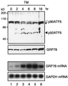
Processing of ATF6 in tunicamycin-treated HeLa cells. HeLa cells cultured in 100-mm dishes until 60% confluency were incubated in the presence of 2 μg/ml tunicamycin (TM) for the indicated periods. Proteins were extracted and analyzed by immunoblotting as described in the legend to Figure 1A. The asterisk denotes an unglycosylated form of p90ATF6. Total RNA was also extracted and analyzed by Northern blot hybridization using radiolabeled probes specific to GRP78 or GAPDH as described previously (Yoshida et al., 1998).
Figure 4
Figure 4
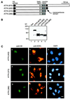
Identification of an internal hydrophobic stretch that anchors ATF6 in the ER membrane. (A) Indirect immunofluorescence analysis. Unstressed HeLa cells were fixed and stained with anti-ATF6 antibody (a), anti-KDEL antibody (b), or DAPI (c) as described in MATERIALS AND METHODS. Bar, 10 μm. (B) Schematic structure of ATF6 consisting of 670 amino acids. The positions of the serine cluster, basic region, and leucine zipper (Zhu et al., 1997; Yoshida et al., 1998) as well as the transmembrane domain identified in this report are indicated. The thick line below the sequence represents the region (amino acids 6–307) fused with E. coli maltose-binding protein to raise anti-ATF6 antibody (Yoshida et al., 1998). Three potential glycosylation sites are also shown schematically (ψ). The hydropathy index was calculated by the method of Kyte and Doolittle (1982).
Figure 5
Figure 5
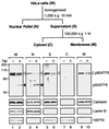
Distribution of p90ATF6 and p50ATF6 in fractions of HeLa cells. HeLa cells cultured in 175-cm2 flasks until 80% confluency were incubated in the absence (−) or presence (+) of 2 μg/ml tunicamycin (TM) for 4 h. Cells were harvested, disrupted using a Dounce-type homogenizer, and then centrifuged at 1000 × g for 10 min to obtain the nuclear pellet (N) essentially as described by Dignam et al. (1983). The resulting supernatant (S) was further centrifuged at 100,000 × g for 1 h to separate the soluble cytosolic fraction (C) from insoluble membrane fraction (M). Aliquots of the indicated fraction as well as unfractionated HeLa cells (whole; W) corresponding to 0.5 × 10⁵ cells were subjected to SDS-PAGE (10% gel) and analyzed by immunoblotting with anti-ATF6 antibody or various other antibodies as indicated. The positions of p90ATF6 and p50ATF6 are marked as in Figure 1.
Figure 6
Figure 6
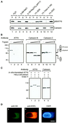
p90ATF6 is an integral membrane glycoprotein with its N terminus located in the cytoplasm. (A) Differential solubilization of p90ATF6. The 1000 × g supernatant fraction prepared from unstressed HeLa cells as described in the legend to Figure 5 was mixed with 0.1 vol of one of the following solutions: H2O, 5 M NaCl, 1 M Na2CO3, pH 11, 10% SDS, 10% Triton X-100, or 10% sodium deoxycholate (DOC). After incubation for 15 min at room temperature, mixtures were centrifuged at 100,000 × g for 1 h to separate supernatant (S) from pellet (P), followed by SDS-PAGE (10% gel) and immunoblotting analysis using anti-ATF6 antibody or anti-N terminus of calnexin antibody. (B) Topology of p90ATF6. The 1000 × g supernatant fraction prepared from unstressed HeLa cells (50 μg of proteins) was incubated with increasing amounts of trypsin (0 μg for lanes 1, 5, and 9; 0.1 μg for lanes 2, 6, and 10; 0.3 μg for lanes 3, 7, and 11; and 1.0 μg for lanes 4, 8, and 12) for 15 min at room temperature. Digestion was terminated by addition of an equal volume of 2× Laemmli’s SDS sample buffer followed by boiling for 5 min. Samples were subjected to SDS-PAGE (10% gel) and analyzed by immunoblotting with anti-ATF6 antibody (lanes 1–4), anti-N terminus of calnexin antibody (Calnexin-N; lanes 5–8), or anti-C terminus of calnexin antibody (Calnexin-C; lanes 9–12). The position of p90ATF6 is marked by the open arrowhead. The positions of full-length calnexin and its truncated form lacking the cytoplasmic domain are shown schematically. (C) Glycosylation of p90ATF6. The 1000 × g supernatant fraction prepared from unstressed HeLa cells (2.5 μg of proteins) was boiled in the presence of 1% SDS and 1% 2-mercaptoethanol for 5 min. After addition of 2 volumes of 150 mM sodium citrate buffer, pH 5.5, the samples were incubated for 20 h at 37°C in the absence (lanes 2 and 4) or presence (lanes 3 and 5) of 0.25 mU endoglycosidase H (Endo H) obtained from ICN (Costa Mesa, CA). Samples as well as in vitro–translated ATF6 (lane 1) prepared as described by Yoshida et al. (1998) were subjected to SDS-PAGE (7.5% gel) and analyzed by immunoblotting with anti-ATF6 antibody (lanes 1–3) or anti-N terminus of calnexin antibody (lanes 4 and 5). (D) Indirect immunofluorescence analysis of transfected cells. HeLa cells on slide glasses transiently transfected with pCGN-ATF6 (670) (see Figure 8A for its schematic structure) were fixed and stained with anti-HA epitope antibody (a), anti-KDEL antibody (b), or DAPI (c). Bar, 10 μm.
Figure 7
Figure 7
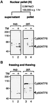
Solubility of p90ATF6 and p50ATF6. (A) Fractionation of nuclear pellet. The nuclear pellet fraction prepared as described in the legend to Figure 5 was washed with PBS three times and resuspended in nuclear extraction buffer (20 mM HEPES-KOH, pH 7.6, 25% glycerol, 0.5 M NaCl, 1.5 mM MgCl2, 1 mM EDTA, 5 μg/ml pepstatin A, 5 μg/ml leupeptin, and 2 μg/ml aprotinin). After rotating for 1 h at 4°C, the samples were centrifuged at 100,000 × g for 1 h to separate the supernatant (sup.) from the pellet (ppt.). Aliquots of the indicated fractions were subjected to SDS-PAGE (10% gel) and analyzed by immunoblotting with anti-ATF6 antibody. The positions of p90ATF6 and p50ATF6 are marked as in Figure 1. (B) Effect of freezing and thawing. HeLa cells cultured in 60-mm dishes until 80% confluency were incubated in the absence (−) or presence (+) of 2 μg/ml tunicamycin (TM) for 4 h. Cells were washed with PBS, scraped with a rubber policeman, and centrifuged at 1000 × g for 5 min. After three cycles of freezing and thawing of cell pellets suspended in 50 μl of PBS, samples were centrifuged at 15,000 × g for 10 min to separate the supernatant (sup.) from the pellet (ppt.), which was then resuspended in 50 μl of PBS. Aliquots of each supernatant and pellet corresponding to 1 × 10⁵ cells were subjected to SDS-PAGE (10% gel) and analyzed by immunoblotting with anti-ATF6 antibody.
Figure 8
Figure 8
Expression and localization of various mutant forms of ATF6 in transfected HeLa cells. (A) Schematic structures of ATF6 derivatives analyzed. Full-length ATF6 cDNA, ATF6 (670), and three C-terminal deletion mutants were inserted into the mammalian expression vector pCGN. The positions of the HA epitope, basic region, leucine zipper, and transmembrane domain are indicated. (B) Immunoblotting analysis of transfected cells. HeLa cells in 60-mm dishes were transiently transfected with 1 μg of pCGN vector alone (Vec) or each of the ATF6 expression plasmids as indicated. Total proteins were extracted from transfected cells directly with 1× Laemmli’s SDS sample buffer followed by boiling for 5 min. Samples were subjected to SDS-PAGE (10% gel) and analyzed by immunoblotting with anti-ATF6 antibody. (C) Indirect immunofluorescence analysis of transfected cells. HeLa cells on slide glasses transiently transfected with pCGN-ATF6 (402) (a–c), pCGN-ATF6 (373) (d–f), and pCGN-ATF6 (366) (g–i) were fixed and stained with anti-HA epitope antibody (a–g), anti-KDEL antibody (b–h), or DAPI (c–i). Bar, 10 μm.
Figure 9
Figure 9
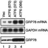
Effects of overexpression of full-length ATF6 and two nuclear localization mutants on the levels of endogenous GRP78. HeLa cells in 60-mm dishes were transiently transfected with 10 μg of pCGN vector alone (Vec) or each of the ATF6-expression plasmids as indicated, the structures of which are shown schematically in Figure 8A. Total RNA was prepared 48 h after transfection and analyzed by Northern blot hybridization using radiolabeled probes specific to GRP78 or GAPDH as described previously (Yoshida et al., 1998). Proteins were extracted 48 h after transfection by repeated freezing and thawing followed by sonication. The amounts of proteins recovered in the supernatant after centrifugation at 15,000 × g for 10 min were determined using a Bio-Rad protein assay kit. Aliquots of 30 μg were subjected to SDS-PAGE (10% gel) and analyzed by immunoblotting with anti-KDEL antibody.
Figure 10
Figure 10
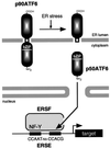
Model for ER stress-induced processing of ATF6. ATF6 is constitutively synthesized as a precursor protein (p90ATF6) that anchors in the ER membrane through the single transmembrane domain near the center of the molecule. ER stress-induced proteolysis of p90ATF6 releases the N-terminal fragment (p50ATF6) containing bZIP, although the precise cleavage site is unknown. p50ATF6 translocates into the nucleus and interacts with the general transcription factor NF-Y to form a complex designated here as ER stress response factor (ERSF). ERSF activates transcription through ERSE (CCAAT-N9-CCACG) present in the promoter regions of mammalian UPR target genes.

Similar articles

Cited by

References

    1. Brown MS, Goldstein JL. The SREBP pathway: regulation of cholesterol metabolism by proteolysis of a membrane-bound transcription factor. Cell. 1997;89:331–340. - PubMed
    1. Chan Y-M, Jan YN. Roles for proteolysis and trafficking in Notch maturation and signal transduction. Cell. 1998;94:423–426. - PubMed
    1. Chapman RE, Walter P. Translational attenuation mediated by an mRNA intron. Curr Biol. 1997;7:850–859. - PubMed
    1. Clark MW, Abelson J. The subnuclear localization of tRNA ligase in yeast. J Cell Biol. 1987;105:1515–1526. - PMC - PubMed
    1. Cox JS, Chapman RE, Walter P. The unfolded protein response coordinates the production of endoplasmic reticulum protein and endoplasmic reticulum membrane. Mol Biol Cell. 1997;8:1805–1814. - PMC - PubMed

MeSH terms