. 1999 Nov;10(11):3675-88.
doi: 10.1091/mbc.10.11.3675.

Differential regulation of secretory compartments containing the insulin-responsive glucose transporter 4 in 3T3-L1 adipocytes

Affiliations
Free PMC article

Differential regulation of secretory compartments containing the insulin-responsive glucose transporter 4 in 3T3-L1 adipocytes

C A Millar et al. Mol Biol Cell. 1999 Nov.
Free PMC article

Abstract

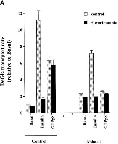
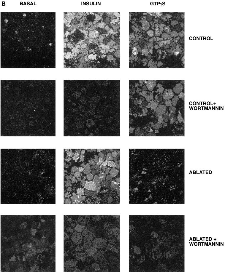
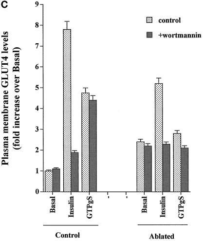
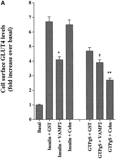
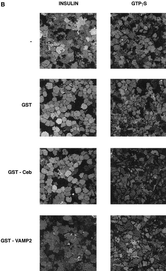
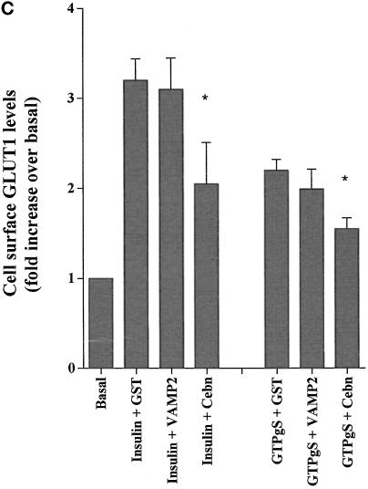
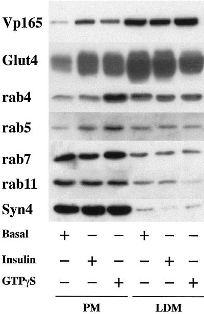
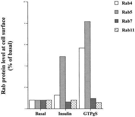
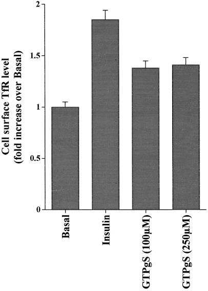
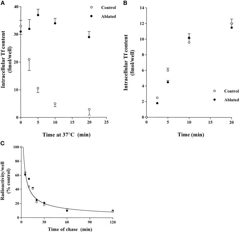
Insulin and guanosine-5'-O-(3-thiotriphosphate) (GTPgammaS) both stimulate glucose transport and translocation of the insulin-responsive glucose transporter 4 (GLUT4) to the plasma membrane in adipocytes. Previous studies suggest that these effects may be mediated by different mechanisms. In this study we have tested the hypothesis that these agonists recruit GLUT4 by distinct trafficking mechanisms, possibly involving mobilization of distinct intracellular compartments. We show that ablation of the endosomal system using transferrin-HRP causes a modest inhibition ( approximately 30%) of insulin-stimulated GLUT4 translocation. In contrast, the GTPgammaS response was significantly attenuated ( approximately 85%) under the same conditions. Introduction of a GST fusion protein encompassing the cytosolic tail of the v-SNARE cellubrevin inhibited GTPgammaS-stimulated GLUT4 translocation by approximately 40% but had no effect on the insulin response. Conversely, a fusion protein encompassing the cytosolic tail of vesicle-associated membrane protein-2 had no significant effect on GTPgammaS-stimulated GLUT4 translocation but inhibited the insulin response by approximately 40%. GTPgammaS- and insulin-stimulated GLUT1 translocation were both partially inhibited by GST-cellubrevin ( approximately 50%) but not by GST-vesicle-associated membrane protein-2. Incubation of streptolysin O-permeabilized 3T3-L1 adipocytes with GTPgammaS caused a marked accumulation of Rab4 and Rab5 at the cell surface, whereas other Rab proteins (Rab7 and Rab11) were unaffected. These data are consistent with the localization of GLUT4 to two distinct intracellular compartments from which it can move to the cell surface independently using distinct sets of trafficking molecules.


Figures

Figure 1
Figure 1
DeGlc uptake and GLUT4 translocation in response to insulin and GTPγS: effects of ablation and wortmannin. In these experiments, 3T3-L1 adipocytes were incubated with TfHRP for 1 h and transferred to ice. After removal of cell surface TfHRP, DAB with or without hydrogen peroxide was added, and the DAB cytochemistry reaction progressed as outlined in MATERIALS AND METHODS. After ablation, cells were washed once in ice-cold PBS and twice in IC buffer at 37°C and then permeabilized with α-toxin as outlined. Cells were stimulated with GTPγS (100 μM) or insulin (1 μM) for 15 min before assay of deGlc uptake (A) or GLUT4 levels in plasma membrane lawns (B and C). In A, each point is the mean of triplicate determinations (± SEM) expressed as fold increase over basal transport rate. (B) Result of a typical experiment assaying GLUT4 levels by plasma membrane lawns. (C) Results of three experiments of this type, quantified as outlined in MATERIALS AND METHODS. In experiments examining the effect of wortmannin, this was added at 100 nM for 5 min before the addition of insulin or GTPγS.
Figure 1
Figure 1
DeGlc uptake and GLUT4 translocation in response to insulin and GTPγS: effects of ablation and wortmannin. In these experiments, 3T3-L1 adipocytes were incubated with TfHRP for 1 h and transferred to ice. After removal of cell surface TfHRP, DAB with or without hydrogen peroxide was added, and the DAB cytochemistry reaction progressed as outlined in MATERIALS AND METHODS. After ablation, cells were washed once in ice-cold PBS and twice in IC buffer at 37°C and then permeabilized with α-toxin as outlined. Cells were stimulated with GTPγS (100 μM) or insulin (1 μM) for 15 min before assay of deGlc uptake (A) or GLUT4 levels in plasma membrane lawns (B and C). In A, each point is the mean of triplicate determinations (± SEM) expressed as fold increase over basal transport rate. (B) Result of a typical experiment assaying GLUT4 levels by plasma membrane lawns. (C) Results of three experiments of this type, quantified as outlined in MATERIALS AND METHODS. In experiments examining the effect of wortmannin, this was added at 100 nM for 5 min before the addition of insulin or GTPγS.
Figure 2
Figure 2
Effects of GST-v-SNARE fusion proteins on insulin- and GTPγS-stimulated GLUT4 and GLUT1 translocation. SLO-permeabilized 3T3-L1 adipocytes were incubated for 10 min with buffer containing GST alone or GST-VAMP2 or GST-cellubrevin fusion proteins, all at a concentration of 15 μg/ml. Cells were then stimulated with GTPγS (100 μM) or insulin (1 μM) for a further 15 min (still in the presence of fusion proteins) before preparation of plasma membrane lawns. (A) Effects of GST fusion proteins on GLUT4 translocation. (B) Representative experiment of this type. In B, cells were stimulated with GTPγS (100 μM) or insulin (1 μM) in the presence of GST alone (GST), GST-cellubrevin (GST-Ceb), or GST-VAMP2 or in the absence of any recombinant protein (−). (C) Effects of the same fusion proteins on GLUT1 translocation. In A and C, data are mean ± SEM of three independent experiments.  No significant difference compared with insulin alone. Significant inhibition is indicated: * p < 0.05; ** p ∼ 0.01.
Figure 2
Figure 2
Effects of GST-v-SNARE fusion proteins on insulin- and GTPγS-stimulated GLUT4 and GLUT1 translocation. SLO-permeabilized 3T3-L1 adipocytes were incubated for 10 min with buffer containing GST alone or GST-VAMP2 or GST-cellubrevin fusion proteins, all at a concentration of 15 μg/ml. Cells were then stimulated with GTPγS (100 μM) or insulin (1 μM) for a further 15 min (still in the presence of fusion proteins) before preparation of plasma membrane lawns. (A) Effects of GST fusion proteins on GLUT4 translocation. (B) Representative experiment of this type. In B, cells were stimulated with GTPγS (100 μM) or insulin (1 μM) in the presence of GST alone (GST), GST-cellubrevin (GST-Ceb), or GST-VAMP2 or in the absence of any recombinant protein (−). (C) Effects of the same fusion proteins on GLUT1 translocation. In A and C, data are mean ± SEM of three independent experiments.  No significant difference compared with insulin alone. Significant inhibition is indicated: * p < 0.05; ** p ∼ 0.01.
Figure 3
Figure 3
Effects of insulin and GTPγS on the subcellular distribution of Rab proteins in 3T3-L1 adipocytes. 3T3-L1 adipocytes were stimulated with GTPγS (100 μM) or insulin (1 μM) for 15 min and subjected to subcellular fractionation as outlined in MATERIALS AND METHODS. Aliquots (10 μg of protein) of isolated plasma membrane (PM) and low-density microsomes (LDM) were separated by SDS PAGE, transferred to nitrocellulose, and immunoblotted with antibodies against vp165, GLUT4, Rab4, Rab5, Rab7, or Rab11 and Syntaxin 4 as indicated. Shown are representative immunoblots from three experiments of this type. Quantification of this data is presented in B.
Figure 3
Figure 3
Effects of insulin and GTPγS on the subcellular distribution of Rab proteins in 3T3-L1 adipocytes. 3T3-L1 adipocytes were stimulated with GTPγS (100 μM) or insulin (1 μM) for 15 min and subjected to subcellular fractionation as outlined in MATERIALS AND METHODS. Aliquots (10 μg of protein) of isolated plasma membrane (PM) and low-density microsomes (LDM) were separated by SDS PAGE, transferred to nitrocellulose, and immunoblotted with antibodies against vp165, GLUT4, Rab4, Rab5, Rab7, or Rab11 and Syntaxin 4 as indicated. Shown are representative immunoblots from three experiments of this type. Quantification of this data is presented in B.
Figure 4
Figure 4
Effects of insulin and GTPγS on cell surface TfR levels. 3T3-L1 adipocytes were permeabilized with α-toxin as outlined in MATERIALS AND METHODS and incubated for 5 min before addition of insulin (1 μM) or GTPγS (100 μM) for 15 min at 37°C. Cells were then rapidly transferred to ice-cold buffer for assay of cell surface TfR levels. Shown are data from a representative experiment; each point is the mean of triplicate determinations (± SEM).
Figure 5
Figure 5
TfR recycling in ablated cells indicates de novo endosome formation. (A) 3T3-L1 adipocytes were incubated with 2.2 μg/ml [125I]transferrin (3 μCi) together with TfHRP at 20 μg/ml. Subsequently, cell surface–attached Tf/TfHRP was removed, and cytochemistry using DAB with or without hydrogen peroxide was performed at 4°C as outlined. After incubation at 4°C for 1 h to complete the ablation reaction, cells were rewarmed to 37°C and incubated for the times shown to allow assay of the release of radiolabeled transferrin from the cells. Cell-associated transferrin was measured at the time shown in control (open circles) and ablated (filled circles) cells. Each point is the mean of three determinations, and data from a representative experiment are shown. (B) In these experiments, cells were loaded with TfHRP as outlined and then transferred to ice-cold buffer to stop membrane trafficking. Subsequently, cell surface–attached Tf/TfHRP was removed, and cytochemistry using DAB with or without hydrogen peroxide was performed at 4°C as outlined. After incubation at 4°C for 1 h to complete the ablation reaction, cells were rewarmed to 37°C, and the rate of uptake of 3 nM [125I]transferrin was measured as described. In these experiments, cells were incubated at 37°C with 3 nM [125I]transferrin for the times shown. Cells were then transferred to ice-cold buffer, cell surface–associated [125I]transferrin was removed by acid washing, and cell-associated transferrin was then determined as outlined. Shown are the data from a typical experiment (each point is the mean of triplicate determina-tions), repeated a further two times. Open circles represent uptake in control cells, and filled circles represent uptake in ablated cells. There was no detectable difference in the rate of Tf uptake under these conditions. (C) The experiment shown in B indicates that the TfR that remains at the cell surface after endosome ablation is still capable of binding ligand and internalizing it. This suggests that an endosomal system may reform de novo. To examine this possibility we next measured TfR recycling under the same conditions. Cells were loaded with TfHRP as outlined and then transferred to ice-cold buffer to stop membrane trafficking. Subsequently, cell surface–attached TfHRP was removed, and cytochemistry using DAB with or without hydrogen peroxide was performed at 4°C as outlined. After incubation at 4°C for 1 h to complete the ablation reaction, cells were rewarmed to 37°C, and 3 nM [125I]transferrin was added to the cells for 30 min at 37°C. Cells were then transferred to ice-cold buffer, and cell surface–associated [125I]transferrin was removed by acid washing. The cells were then rewarmed to 37°C, and the rate of externalization of transferrin was determined. Shown is the result of a typical experiment; each point is the mean of triplicate determinations at each condition. Open circles represent uptake in control cells, and filled circles represent uptake in ablated cells. There was no detectable difference in the rate of Tf release under these conditions.

Similar articles

Cited by

References

    1. Aledo JC, Lavoie L, Volchuck A, Keller SR, Klip A, Hundal HS. Identification and characterization of two distinct intracellular GLUT4 pools in rat skeletal muscle: evidence for an endosomal and an insulin-sensitive GLUT4 compartment. Biochem J, 1997;325:727–732. - PMC - PubMed
    1. Baldini G, Hohman R, Charron MJ, Lodish HF. Insulin and nonhydrolyzable GTP analogs induce translocation of GLUT4 to the plasma membrane in a-toxin-permeabilized rat adipose cells J. Biol Chem, 1991;266:4037–4040. - PubMed
    1. Bottger G, Nagelkerken B, van der Sluijs P. Rab4 and Rab7 define distinct nonoverlapping endosomal compartments J. Biol Chem, 1996;271:29191–29197. - PubMed
    1. Calderhead DM, Kitagawa K, Tanner LI, Holman GD, Lienhard GE. Insulin regulation of the two glucose transporters in 3T3–L1 adipocytes J. Biol Chem, 1990;265:13800–13808. - PubMed
    1. Chen D, Elmendorf JS, Olson AL, Li X, Earp HS, Pessin JE. Osmotic shock stimulates GLUT4 translocation in 3T3–L1 adipocytes by a novel tyrosine kinase pathway J. Biol Chem. 1997;272:27401–27410. - PubMed

Publication types

MeSH terms