. 1999 Nov 15;19(22):9803-12.
doi: 10.1523/JNEUROSCI.19-22-09803.1999.

Subcellular localization of tetanus neurotoxin-insensitive vesicle-associated membrane protein (VAMP)/VAMP7 in neuronal cells: evidence for a novel membrane compartment

Affiliations

Subcellular localization of tetanus neurotoxin-insensitive vesicle-associated membrane protein (VAMP)/VAMP7 in neuronal cells: evidence for a novel membrane compartment

S Coco et al. J Neurosci. .

Abstract

The clostridial neurotoxin-insensitive soluble N-ethylmaleimide-sensitive factor attachment protein (SNAP) receptors, tetanus neurotoxin-insensitive (TI)-vesicle-associated membrane protein (VAMP)/VAMP7, SNAP23, and syntaxin 3 have recently been implicated in transport of exocytotic vesicles to the apical plasma membrane of epithelial cells. This pathway had been shown previously to be insensitive to tetanus neurotoxin and botulinum neurotoxin F. TI-VAMP/VAMP7 is also a good candidate to be implicated in an exocytotic pathway involved in neurite outgrowth because tetanus neurotoxin does not inhibit this process in conditions in which it abolishes neurotransmitter release. We have now found that TI-VAMP/VAMP7 has a widespread distribution in the adult rat brain in which its localization strikingly differs from that of nerve terminal markers. TI-VAMP/VAMP7 does not enrich in synaptic vesicles nor in large dense-core granules but is associated with light membranes. In hippocampal neurons developing in vitro, TI-VAMP/VAMP7 localizes to vesicles in the axonal and dendritic outgrowths and concentrates into the leading edge of the growth cone, a region devoid of synaptobrevin 2, before synaptogenesis. After the onset of synaptogenesis, TI-VAMP/VAMP7 is found predominantly in the somatodendritic domain. In PC12 cells, TI-VAMP/VAMP7 does not colocalize with synaptobrevin 2, chromogranin B, or several markers of endocytic compartments. At the electron microscopic level, TI-VAMP/VAMP7 is mainly associated with tubules and vesicles. Altogether, these results suggest that TI-VAMP/VAMP7 defines a novel membrane compartment in neurite outgrowths and in the somatodendritic domain.


Figures
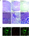
Fig. 1.
Fig. 1.
A widespread distribution of TI-VAMP/VAMP7 in the rat brain. Rat brain paraffin sections were stained for TI-VAMP/VAMP7 and slightly counterstained with hematoxylin (blue staining of nuclei). (Control slides were obtained on serial sections by omitting the first antibody.) Digital pictures were obtained on a DMR HC microscope (Leica) equipped with a tri-CCD color video camera Power HAD (Sony). [Scale bars: top left, 600 μm; top middle, top right, 200 μm; bottom left (hippocampus CA1 region), 30 μm; bottom middle, bottom right (reticular and Purkinje cells), 10 μm.] TI-VAMP/VAMP7 is present in most neurons. It is concentrated in cell bodies.B, TI-VAMP/VAMP7 does not localize in nerve terminals in reticular cells. Double immunofluorescence confocal images showing the lack of colocalization of TI-VAMP/VAMP7 and SV2, an SV protein in the deep nuclei of the cerebellum. Scale bar, 25 μm.
Fig. 2.
Fig. 2.
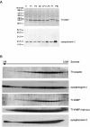
A TI-VAMP/VAMP7 is not a typical SV protein. Brain synaptic vesicles were prepared according to the procedure of Hell and Jahn (1994). The fractions are as follows. H, Homogenate; P1, 1000 gm pellet; P2, synaptosomal fraction, 12,000 gm pellet; S2, microsomes and cytosol, 12,000 gm supernatant; LP1, 33,000 gm pellet of lysed synaptosomes; LP2, crude SV fraction, 260,000 gm pellet of lysed synaptosomes; PI, controlled pore glass fraction corresponding mostly to plasma membrane and microsomes; PIII, controlled pore glass purified SVs. Twenty micrograms of proteins were loaded in each lane. Note that synaptobrevin 2 enriches greatly in LP2 and PIII fractions, whereas TI-VAMP/VAMP7 does not enrich in these fractions. B, TI-VAMP/VAMP7 does not enrich in large dense core vesicles. Distribution of secretory vesicle markers from a low-speed supernatant of bovine adrenal medulla homogenate by isopycnic centrifugation on a 0.4–2 m sucrose gradient. Equal volumes of gradient fractions were analyzed by SDS-PAGE and Western blotting for the proteins indicated. Note that TI-VAMP/VAMP7 is not found in the membrane fractions at the bottom of the gradient in which synaptotagmin 1 and synaptobrevin 2 concentrates. TI-VAMP/VAMP7 and transferrin receptor are mainly concentrated in light fractions. A high exposure of the autoradiogram reveals a low concentration of TI-VAMP/VAMP7 in the heavy fractions corresponding to LDCVs.
Fig. 3.
Fig. 3.
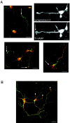
Localization of TI-VAMP/VAMP7 in axonal and dendritic outgrowths. A, TI-VAMP/VAMP7 in axonal outgrowths at 2 div. Rat hippocampal neurons in primary culture were fixed, stained at 2 div for TI-VAMP/VAMP7 (red) and synaptobrevin 2 (green), and observed by confocal microcopy (color plates) or conventional microscopy (black and white plates). TI-VAMP/VAMP7 localized to vesicles scattered throughout the cell body, the neurites, and the growing axon. A significant amount of the protein was found at the leading edge of the growing axon (arrows) in which synaptobrevin 2 was not found. Distinct hot spots of TI-VAMP/VAMP7 and synaptobrevin 2 were observed along the axon. Very high concentrations of both proteins were found in the cell bodies. Scale bars: left color micrograph, 25 μm; right color micrograph, 10 μm; black and white micrographs, 12 μm. B, TI-VAMP/VAMP7 in dendritic outgrowths at 7 div. Rat hippocampal neurons in primary culture were fixed, stained at 7 div for TI-VAMP/VAMP7 (red) and synaptobrevin 2 (green), and observed by confocal microcopy. Synaptobrevin 2 was found mainly in nerve terminals, whereas most of TI-VAMP/VAMP7 was localized in dendrites and concentrated in the leading edge of dendrites. Scale bar, 25 μm.
Fig. 4.
Fig. 4.
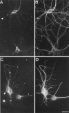
TI-VAMP/VAMP7 enriches in dendrites in mature hippocampal neuron. Rat hippocampal neurons were differentiated in primary culture and stained at 14 div. TI-VAMP/VAMP7 is present in neuronal cell bodies, and it is concentrated in a subpopulation of processes that correspond to dendrites. A,B, Double immunostaining for TI-VAMP/VAMP7 (A) and microtubules (B). Note that TI-VAMP/VAMP7 immunoreactivity is enriched in a subset of short and tapered processes (putative dendrites). C,D, Double immunostaining for TI-VAMP/VAMP7 (C) and MAP2 (D). Note that TI-VAMP/VAMP7 concentrates in MAP2-positive cell extensions. Scale bar:A, B, 28 μm; C,D, 21 μm.
Fig. 5.
Fig. 5.
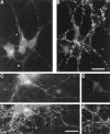
TI-VAMP/VAMP7 does not concentrate in nerve terminals in mature hippocampal neuron. Rat hippocampal neurons were differentiated in primary culture and stained at 14 div.A, B, Double immunostaining for TI-VAMP/VAMP7 (A) and synaptobrevin 2 (B). Note that TI-VAMP/VAMP7 immunoreactivity does not coincide with synaptobrevin 2 but is sometimes adjacent.C, D, Double immunostaining for TI-VAMP/VAMP7 (C) and synaptotagmin 1 (D) in hippocampal neurons that had been treated with TeNT. TeNT induces the loss of synaptobrevin 2 staining (E) but not synaptophysin (F). Note that TeNT has no effect on TI-VAMP/VAMP7 subcellular localization. Scale bars: A,B, 42 μm; C–F, 36 μm.
Fig. 6.
Fig. 6.
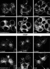
TI-VAMP/VAMP7 has an original localization in control and NGF-treated PC12 cells. PC12 cells were grown on collagen-coated glass and treated with (bottom panels) or without (top panels) 50 ng/ml NGF for 7 d. The cells were then stained for TI-VAMP/VAMP7 and several membrane markers and observed by confocal microscopy. We found that TI-VAMP/VAMP7 does not colocalize with synaptobrevin 2 or chromogranin B but overlaps to a low extent with CD63, a protein known to recycle between lysosomes or granules and the plasma membrane (arrows in top right micrographs). TI-VAMP/VAMP7 does not enrich in varicosities (arrowheads in bottom left micrographs) in which synaptobrevin 2 concentrates after NGF treatment. Scale bars, 5 μm.
Fig. 7.
Fig. 7.
TI-VAMP/VAMP7 does not localize into endocytic compartments in NGF-treated PC12 cells. PC12 cells were grown on collagen-coated glass and treated with 50 ng/ml NGF for 6 d.Top row, The cells were starved for 30 min in serum-free medium in the presence of NGF and then incubated in the same medium containing 5 mg/ml Texas Red-coupled BSA for 2 hr, and the fluid phase marker was subsequently chased in serum-free medium for 15 min. The cells were then stained for TI-VAMP/VAMP7 and observed by confocal microscopy. Note that TI-VAMP/VAMP7 does not colocalize with the BSA-positive endocytic structures. Bottom row, The cells were stained for TI-VAMP/VAMP7 and the early endosome marker EEA1 and observed by confocal microscopy. TI-VAMP/VAMP7 does not colocalize with EEA1. In both cases, note the great differences in the pattern of TI-VAMP staining and the localization of endocytic structures. Scale bar, 10 μm.
Fig. 8.
Fig. 8.
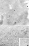
TI-VAMP/VAMP7 is present in tubules and vesicles in PC12 cells. Ultrathin cryosections of PC12 cells were labeled with antibodies directed against TI-VAMP/VAMP7, followed by protein A coupled to 10 nm gold particles (PAG10). Labeling for TI-VAMP/VAMP7 is detected mainly on the cytoplasmic side of vesicles and tubules (arrowheads), LDCVs, and occasionally in Golgi stacks. Note the labeling on a bud from LDCV (inset) and the lack of staining of lysosomes (open arrowhead indicates a labeled tubule nearby an unlabeled lysosome). Note that the topmicrograph is particularly poor in LDCVs compared with the bottom micrograph. This is because of the fact that the distribution of LDCVs is not homogeneous within a PC12 cell in which these heavy organelles tend to enrich at the bottom of the cell. The distribution of gold particles of these micrographs qualitatively but not quantitatively represents the cell profiles that were used to quantitate the data expressed in Results. Scale bars, 104 nm.PM, Plasma membrane; M, mitochondria,GA, Golgi apparatus; L, lysosome.

Similar articles

Cited by

References

    1. Advani RJ, Bae HR, Bock JB, Chao DS, Doung YC, Prekeris R, Yoo JS, Scheller RH. Seven novel mammalian SNARE proteins localize to distinct membrane compartments. J Biol Chem. 1998;273:10317–10324. - PubMed
    1. Banker GA, Cowan WM. Rat hippocampal neurons in dispersed cell culture. Brain Res. 1977;126:397–425. - PubMed
    1. Bartlett WP, Banker GA. An electron microscopic study of the development of axons and dendrites by hippocampal neurons in culture. I. Cells which develop without intercellular contacts. J Neurosci. 1984;4:1944–1953. - PMC - PubMed
    1. Berditchevski F, Bazzoni G, Hemler ME. Specific association of CD63 with the VLA-3 and VLA-6 integrins. J Biol Chem. 1995;270:17784–17790. - PubMed
    1. Bock JB, Scheller RH. Protein transport—a fusion of new ideas. Nature. 1997;387:133–135. - PubMed

Publication types

MeSH terms

Grants and funding

LinkOut - more resources