Infection of neonatal mice with sindbis virus results in a systemic inflammatory response syndrome
- PMID: 10559357
- PMCID: PMC113094
- DOI: 10.1128/JVI.73.12.10387-10398.1999
Infection of neonatal mice with sindbis virus results in a systemic inflammatory response syndrome
Abstract
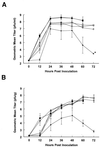
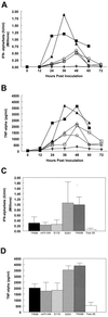
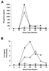
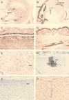
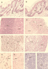
Laboratory strains of viruses may contain cell culture-adaptive mutations which result in significant quantitative and qualitative alterations in pathogenesis compared to natural virus isolates. This report suggests that this is the case with Sindbis virus strain AR339. A cDNA clone comprising a consensus sequence of Sindbis virus strain AR339 has been constructed (W. B. Klimstra, K. D. Ryman, and R. E. Johnston, J. Virol. 72:7357-7366, 1998). This clone (pTR339) regenerates a sequence predicted to be very close to that of the original AR339 isolate by eliminating several cell culture-adaptive mutations present in individual laboratory strains of the virus (K. L. McKnight et al., J. Virol. 70:1981-1989, 1996). It thus provides a unique reagent for study of the pathogenesis of Sindbis virus strain AR339 in mice. Neonatal mouse pathogenesis of virus (TR339) generated from the pTR339 clone was compared with that of virus from a cDNA clone of the cell culture-passaged laboratory AR339 strain, TRSB, and virus from a clone of a more highly cell culture-adapted strain, HR(sp) (Toto 50). The sequence of TRSB differs from the consensus at three coding positions, while Toto 50 differs at eight codons and one nucleotide in the 5' nontranslated region. Both cell culture-adapted strains contain mutations associated with heparan sulfate (HS)-dependent attachment to cells (W.B. Klimstra, K. D. Ryman, and R. E. Johnston, J. Virol. 72:7357-7366, 1998). TR339 caused 100% mortality with an average survival time (AST) of 1.7 +/- 0.25 days. While TRSB also caused 100% mortality, the AST was extended to 2.9 +/- 0.52 days. The more extensively cell culture-adapted virus Toto 50 caused only 30% mortality with an AST extended to 11.0 +/- 4.8 days. TRSB and TR339 induced high serum levels of alpha/beta interferon, gamma interferon, tumor necrosis factor alpha, interleukin-6, and corticosterone and induced pathology reminiscent of lipopolysaccharide-induced endotoxic shock, a type of systemic inflammatory response syndrome. However, the reduced intensity of this response in TRSB-infected mice correlated with the increased AST. Toto 50 failed to induce the shock-like cytokine cascade. In situ hybridization studies indicated that TR339 and TRSB replicated in identical tissues, but the TRSB signal was less widespread at early times postinfection. While Toto 50 also replicated in similar tissues, the extent of replication was severely restricted and mice developed lesions characteristic of encephalitis. A single mutation in TRSB at E2 position 1 (Arg) conferred HS-dependent attachment to cells and was associated with reduced cytokine induction and extended AST in vivo.
Figures



Similar articles
-
Adaptation of Sindbis virus to BHK cells selects for use of heparan sulfate as an attachment receptor.J Virol. 1998 Sep;72(9):7357-66. doi: 10.1128/JVI.72.9.7357-7366.1998. J Virol. 1998. PMID: 9696832 Free PMC article.
-
Deduced consensus sequence of Sindbis virus strain AR339: mutations contained in laboratory strains which affect cell culture and in vivo phenotypes.J Virol. 1996 Mar;70(3):1981-9. doi: 10.1128/JVI.70.3.1981-1989.1996. J Virol. 1996. PMID: 8627724 Free PMC article.
-
Fatal Sindbis virus infection of neonatal mice in the absence of encephalitis.Virology. 1996 Oct 1;224(1):73-83. doi: 10.1006/viro.1996.0508. Virology. 1996. PMID: 8862401
-
Age-dependent susceptibility to fatal encephalitis: alphavirus infection of neurons.Arch Virol Suppl. 1994;9:31-9. doi: 10.1007/978-3-7091-9326-6_4. Arch Virol Suppl. 1994. PMID: 8032263 Review.
-
The role of antibody in recovery from alphavirus encephalitis.Immunol Rev. 1997 Oct;159:155-61. doi: 10.1111/j.1600-065x.1997.tb01013.x. Immunol Rev. 1997. PMID: 9416509 Review.
Cited by
-
Fermented Glutinous Rice Extract Mitigates DSS-Induced Ulcerative Colitis by Alleviating Intestinal Barrier Function and Improving Gut Microbiota and Inflammation.Antioxidants (Basel). 2023 Jan 31;12(2):336. doi: 10.3390/antiox12020336. Antioxidants (Basel). 2023. PMID: 36829894 Free PMC article.
-
Hepatic Disorders and COVID-19: From Pathophysiology to Treatment Strategy.Can J Gastroenterol Hepatol. 2022 Dec 8;2022:4291758. doi: 10.1155/2022/4291758. eCollection 2022. Can J Gastroenterol Hepatol. 2022. PMID: 36531832 Free PMC article. Review.
-
The life cycle of the alphaviruses: From an antiviral perspective.Antiviral Res. 2023 Jan;209:105476. doi: 10.1016/j.antiviral.2022.105476. Epub 2022 Nov 25. Antiviral Res. 2023. PMID: 36436722 Free PMC article. Review.
-
Requirement of a functional ion channel for Sindbis virus glycoprotein transport, CPV-II formation, and efficient virus budding.PLoS Pathog. 2022 Oct 3;18(10):e1010892. doi: 10.1371/journal.ppat.1010892. eCollection 2022 Oct. PLoS Pathog. 2022. PMID: 36191050 Free PMC article.
-
Myricetin inhibits pseudorabies virus infection through direct inactivation and activating host antiviral defense.Front Microbiol. 2022 Sep 15;13:985108. doi: 10.3389/fmicb.2022.985108. eCollection 2022. Front Microbiol. 2022. PMID: 36187970 Free PMC article.
References
-
- American College of Chest Physicians/Society of Critical Care Medicine. Definitions for sepsis and organ failure and guidelines for the use of innovative therapies in sepsis. Crit Care Med. 1992;1992:864–874. - PubMed
-
- Billiau A, Vandekerckhove F. Cytokines and their interactions with other inflammatory mediators in the pathogenesis of sepsis and septic shock. Eur J Clin Investig. 1991;21:559–573. - PubMed
-
- Calandra T, Baumgartner J-D, Grau G E, Wu M-M, Lambert P-H, Schellekens J, Verhoef J, Glauser M P The Swiss-Dutch J5 Immunoglobulin Study Group. Prognostic values of tumor necrosis factor/cachectin, interleukin-1, interferon-alpha, and interferon-gamma in the serum of patients with septic shock. J Infect Dis. 1990;161:982–987. - PubMed
-
- Davies M G, Hagen P-O. Systemic inflammatory response syndrome. Br J Surg. 1997;84:920–935. - PubMed
Publication types
MeSH terms
Substances
Grants and funding
LinkOut - more resources
Full Text Sources
Other Literature Sources

