. 1999 Dec;73(12):10329-38.
doi: 10.1128/JVI.73.12.10329-10338.1999.

Human immunodeficiency virus replication in a primary effusion lymphoma cell line stimulates lytic-phase replication of Kaposi's sarcoma-associated herpesvirus

Affiliations

Human immunodeficiency virus replication in a primary effusion lymphoma cell line stimulates lytic-phase replication of Kaposi's sarcoma-associated herpesvirus

V Varthakavi et al. J Virol. 1999 Dec.

Abstract

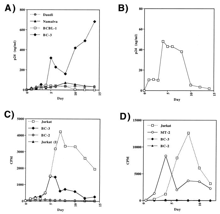
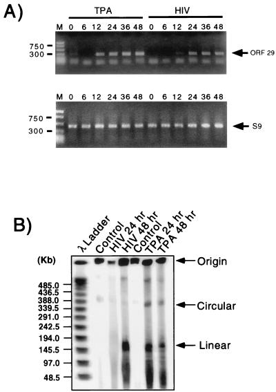
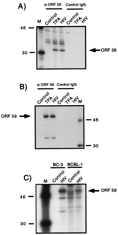
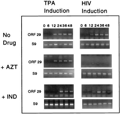
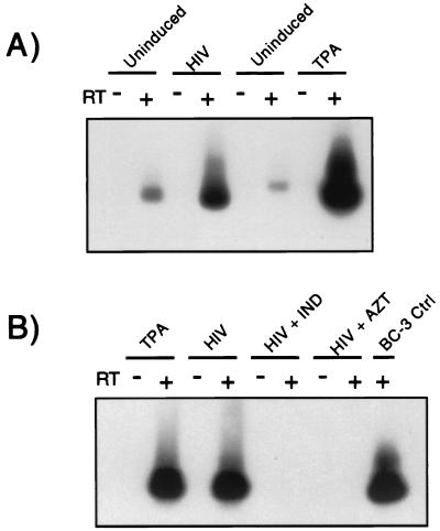
Human immunodeficiency virus (HIV) and Kaposi's sarcoma-associated herpesvirus (KSHV) coinfect many individuals in North America and in parts of Africa. Infection with HIV is a leading risk factor for the development of Kaposi's sarcoma (KS). In this study, we tested the hypothesis that HIV infection of common or adjacent cells would stimulate replication and spread of KSHV. Infection of a primary effusion lymphoma cell line by vesicular stomatitis virus type G-pseudotyped HIV type 1 led to a rapid induction of lytic-phase KSHV replication. Induction of lytic KSHV replication by HIV required active replication of HIV. The addition of the nucleoside reverse transcriptase inhibitor azidothymidine or the protease inhibitor indinavir to the culture prevented HIV spread and inhibited the associated induction of KSHV lytic replication. Lytic replication occurred in both HIV-infected and HIV-uninfected cells within the culture, and could be induced in uninfected cells via a soluble factor released from the HIV-infected cells. Transmission of infectious KSHV to an uninfected target cell was enhanced by HIV replication and was inhibited by antiretroviral drugs. These results may have implications for the pathogenesis and treatment of KS in individuals coinfected with KSHV and HIV.


Figures

FIG. 1
FIG. 1
Productive replication of HIV-1 in PEL cells. (A) Release of p24 antigen from BC-3 and control cell lines infected with VSV-G-pseudotyped HIV-1. p24 release in the cellular supernatants was monitored by p24 antigen capture ELISA. (B) Release of p24 antigen from BCBL-1 cells. Data are from the same experiment as in panel A. Note different scale for p24 values. (C) Cocultivation of PEL cell lines with irradiated, HIV-infected Jurkat cells. A total of 106 of the indicated target cells were cocultivated with 106 gamma-irradiated and HIV-infected producer cells. The release of virus was measured by RT assay in counts per minute (cpm). Jurkat (I) indicates irradiated, HIV-infected controls alone. (D) Transwell experiment examining transmission of cell-free virus from HIV-infected BC-3 cells to target cells separated by a 0.4 μm membrane. Replication of HIV from susceptible or nonsusceptible target cells is indicated by RT activity (cpm).
FIG. 2
FIG. 2
Induction of lytic-phase KSHV replication by HIV infection. (A) Detection of KSHV ORF29 transcripts by RT-PCR. BC-3 cells were treated with TPA (left) or infected with pseudotyped HIV (right), and cells were harvested at 0, 6, 12, 24, 36, and 48 h posttreatment or postinfection. RNA was prepared from cell lysates, and a standard amount of total cellular RNA subjected to reverse transcription, followed by 32 cycles of PCR with ORF29-specific primers. The predicted ORF29 product is 300 bp and represents a spliced RNA product present only upon lytic replication of KSHV. Control RT-PCR reactions employing S9 rRNA primers are shown below. (B) Induction of linear KSHV DNA upon HIV infection. BC-3 cells were infected by pseudotyped HIV or treated with TPA as already described. Cells harvested at time 0 (control) or 24 or 48 h after treatment were molded into agarose plugs, separated on a native agarose gel by the method of Gardella (24), and probed for KSHV sequences by using a 32P-labelled probe specific for the KSHV cyclin D homolog. The positions of linear and circular forms of KSHV DNA are indicated, with size markers (in kilobases) given on the left.
FIG. 3
FIG. 3
Induction of KSHV lytic-phase protein synthesis by HIV infection. BC-3 or BCBL-1 cells were labelled with [35S]cysteine-[35S]methionine for 20 h and then harvested to generate control uninduced lysates for immunoprecipitation. Treatment with TPA or infection with pseudotyped HIV was carried out for 24 h prior to harvesting of samples represented in the TPA and HIV lanes. Cells were lysed, the nuclei were removed, and large nucleic acid fragments were pelleted by centrifugation prior to immunoprecipitation with antibodies specific for KSHV lytic gene products. Control antibodies matched for species and isotype but lacking specificity for KSHV products were utilized in parallel reactions to assess the specificity of the immunoprecipitation. (A) Induction of ORF26 gene product in BC-3 cells. Immunoprecipitation with rabbit antisera directed against the KSHV ORF26 gene product (minor capsid) is shown on the left. The results of immunoprecipitation with control rabbit IgG are shown on the right. Molecular mass markers are indicated on the left of the gel in kilodaltons. (B) Induction of ORF59 gene product in BC-3 cells. Immunoprecipitation was done with murine monoclonal antibody directed against the ORF59 gene product (left) or with control murine IgG (right). Molecular mass markers are indicated on the right in kilodaltons. (C) Induction of ORF59 gene product in BCBL-1 cells. Immunoprecipitation with the ORF59 monoclonal antibody was performed in BCBL-1 cells infected with VSV-G-pseudotyped HIV. HIV-infected BC-3 cells were immunoprecipitated identically and are presented on the left. Control lanes indicate immunoprecipitation of HIV-uninfected BC-3 and BCBL-1 cells. Molecular mass markers are indicated at the left in kilodaltons (lane M).
FIG. 4
FIG. 4
Immunofluorescence microscopy of HIV-infected BC-3 cells. Cells were fixed on glass coverslips, stained with primary antibodies and secondary antibodies as indicated, and photographed on a Zeiss epifluorescence microscope. Secondary antibodies used with ORF59 primary antibody are shown in red (Cy3); antimatrix antibodies were detected by Cy2 secondary antibodies and are shown in green. Panels A to C and G to L were photographed under the low-power (20×) objective; panels D to F were photographed under the high-power (100×) objective. (A) Untreated cells stained with murine anti-ORF59 antibody. (B) Cells collected after 24 h of TPA treatment and stained with murine anti-ORF59 antibody. (C) Cells collected 24 h postinfection with pseudotyped HIV and stained with anti-ORF59 antibody. (D) Detection of ORF59 in a high-power field; arrows indicate cells which are dually stained and are present in panels D, E, and F. (E) Detection of HIV matrix protein in the same high-power field. (F) Overlay of images from panels D and E reveals dually stained (arrows) and individually stained cells in the culture. (G) Detection of HIV matrix protein in a spreading infection of BC-3 cells at day 1 postinfection. (H) HIV matrix protein detection at day 3 postinfection. (I) HIV matrix protein detection at day 7 postinfection. (J) ORF59 detection in the same spreading HIV infection of BC-3 cells at day 1 postinfection. (K) ORF59 detection at day 3 postinfection. (L) ORF59 detection at day 7 postinfection.
FIG. 5
FIG. 5
Induction of KSHV lytic replication by a soluble factor released from HIV-infected PEL cells. BC-3 cells were infected with VSV-G-pseudotyped HIV and placed in the upper chamber of a culture plate insert with a 0.45-μm filter. MT-2 cells infected with HIV-1NL4-3 were placed in the upper chamber of a separate insert. Untreated BC-3 cells were placed in the lower chambers. After 24 h, the inserts were removed and cell lysates were prepared from the cells in the lower chamber. Immunoprecipitation was performed with ORF59 monoclonal antibody as for Fig. 3. The cells in each upper chamber are indicated at the top of the lanes above the bar, with the target (BC-3 cells) below the bar. Minus signs indicate uninfected cells in the upper chamber; plus signs indicate HIV-infected cells. A control BC-3 well stimulated with TPA is shown in the far right lane. Molecular mass markers are indicated on the left of the figure in kilodaltons.
FIG. 6
FIG. 6
Inhibition of HIV-induced but not TPA-induced KSHV lytic replication by antiretroviral drugs. VSV-G-pseudotyped HIV or treatment with TPA was utilized to induce KSHV lytic-phase replication, and results were monitored by ORF29 RT-PCR as previously described. Cells were harvested at 0, 6, 12, 24, 36, or 48 h posttreatment or postinfection. S9 rRNA primers were used as a control for the reverse transcription and PCR reactions. Shown are results after treatment with no antiretroviral drug (top panels), with AZT (2 μM) (middle panels), or with indinavir (50 nM) (bottom panels). In the drug-treated experiments, the drug was added at the time of TPA induction or HIV infection (time zero).
FIG. 7
FIG. 7
Analysis of KSHV transmission and the effect of antiretroviral drugs. KSHV lytic replication in BC-3 cells was uninduced or was induced by TPA treatment or HIV infection. At 24 h postinduction, cellular supernatants were harvested and filtered through a 0.45-μm filter. Viruses were pelleted at 100,000 × g for 30 min, resuspended in DMEM, and applied to 293GN cells. After 48 h of incubation with virus, 293GN cells were treated with TPA. Cell lysates were prepared, and an RT-PCR reaction for KSHV ORF29 was performed. RT-PCR products were separated on agarose gels, transferred to nylon membranes, and probed with a 32P-labelled probe specific for ORF29. Pluses indicate RT-PCR reactions performed in the presence of RT, and minuses indicate control reactions performed in the absence of RT. (A) Transmission experiment indicating transmission by virus from uninduced, HIV-infected, and TPA-treated cells. (B) Transmission in the presence or absence of antiretroviral drugs. TPA and HIV lanes represent experiments performed as in panel A. “HIV + IND” indicates results of the addition of indinavir to BC-3 cells at the time of HIV infection at the same concentrations as those in Fig. 6. “HIV + AZT” indicates results of the addition of AZT to BC-3 cells. The rightmost lane is a probe of ORF29 RT-PCR product from TPA-induced BC-3 cells as a marker.

Similar articles

Cited by

References

    1. Aiken C. Pseudotyping human immunodeficiency virus type 1 (HIV-1) by the glycoprotein of vesicular stomatitis virus targets HIV-1 entry to an endocytic pathway and suppresses both the requirement for Nef and the sensitivity to cyclosporin A. J Virol. 1997;71:5871–5877. - PMC - PubMed
    1. Aluigi M G, Albini A, Carlone S, Repetto L, De Marchi R, Icardi A, Moro M, Noonan D, Benelli R. KSHV sequences in biopsies and cultured spindle cells of epidemic, iatrogenic and Mediterranean forms of Kaposi’s sarcoma. Res Virol. 1996;147:267–275. - PubMed
    1. Ambroziak J A, Blackbourn D J, Herndier B G, Glogau R G, Gullett J H, McDonald A R, Lennette E T, Levy J A. Herpes-like sequences in HIV-infected and uninfected Kaposi’s sarcoma patients. Science. 1995;268:582–583. - PubMed
    1. Arvanitakis L, Mesri E A, Nador R G, Said J W, Asch A S, Knowles D M, Cesarman E. Establishment and characterization of a primary effusion (body cavity-based) lymphoma cell line (BC-3) harboring Kaposi’s sarcoma-associated herpesvirus (KSHV/HHV-8) in the absence of Epstein-Barr virus. Blood. 1996;88:2648–2654. - PubMed
    1. Belec L, Tevi-Benissan C, Mohamed A S, Carbonel N, Matta M, Gresenguet G. Enhanced detection of human herpesvirus-8 and cytomegalovirus in semen of HIV-seropositive asymptomatic heterosexual men living in Central Africa. AIDS. 1998;12:674–676. - PubMed

Publication types

MeSH terms

LinkOut - more resources