. 1999 Nov 1;147(3):599-610.
doi: 10.1083/jcb.147.3.599.

Molecular characterization of dendritic cell-derived exosomes. Selective accumulation of the heat shock protein hsc73

Affiliations

Molecular characterization of dendritic cell-derived exosomes. Selective accumulation of the heat shock protein hsc73

C Théry et al. J Cell Biol. .

Abstract

Exosomes are membrane vesicles secreted by hematopoietic cells upon fusion of late multivesicular endosomes with the plasma membrane. Dendritic cell (DC)-derived exosomes induce potent antitumor immune responses in mice, resulting in the regression of established tumors (Zitvogel, L., A. Regnault, A. Lozier, J. Wolfers, C. Flament, D. Tenza, P. Ricciardi-Castagnoli, G. Raposo, and S. Amigorena. 1998. Nat. Med. 4:594-600). To unravel the molecular basis of exosome-induced immune stimulation, we now analyze the regulation of their production during DC maturation and characterize extensively their protein composition by peptide mass mapping. Exosomes contain several cytosolic proteins (including annexin II, heat shock cognate protein hsc73, and heteromeric G protein Gi2alpha), as well as different integral or peripherally associated membrane proteins (major histocompatibility complex class II, Mac-1 integrin, CD9, milk fat globule-EGF-factor VIII [MFG-E8]). MFG-E8, the major exosomal component, binds integrins expressed by DCs and macrophages, suggesting that it may be involved in exosome targeting to these professional antigen-presenting cells. Another exosome component is hsc73, a cytosolic heat shock protein (hsp) also present in DC endocytic compartments. hsc73 was shown to induce antitumor immune responses in vivo, and therefore could be involved in the exosome's potent antitumor effects. Finally, exosome production is downregulated upon DC maturation, indicating that in vivo, exosomes are produced by immature DCs in peripheral tissues. Thus, DC-derived exosomes accumulate a defined subset of cellular proteins reflecting their endosomal biogenesis and accounting for their biological function.


Figures
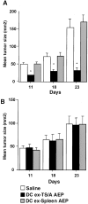
Figure 1
Figure 1
In vivo effects of BM-DC–derived exosomes. Immunocompetent (A) or nude (B) mice bearing 4-d-old TS/A mammary tumors were injected intradermally with exosomes (5 μg/mouse) produced by BM-DCs pulsed with AEP (i.e., MHC-associated peptides) from either TS/A tumor cells (DC ex-TS/A AEP) or splenic cells (DC ex-Spleen AEP). Tumor size was monitored weekly. Tumor growth was significantly delayed in tumor-AEP exosome–injected mice, compared with splenic-AEP exosome– or saline-injected mice (A). In nude mice lacking T lymphocytes, TS/A tumor growth was not affected by the same treatments (B). Each experimental group included five mice. Experiments were reproduced twice with similar results. Asterisks represent significant results at 95% using Fisher's exact method compared with injection of saline.
Figure 2
Figure 2
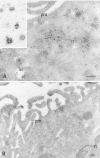
Immunoelectron microscopy of D1 cells and exosomes. Ultrathin cryosections of immature (A) and mature (B) D1 cells were single immunogold-labeled with the rat anti–MHC class II mAb M5114 as described in Materials and Methods. (A) Immature D1 cells show numerous MHC class II–positive compartments with intraluminal vesicles. MHC class II are also detected at the plasma membrane (pm). (Insert) Whole-mounted exosomes immunogold-labeled with M5114. (B) In mature D1 cells (24 h in the presence of LPS), the plasma membrane (pm) shows numerous processes and is intensely labeled with the M5114 antibody. Very few intracellular compartments containing MHC class II molecules are observed. n, nucleus. Bars, 200 nm.
Figure 3
Figure 3
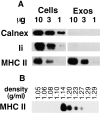
Western blot analysis of MHC class II, Ii, and calnexin in whole D1 cells and exosomes. (A) 10, 3, or 1 μg of proteins from whole cell lysate (Cells) or exosomes (Exos) was loaded on a 12% SDS gel followed by Western blotting using antibodies against MHC class II molecules (MHC II), Ii, and calnexin (Calnex). MHC class II are enriched in exosomes compared with whole cells, whereas Ii and calnexin are absent from exosomes. (B) 90 μg of exosomes was loaded on a continuous sucrose gradient (0.25–2 M sucrose, resulting in densities ranging 1–1.5 g/ml), and the fractions were analyzed by Western blot using the anti–MHC class II antibody. Exosome-associated MHC class II molecules float at an average density of 1.14 g/ml.
Figure 4
Figure 4
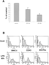
Exosomes production by D1 cells upon maturation. (A) Exosomes were purified from a 24-h supernatant of D1 cells in control culture medium (control), or in the presence of 20 μg/ml LPS (LPS 24h), or in the presence of LPS and after a 16-h pretreatment with LPS (LPS 40h). The same number of D1 cells was used in each condition. Exosome production in the presence of LPS, as quantified by the Bradford assay, was rapported to the production by control cells. This graph represents the average of seven (LPS 24h) or two (LPS 40h) independent experiments. LPS treatment reduces the production of exosomes by D1 cells. (B) D1 cells in control conditions (Cont) or after 40 h in the presence of LPS (LPS 40h) were analyzed by FACS® after staining with antibodies specific for MHC class II molecules (MHC II) and CD40 (grey histograms; white histograms are negative controls obtained without primary antibodies), or after 15 min internalization at 37°C of FITC-coupled 40,000-Dalton dextran (DX-FITC) (grey histogram; white histogram is a negative control obtained after 15 min internalization at 4°C). As expected from mature D1 cells, both MHC class II and CD40 are upregulated at the surface of LPS-treated cells, whereas their endocytosis ability (as measured by the capacity to internalize dextran) is reduced compared with control cells.
Figure 5
Figure 5
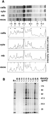
Overall protein composition of metabolically labeled exosomes and D1 cells. (A) Exosomes purified from the supernatant of metabolically labeled D1 cells were run on a 12% SDS gel (exos), together with lysate (including both cytosolic and membrane components) obtained from the whole cells (cells), cytosol (cyto), or total membranes (mb) prepared from the same cells in the absence of detergent. The dried gel was autoradiographed for 24 h (proteins >45 kD) or 48 h (proteins <45 kD). A pic profile of each lane was obtained with the NIH Image software. Stars point to proteins abundant in the cells and absent in exosomes, dots to proteins enriched in exosomes. (B) Metabolically labeled exosomes were subjected to flotation on a continuous sucrose gradient. The fractions were collected (density 1.08–1.28 g/ml), run on a 10% SDS-PAGE, and compared with exosomes that had not been subjected to flotation (Ex). All the proteins present in exosomes comigrate at the expected density (1.15 g/ml) (see Fig. 2 B; Raposo et al. 1996).
Figure 6
Figure 6
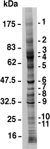
Protein profile of D1 exosomes as seen by Coomassie blue staining. 30 μg of exosomes was separated on an 8–15% gradient SDS gel and stained with Coomassie brilliant blue. The band pattern is similar to that obtained with metabolically labeled exosomes. The major bands (1–11) were analyzed by trypsin digestion and MALDI-TOF-MS (see Table ).
Figure 7
Figure 7
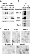
Analysis of five of the identified proteins in D1- and fresh BM-DC–derived exosomes. (A) 6 and 2 μg of proteins from whole cells (Cells) or exosomes (Exos) was separated on a 10% SDS gel and analyzed by Western blot using antibodies specific for annexin II, MHC II, CD9, and hsc73. (B) 3 × 10⁶ cpm from metabolically labeled D1 cells (Cell) or exosomes (Ex) were immunoprecipitated with antibodies specific for annexin II, MHC II, CD9, hsc73, and Mac-1 (Ab), or with the corresponding isotype-matched control antibodies (neg). Immunoprecipitates were run on 10 or 8% SDS gels and autoradiographed for 1 mo (the gel corresponding to hsc73 was only exposed for 1 wk to better distinguish hsc73 from a nonspecific band that also precipitates in cell lysates, but not exosomes, with protein G alone). (C) Immunoelectron microscopy was performed on whole-mounted D1-derived exosomes. Due to the small size of exosomes and the potentially low number of molecules on each vesicle, not all vesicles are positive for each antibody. However, exosomes are distinctly positive for MHC II, CD9, and Mac-1. hsc73 and annexin II, on the other hand, are not detected in these preparations, suggesting that they are either present at a level below the detection threshold of the technique or not accessible to antibodies, i.e., contained within the lumen of exosomes.
Figure 8
Figure 8
Analysis of the hsps73, hsp84, and gp96 in D1 cells and exosomes. (A) 2 μg of proteins from whole cells (Cells) or exosomes (Exos) was analyzed by Western blot using antibodies against the 90-kD hsp family members gp96 and hsp84, or against hsc73 and MHC class II. Only hsc73 is enriched in exosomes. (B) D1 cells were fractionated on a discontinuous sucrose gradient to obtain an HDM fraction containing ER, Golgi, and plasma membranes, and an LDM fraction containing most endosomal and lysosomal membranes as well as ER, Golgi, and plasma membranes. These membranes and cytosol were analyzed by Western blot using antibodies against gp96, hsp84, hsc73, and MHC class II. (C) After mild trypsin digestion, LDMs were fractionated by FFE to separate negatively charged endosomes and lysosomes from the ER, Golgi, and plasma membranes (Marsh et al. 1987). Protein concentration in each fraction was measured by the Bradford assay (lower panel, Bradford curve). Activity of the lysosomal enzyme β-hexosaminidase was measured in each fraction (lower panel, β Hex curve). Fractions were then analyzed by Western blot using antibodies against MHC class II (known to be present both at the surface and in lysosomes in immature DCs), gp96, and hsc73 (lower panel). gp96 and the 80-kD band generated by trypsin digestion (see upper panel, LDM − or + trypsin) are only localized in the noncharged ER/Golgi/plasma membrane fractions, whereas the 60-kD band generated from hsc73 upon trypsin digestion accumulates in negatively charged endosomo-lysosomal fractions.
Figure 9
Figure 9
A speculative model for the molecular structure of DC-derived exosomes. The proteins represented have been found in DC exosomes, here (MFG-E8, Mac-1, CD9, hsc73, annexin II, Gi2α, gag) or in our previous studies (MHC class I and II, B7.2) (Zitvogel et al. 1998). The topology of membrane proteins is based on previous studies on exosome membrane orientation (Pan et al. 1985; Raposo et al., 1997), and on EM observations of whole-mounted exosomes reported here (Fig. 7). MHC I and II and B7.2 may be involved in the induction of immune responses by exosomes (Fig. 1). MFG-E8, Mac-1, and CD9 could play a role in addressing exosomes to target cells expressing their respective ligands (integrins αvβ3 and αvβ5, ICAM-1, and -2). Annexin II, Gi2α, gag, and hsc73 may play a role in exosomes biogenesis and/or biological functions.

Similar articles

Cited by

References

    1. Agarraberes F.A., Terlecky S.R., Dice J.F. An intralysosomal hsp70 is required for a selective pathway of lysosomal protein degradation. J. Cell Biol. 1997;137:825–834. - PMC - PubMed
    1. Albert M.L., Sauter B., Bhardwaj N. Dendritic cells acquire antigen from apoptotic cells and induce class I-restricted CTLs. Nature. 1998;392:86–89. - PubMed
    1. Amigorena S., Drake J.R., Webster P., Mellman I. Transient accumulation of new class II MHC molecules in a novel endocytic compartment in B lymphocytes. Nature. 1994;369:113–120. - PubMed
    1. Andersen M.H., Berglund L., Rasmussen J.T., Petersen T.E. Bovine PAS-6/7 binds alpha v beta 5 integrins and anionic phospholipids through two domains. Biochemistry. 1997;36:5441–5446. - PubMed
    1. Aoki N., Kishi M., Taniguchi Y., Adachi T., Nakamura R., Matsuda T. Molecular cloning of glycoprotein antigens MGP57/53 recognized by monoclonal antibodies raised against bovine milk fat globule membrane. Biochim. Biophys. Acta. 1995;1245:385–391. - PubMed

Publication types

MeSH terms