. 1999 Oct 18;147(2):435-46.
doi: 10.1083/jcb.147.2.435.

Formation process of autophagosome is traced with Apg8/Aut7p in yeast

Affiliations

Formation process of autophagosome is traced with Apg8/Aut7p in yeast

T Kirisako et al. J Cell Biol. .

Abstract

We characterized Apg8/Aut7p essential for autophagy in yeast. Apg8p was transcriptionally upregulated in response to starvation and mostly existed as a protein bound to membrane under both growing and starvation conditions. Immunofluorescence microscopy revealed that the intracellular localization of Apg8p changed drastically after shift to starvation. Apg8p resided on unidentified tiny dot structures dispersed in the cytoplasm at growing phase. During starvation, it was localized on large punctate structures, some of which were confirmed to be autophagosomes and autophagic bodies by immuno-EM. Besides these structures, we found that Apg8p was enriched on isolation membranes and in electron less-dense regions, which should contain Apg8p-localized membrane- or lipid-containing structures. These structures would represent intermediate structures during autophagosome formation. Here, we also showed that microtubule does not play an essential role in the autophagy in yeast. The result does not match with the previously proposed role of Apg8/Aut7p, delivery of autophagosome to the vacuole along microtubule. Moreover, it is revealed that autophagosome formation is severely impaired in the apg8 null mutant. Apg8p would play an important role in the autophagosome formation.


Figures
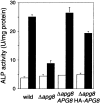
Figure 1
Figure 1
ALP assay. The cells expressing Pho8Δ60p were cultured in SD+CA medium (open bars) and shifted to SD(−N) medium for three hours (filled bars). The ALP activity of the lysates was measured to estimate autophagic activity. Wild-type cells, TN124; Δapg8 mutant harboring vector, TK303; Δapg8 mutant harboring APG8 on a centromeric vector, TK301; Δapg8 mutant harboring 3 × HA-tagged APG8 plasmid, TK307.
Figure 2
Figure 2
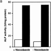
Effect of nocodazole on autophagy. Vegetatively growing cells, YW5-1B and TN124, were treated with 10 μg/ml nocodazole in YEPD medium for three hours. A, YW5-1B cells were transferred to 0.17% yeast nitrogen base without amino acid and ammonium sulfate containing 1 mM PMSF and 10 μg/ml nocodazole, and further incubated for 4.5 h. The accumulation of autophagic bodies was examined under a light microscope. Arrows indicate autophagic bodies. B, TN124 cells were starved in SD(−N) containing 10 μg/ml nocodazole for 4.5 h. ALP activity was measured before (open bars) and after (filled bars) starvation.
Figure 2
Figure 2
Effect of nocodazole on autophagy. Vegetatively growing cells, YW5-1B and TN124, were treated with 10 μg/ml nocodazole in YEPD medium for three hours. A, YW5-1B cells were transferred to 0.17% yeast nitrogen base without amino acid and ammonium sulfate containing 1 mM PMSF and 10 μg/ml nocodazole, and further incubated for 4.5 h. The accumulation of autophagic bodies was examined under a light microscope. Arrows indicate autophagic bodies. B, TN124 cells were starved in SD(−N) containing 10 μg/ml nocodazole for 4.5 h. ALP activity was measured before (open bars) and after (filled bars) starvation.
Figure 3
Figure 3
Expression of APG8. A, The lysates prepared from the growing cells were subjected to immunoblotting with the anti-Apg8p antibody. Lane 1, wild-type cells (YW5-1B); lane 2, Δapg8 cells harboring APG8 on a multicopy vector (TK201); lane 3, Δapg8 cells (TK404). B, The wild-type (YW5-1B) cells were cultured in YEPD medium until 1–2 × 10⁷ cells/ml (0 h) and shifted to SD(−N) medium for 0.5, 1, 2, 3, 4.5, and 6 h at 30°C. The amount of Apg8p was estimated by immunoblotting with anti-Apg8p antibody. Each lane has 10 μg of total protein. C, YW5-1B cells were cultured in YEPD medium until 1–2 × 10⁷ cells/ml (0 h) and shifted to SD(−N) medium for 0.5, 1, 2, and 6 h at 30°C, and mRNA was prepared from each culture as described in Materials and Methods. APG8 mRNA and ACT1 mRNA were detected by Northern blotting with each specific probe. Each lane has 4 μg of total mRNA. D, YW5-1B cells were cultured in YEPD medium until 1 × 10⁷ cells/ml at 30°C. Rapamycin was added to the culture at a final concentration of 0.2 μg/ml and the cells were further incubated in YEPD medium for two hours at 30°C. Western blotting was performed with the anti-Apg8p antibody. Each lane has 20 μg of total protein.
Figure 4
Figure 4
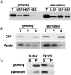
Subcellular fractionation and solubilization of Apg8p. The wild-type cells (YW5-1B) growing logarithmically in YEPD medium or starved in SD(−N) medium for three hours were lysed. A, The lysates (T) were centrifuged at 13,000 g for 15 min to generate pellet (LSP) and supernatant (LSS). LSS fraction was further centrifuged at 100,000 g for one hour and separated to pellet (HSP) and supernatant (HSS). LSP and HSP were resuspended in an equal volume of the lysis buffer to the original lysates. Equal volume of each sample was applied to each well, and immunoblotting was performed with anti-Apg8p antibody. B, The lysates (T) were sonicated and centrifuged at 100,000 g for one hour to separate pellet (P) and supernatant (S). The distribution of CPY and Apg8p was examined by immunoblotting with each specific antibody, respectively. C, The sonicated lysates were centrifuged at 100,000 g for one hour to generate pellet fraction. The pellet fraction was treated with 2% Triton X-100 (TX-100) for 30 min on ice. The sample was centrifuged at 100,000 g for one hour and separated to supernatant (S) and pellet (P) fractions. The distribution of Apg8p was examined by immunoblotting with the anti-Apg8p antibody.
Figure 5
Figure 5
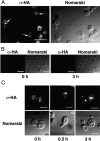
Immunofluores-cent staining of Δapg8 cells expressing 3 × HA–Apg8p. A, The Δapg8 cells harboring 3 × HA-tagged APG8 plasmid, TK114 cells, were grown until logarithmic phase. The cells were fixed by formaldehyde and were treated with Zymolyase 100T to generate spheroplasts. The spheroplasts were permeabilized by 0.5% Triton X-100, and incubated with the anti-HA antibody, 16B12, followed with the FITC-conjugated anti–mouse IgG. Left, Fluorescence image of 3 × HA–Apg8p; right, Nomarski image of the cells. Arrows show punctate signals that are a little larger than tiny dot signals. Bars, 10 μm. B, Immunofluorescent staining of YW5-1B cells before (0 h) and after (3 h) shift to starvation (negative control). Left, Fluorescence image; right, Nomarski image. Bars, 10 μm. C, The cells were shifted to starvation for 0, 0.5, and 3 h. Upper panels, Fluorescence images of 3 × HA–Apg8p; lower panels, Nomarski images of the cell. Bars, 5 μm.
Figure 6
Figure 6
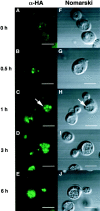
Change of Apg8p localization in Δpep4 cells after shift to starvation. The Δapg8Δpep4 cells harboring 3 × HA-tagged APG8 plasmid, TK116 cells, were shifted to starvation for 0, 0.5, 1, 3, and 6 h. Immunofluorescence microscopy was performed as described in Fig. 5. A–E, FITC staining of 3 × HA–Apg8p. F–J, Nomarski images of the cells. Arrows show an autophagic body. Bars, 5 μm.
Figure 7
Figure 7
Immunostaining image of a cell containing autophagic bodies in the vacuole. The Δapg8Δpep4 cells expressing 3 × HA–Apg8p, TK116 cells, were shifted to SD(−N) medium for two hours. Cells were immunolabeled with anti-HA mAb, 16B12, followed by 10-nm gold-conjugated goat anti–mouse IgG. AB, autophagic body; N, nucleus; V, vacuole.
Figure 8
Figure 8
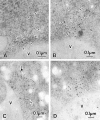
Immuno-EM of autophagosomes. A and B, Mature autophagosome. C and D, Premature autophagosome. Arrows show expanded regions of the intramembrane space of the premature autophagosomes. TK116 cells were incubated in SD(−N) medium for one hour (D), two hours (B and C), or three hours (A). The localization of 3 × HA–Apg8p was detected with anti-HA antibody, followed by the incubation with 5-nm gold- (A and D) or 10-nm gold- (B and C) conjugated goat anti–mouse IgG. AP, autophagosome; N, nucleus; V, vacuole.
Figure 9
Figure 9
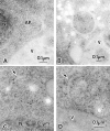
Possible intermediate structures of autophagosome. A, A membrane sac under construction. Arrows show gold particles. B and C, Isolation membranes. B, Isolation membrane is detected along the arrows. C, The small arrow shows a semicircular isolation membrane and a large arrow marks its open area. D, Apg8p-residing structures gathered in the area close to the vacuole. TK116 cells were incubated in SD(−N) medium for one hour (A and C), or two hours (B and D). The localization of 3 × HA–Apg8p was detected with anti-HA antibody, followed by the incubation with 5-nm gold- (A) or 10-nm gold- (B–D) conjugated goat anti–mouse IgG. V, vacuole.
Figure 10
Figure 10
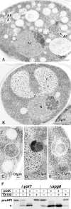
Fine morphology of Δypt7 and Δapg8 cells and proteinase-K accession to proAPI in these mutants. A, EM image of Δypt7 cell starved in SD(−N) medium for four hours. Autophagosomes (AP, arrows) were accumulated. B, EM image of the starved Δapg8 cell. C–E, The representatives of the membrane structures detected in the starved Δapg8 cells. C, Autophagosome-like structure indistinguishable from autophagosome. D and E, Aberrant multivesicular structures. N, nucleus; V, vacuole. F, Proteinase K-sensitivity of proAPI. Cell lysates were prepared from Δypt7 and Δapg8 cells starved in SD(−N) medium for 4.5 h. The lysates were treated with 100 μg/ml proteinase K in the presence or absence of Triton X-100. Asterisk shows mature API and digestion product of proAPI by proteinase K.

Similar articles

Cited by

References

    1. Baba M., Takeshige K., Baba N., Ohsumi Y. Ultrastructural analysis of the autophagic process in yeastdetection of autophagosomes and their characterization. J. Cell Biol. 1994;124:903–913. - PMC - PubMed
    1. Baba M., Osumi M., Ohsumi Y. Analysis of the membrane structures involved in autophagy in yeast by freeze-replica method. Cell Struct. Funct. 1995;20:465–471. - PubMed
    1. Baba M., Osumi M., Scott S.V., Klionsky D.J., Ohsumi Y. Two distinct pathways for targeting proteins from the cytoplasm to the vacuole/lysosome. J. Cell Biol. 1997;139:1687–1695. - PMC - PubMed
    1. Belazzi T., Wagner A., Wieser R., Schanz M., Adam G., Hartig A., Ruis H. Negative regulation of transcription of the Saccharomyces cerevisiae catalase T (CTT1) gene by cAMP is mediated by a positive control element. EMBO (Eur. Mol. Biol. Organ.) J. 1991;10:585–592. - PMC - PubMed
    1. Darsow T., Rieder S.E., Emr S.D. A multispecificity syntaxin homologue, Vam3p, essential for autophagic and biosynthetic protein transport to the vacuole. J. Cell Biol. 1997;138:517–529. - PMC - PubMed

Publication types

MeSH terms

Substances