. 1999 Oct 18;147(2):417-34.
doi: 10.1083/jcb.147.2.417.

Binding of integrin alpha6beta4 to plectin prevents plectin association with F-actin but does not interfere with intermediate filament binding

Affiliations

Binding of integrin alpha6beta4 to plectin prevents plectin association with F-actin but does not interfere with intermediate filament binding

D Geerts et al. J Cell Biol. .

Abstract

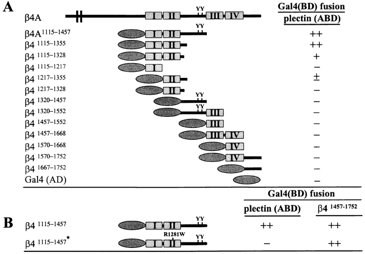
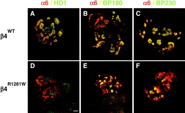
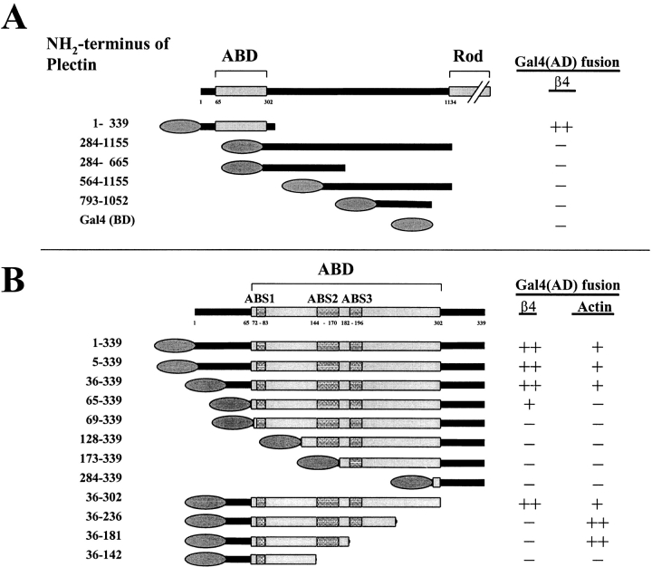
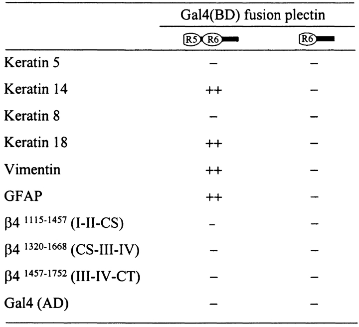
Hemidesmosomes are stable adhesion complexes in basal epithelial cells that provide a link between the intermediate filament network and the extracellular matrix. We have investigated the recruitment of plectin into hemidesmosomes by the alpha6beta4 integrin and have shown that the cytoplasmic domain of the beta4 subunit associates with an NH(2)-terminal fragment of plectin that contains the actin-binding domain (ABD). When expressed in immortalized plectin-deficient keratinocytes from human patients with epidermol- ysis bullosa (EB) simplex with muscular dystrophy (MD-EBS), this fragment is colocalized with alpha6beta4 in basal hemidesmosome-like clusters or associated with F-actin in stress fibers or focal contacts. We used a yeast two-hybrid binding assay in combination with an in vitro dot blot overlay assay to demonstrate that beta4 interacts directly with plectin, and identified a major plectin-binding site on the second fibronectin type III repeat of the beta4 cytoplasmic domain. Mapping of the beta4 and actin-binding sites on plectin showed that the binding sites overlap and are both located in the plectin ABD. Using an in vitro competition assay, we could show that beta4 can compete out the plectin ABD fragment from its association with F-actin. The ability of beta4 to prevent binding of F-actin to plectin explains why F-actin has never been found in association with hemidesmosomes, and provides a molecular mechanism for a switch in plectin localization from actin filaments to basal intermediate filament-anchoring hemidesmosomes when beta4 is expressed. Finally, by mapping of the COOH-terminally located binding site for several different intermediate filament proteins on plectin using yeast two-hybrid assays and cell transfection experiments with MD-EBS keratinocytes, we confirm that plectin interacts with different cytoskeletal networks.


Figures

Figure 1
Figure 1
Expression of hemidesmosomal proteins in immortalized MD-EBS keratinocytes. Western blot of lysates of normal (NHK; lanes 1) and plectin-deficient (MD-EBS; lanes 2) human keratinocytes, probed with mAb 450-11A against integrin β4 (200 kD), mAb 7A8 against plectin (>500 kD), polyclonal antiserum against BP230 (230 kD), or polyclonal antiserum against BP180 (180 kD). All hemidesmosome proteins tested, except plectin, were present in MD-EBS keratinocytes.
Figure 2
Figure 2
Localization of hemidesmosome proteins in immortalized MD-EBS keratinocytes. Cells were fixed, immunolabeled, and subjected to confocal immunofluorescence microscopy using polyclonal antiserum against α6 (A) or β4 (B), mAb 233 against BP180 (C), mAb 6F12 against laminin-5 (D), mAb 121 against plectin/HD1 (E), or polyclonal antiserum against BP230 (F). Results obtained for cell lines from two different, unrelated human patients were similar; shown here are the results for one patient. All hemidesmosome proteins tested, as well as the extracellular matrix protein laminin-5 (a ligand for the integrin α6β4), are concentrated at sites of cell–substrate contact in patches characteristic for hemidesmosome-like clusters, except plectin, which is not expressed. Sections were focused at the cell–substrate interface. Bar, 10 μm.
Figure 3
Figure 3
Subcellular distribution of HA-tagged plectin-ABD fragment in transfected MD-EBS keratinocytes. 48 h after transfection, cells were fixed, double-immunolabeled using mAb 12C05 against the HA epitope (green; A, D, and G) and polyclonal antiserum against β4 (red; H), and processed for immunofluorescence. TRITC-labeled phalloidin was used to stain F-actin (red; B and E). Composite images (C, F, and I) show the superimposition of the green and red signals, with areas of overlap appearing yellow. Cells were visualized by confocal microscopy. The plectin protein fragment is colocalized with actin stress fibers, or with α6β4-containing hemidesmosome-like basal clusters. Sections were focused at the cell–substrate interface. Bar, 10 μm.
Figure 4
Figure 4
Distribution of plectin compared with F-actin in PA-JEB cells. PA-JEB keratinocytes and PA-JEB cells stably expressing the integrin α6β4 were fixed, double-immunolabeled using mAb 121 against plectin (green; A, D, and G) or an affinity-purified polyclonal antiserum against the plectin-ABD (green; H and J) and F-actin (red; B, E, and K), and processed for immunofluorescence. Composite images (C, F, I, and L) were generated by superimposition of the green and red signals, with areas of overlap appearing yellow. Cells were visualized by confocal microscopy. In PA-JEB cells (A–C), the staining patterns of plectin and F-actin partly overlapped, whereas in the α6β4-positive cells (D–F) plectin was exclusively localized in hemidesmosome-like basal clusters that did not stain for F-actin. These sections (A–F) were taken at the optical plane corresponding to the cell–substrate interface. In another plane of focus (G–I), it is shown that the two anti-plectin antibodies produce a similar pattern of staining of the cytoskeleton in PA-JEB keratinocytes. (J–L) Higher magnification shows plectin associated with F-actin filaments in PA-JEB keratinocytes (arrows). Bars: 10 μm (A, D, and G); and 5 μm (J).
Figure 5
Figure 5
Mapping of the binding site between the NH2-terminal part of plectin and the cytoplasmic domain of β4 and of the internal folding of the β4 cytoplasmic domain by yeast two-hybrid analysis. (A) Cotransformation of yeast host strain PJ69-4A with pAS2-plectin1–339 and one each of the listed pACT2-β4 constructs or empty pACT2 vector to determine the plectin-β4 interaction site. (B) Cotransformation with pAS2-plectin1–339 or pAS2-β41457–1752, and pACT2-β41115–1457 (wild-type) or -β41115–1457* (R1281W) to characterize plectin-β4 binding and the internal folding of the β4 cytoplasmic domain. Transformation mixtures were spread on SC-LT and SC-LTHA plates and grown for 12 d at 30°C. Plating efficiency on selective SC-LTHA plates is expressed as a percentage of plating efficiency on nonselective SC-LT plates of the same transformation. ++, >50%; +, >50% (slow-growing colonies); ±, 5–25%; −, 0%. Plates were scored after 6 and 12 d of growth. Slow-growing colonies could only be scored after 12 d of growth. Plating efficiencies of <25% always represented slow-growing colonies. All efficiencies listed represent an average of multiple independent transformations on at least three separate occasions.
Figure 6
Figure 6
Localization of hemidesmosomal components in PA-JEB keratinocytes transiently transfected with cDNAs encoding full-length wild-type β4 or β4R1281W. Cells were fixed, double-immunolabeled using polyclonal antiserum against α6 (red; A–F) and mAb 121 against plectin/HD1 (green; A and D), mAb 233 against BP180 (green; B and E), or polyclonal antiserum against BP230 (green; C and F), and processed for immunofluorescence. Cells were visualized by confocal microscopy. Expression of β4 results in the formation of hemidesmosome-like structures at sites of cell–substrate contact, in which α6β4 is concentrated, as shown by staining for α6. Similar results were obtained when instead of the anti-α6 antiserum, a polyclonal antiserum against β4 was used. The wild-type integrin α6β4 (A–C) is colocalized with plectin, BP180, and BP230. The mutant integrin α6β4R1281W is not colocalized with plectin (D), but is colocalized with BP180 (E) and BP230 (F). Sections were focused at the cell–substrate interface. Bar, 10 μm.
Figure 7
Figure 7
Mapping of the interaction site for β4 and for actin on the NH2 terminus of plectin by yeast two-hybrid analysis. (A) Cotransformation of yeast host strain PJ69-4A with one each of the pAS2-plectin subclones listed together with pACT2-β41115–1457 to determine the interaction site for β4 on plectin (A) and with one each of the pAS2-plectin ABD subclones listed together with pACT2-β41115–1457 or actin for a comparison of the β4- and actin-binding sites on plectin (B). The results shown for plectin36–302, plectin36–236, plectin36–181, and plectin36–142 are identical for those obtained with two sets of four similar subclones starting at residues 1 or 5, respectively. The results shown are for β cytoplasmic actin. No binding was found between pACT2-β41115–1457 and pAS2-actin (data not shown). Details are as for Fig. 5.
Figure 8
Figure 8
Dot blot overlay and actin sedimentation assay of recombinant purified β4 protein fragments with radiolabeled plectin-ABD fragment. (A) SDS-PAGE of 4 ng in vitro translated [35S]-methionine/cysteine-labeled HA-tagged plectin-ABD protein (40 kD). Autoradiogram. (B) Recombinant purified GST fusion proteins encoding β41115–1328, β41115–1382 (wild-type sequence), β41115–1382* (R1281W), β41457–1752 (3 μg each), or GST alone (2 μg), and purified F-actin (1, 2, or 3 μg) were immobilized on nitrocellulose by dot blotting and incubated with 100 ng [35S]methionine/cysteine-labeled plectin-ABD in 3 ml APB or AGB (upper and middle lanes, respectively). Protein loading was verified by amido-black staining (bottom lanes). The GST protein alone does not bind to plectin-ABD. (C) Actin filaments polymerized in the presence of and complexed with MBP-plectin ABD fusion protein were incubated with a 10-fold molar excess of GST fusion proteins encoding β41115–1382 (lanes 1 and 2), β41115–1382* (lanes 3 and 4), β41115–1328 (lanes 5 and 6), or no GST fusion protein (lanes 7 and 8). The F-actin complexes were then isolated by centrifugation, after which supernatant (S; odd-numbered lanes) and pellet (P; even-numbered lanes) were isolated and analyzed using SDS-PAGE. Proteins were visualized using Coomassie brilliant blue staining.
Figure 9
Figure 9
Mapping of the interaction site between the COOH terminus of plectin and the cytoplasmic domain of β4 and of full-length intermediate filament proteins by yeast two-hybrid analysis. Yeast host strain PJ69-4A was cotransformed with pAS2-plectin and one each of the listed pACT2-IF or pACT2-β4 subclone constructs, or with empty pACT2 vector to determine the interaction sites between plectin and β4 or intermediate filament proteins. The plectin R5-R6 clone encodes amino acid residues 4068–4687 of mouse plectin (containing most of repeat R5, repeat R6, and the COOH tail), the plectin R6 clone encodes residues 4351–4574 of human plectin (most of repeat R6 and the COOH tail). Similar results as for the human plectin R6 clone were obtained with a mouse plectin clone encoding residues 4206–4687 (part of repeat R5, repeat R6, and the COOH tail; data not shown). Details are as for Fig. 5.
Figure 10
Figure 10
Subcellular distribution of HA-tagged plectin-IFBD fragment in MD-EBS keratinocytes after transient transfection. Cells were transiently transfected with an HA-tagged plectin R5-R6 cDNA construct. Cells were fixed, double-immunolabeled using polyclonal antiserum HA-11 against the HA tag (green; A, D, and G) and a mixture of mAbs 58XB4 and 4.3E1 against β4 (red; B), a mixture of mAbs SPK-14 and KL1 against keratins (red; E), or mAb V9 against vimentin (red; H), and processed for immunofluorescence. Composite images (C, F, and I) were generated by superimposition of the green and red signals, with areas of overlap appearing yellow. Cells were visualized by confocal microscopy. The plectin-IFBD protein fragment is exclusively found in cytoplasmic filament structures (A, D, and G). α6β4 is present in basally located hemidesmosome-like clusters (B), together with the plectin-IFBD (C). Keratins are observed in a dense cytoplasmic filament network (E), which is decorated by the plectin-IFBD (F). Expression of the IFBD-fragment of plectin causes a collapse of the vimentin filament network (H and I). Bar, 10 μm.
Figure 11
Figure 11
Subcellular distribution of wild-type and mutant β4 and study of the effect of β4 expression on the vimentin intermediate filament network in transfected PA-JEB keratinocytes. PA-JEB cells were transfected with cDNA encoding full-length β4 (A–C), COOH-terminal deletion mutants β41355 (D–F), and β41328 (G–I). Cells were fixed, double-immunolabeled, and processed for confocal immunofluorescence microscopy using polyclonal antiserum against the α6 subunit (green; A, D, and G), or mAb V9 against vimentin (red; B, E, and H). Composite images (C, F, and I) were generated by superimposition of the green and red signals, with areas of overlap appearing yellow. Expression of wild-type full-length β4 results in the formation of hemidesmosome-like structures, in which α6β4 is concentrated at sites of cell–substrate contact, as is shown by staining for α6, and codistributes with the vimentin network, most probably dependent upon its binding to plectin (A–C). The β41355 mutant protein, which efficiently binds to plectin, is found colocalized with the vimentin network (D–F). The β41328 mutant protein, which can only bind plectin with low efficiency in vitro, was only very occasionally found to be colocalized with vimentin (G–I). Bar, 10 μm.

Similar articles

Cited by

References

    1. Aho S., Uitto J. Basement membrane zone protein-protein interactions disclosed by yeast two-hybrid system. J. Invest. Dermatol. 1997;108:546a.
    1. Aho S., Uitto J. Direct interaction between the intracellular domain of bullous pemphigoid antigen 2 (BP180) and β4 integrin, hemidesmosomal components of basal keratinocytes. Biochem. Biophys. Res. Commun. 1998;243:694–699. - PubMed
    1. Aho S., Uitto J. 180-kD bullous pemphigoid antigen/type VXII collagentissue-specific expression and molecular interactions with keratin 18. J. Cell. Biochem. 1999;72:356–367. - PubMed
    1. Andrä K., Lassman H., Bittner R., Shorny S., Fässler R., Propst F., Wiche G. Targeted inactivation of plectin reveals essential function in maintaining the integrity of skin, muscle, and heart cytoarchitecture. Genes Dev. 1997;11:3143–3156. - PMC - PubMed
    1. Andrä K., Nikolic B., Stöcher M., Drenckhahn D., Wiche G. Not just scaffoldingplectin regulates actin dynamics in cultured cells. Genes Dev. 1998;12:3442–3451. - PMC - PubMed

Publication types