. 1999 Oct 18;147(2):277-94.
doi: 10.1083/jcb.147.2.277.

Presenilin 1 controls gamma-secretase processing of amyloid precursor protein in pre-golgi compartments of hippocampal neurons

Affiliations

Presenilin 1 controls gamma-secretase processing of amyloid precursor protein in pre-golgi compartments of hippocampal neurons

W G Annaert et al. J Cell Biol. .

Abstract

Mutations of presenilin 1 (PS1) causing Alzheimer's disease selectively increase the secretion of the amyloidogenic betaA4(1-42), whereas knocking out the gene results in decreased production of both betaA4(1-40) and (1-42) amyloid peptides (De Strooper et al. 1998). Therefore, PS1 function is closely linked to the gamma-secretase processing of the amyloid precursor protein (APP). Given the ongoing controversy on the subcellular localization of PS1, it remains unclear at what level of the secretory and endocytic pathways PS1 exerts its activity on APP and on the APP carboxy-terminal fragments that are the direct substrates for gamma-secretase. Therefore, we have reinvestigated the subcellular localization of endogenously expressed PS1 in neurons in vitro and in vivo using confocal microscopy and fine-tuned subcellular fractionation. We show that uncleaved PS1 holoprotein is recovered in the nuclear envelope fraction, whereas the cleaved PS fragments are found mainly in post-ER membranes including the intermediate compartment (IC). PS1 is concentrated in discrete sec23p- and p58/ERGIC-53-positive patches, suggesting its localization in subdomains involved in ER export. PS1 is not found to significant amounts beyond the cis-Golgi. Surprisingly, we found that APP carboxy-terminal fragments also coenrich in the pre-Golgi membrane fractions, consistent with the idea that these fragments are the real substrates for gamma-secretase. Functional evidence that PS1 exerts its effects on gamma-secretase processing of APP in the ER/IC was obtained using a series of APP trafficking mutants. These mutants were investigated in hippocampal neurons derived from transgenic mice expressing PS1wt or PS1 containing clinical mutations (PS1(M146L) and PS1(L286V)) at physiologically relevant levels. We demonstrate that the APP-London and PS1 mutations have additive effects on the increased secretion of betaA4(1-42) relative to betaA4(1-40), indicating that both mutations operate independently. Overall, our data clearly establish that PS1 controls gamma(42)-secretase activity in pre-Golgi compartments. We discuss models that reconcile this conclusion with the effects of PS1 deficiency on the generation of betaA4(1-40) peptide in the late biosynthetic and endocytic pathways.


Figures
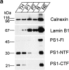
Figure 1
Figure 1
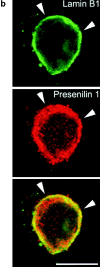
(a) Purification of nuclei (N) and nuclear envelopes (NE) from cortices of 5–7-d-old mice by differential centrifugation (Otto et al. 1992). Brains were homogenized in 0.32 M sucrose containing 1 mM MgCl2 and 20 mM Hepes, pH 7.4. The total homogenate (TH) was centrifuged (at 200 g av for 20 min), the resulting pellet (P1) resuspended to a final concentration of 1.8 M sucrose and centrifuged again (at 67,000 g av for 1 h). The pellet P2 was resuspended to a final concentration of 2.15 M sucrose, layered on a 2.2 M sucrose cushion and centrifuged (at 67,000 g av for 1 h). The pellet of purified nuclei (N) was, after DNAse digestion, centrifuged (at 9,500 g av for 20 min) to obtain nuclear envelopes (NE). Equal amounts of protein of each intermediate step was analyzed by SDS-PAGE and Western blotting. The final fraction (NE), which is highly enriched for lamin B1, also contains low amounts of PS1 fragments and full-length PS1. (b) Double immunofluorescent staining of fully polarized hippocampal neurons (16 d of culture) derived from nontransgenic embryos for PS1 (red) and lamin B1 (green). A merged picture shows considerable overlap of both stainings (yellow). Anti-PS1 pAb B17.2 was detected with CY3 anti–rabbit antibodies, whereas anti–lamin B1 mAb was detected with CY2-conjugated anti–mouse antibodies. Note that the focal plane is through the middle of the nucleus. The bulk of PS1 immunoreactive staining is localized close to the bottom of the cell and in the dendritic arbor (see next figure). Arrowheads point to rims of the nuclear envelope that costains for PS1-CTF (see also yellow color in overlay). Bar, 20 μm.
Figure 1
Figure 1
(a) Purification of nuclei (N) and nuclear envelopes (NE) from cortices of 5–7-d-old mice by differential centrifugation (Otto et al. 1992). Brains were homogenized in 0.32 M sucrose containing 1 mM MgCl2 and 20 mM Hepes, pH 7.4. The total homogenate (TH) was centrifuged (at 200 g av for 20 min), the resulting pellet (P1) resuspended to a final concentration of 1.8 M sucrose and centrifuged again (at 67,000 g av for 1 h). The pellet P2 was resuspended to a final concentration of 2.15 M sucrose, layered on a 2.2 M sucrose cushion and centrifuged (at 67,000 g av for 1 h). The pellet of purified nuclei (N) was, after DNAse digestion, centrifuged (at 9,500 g av for 20 min) to obtain nuclear envelopes (NE). Equal amounts of protein of each intermediate step was analyzed by SDS-PAGE and Western blotting. The final fraction (NE), which is highly enriched for lamin B1, also contains low amounts of PS1 fragments and full-length PS1. (b) Double immunofluorescent staining of fully polarized hippocampal neurons (16 d of culture) derived from nontransgenic embryos for PS1 (red) and lamin B1 (green). A merged picture shows considerable overlap of both stainings (yellow). Anti-PS1 pAb B17.2 was detected with CY3 anti–rabbit antibodies, whereas anti–lamin B1 mAb was detected with CY2-conjugated anti–mouse antibodies. Note that the focal plane is through the middle of the nucleus. The bulk of PS1 immunoreactive staining is localized close to the bottom of the cell and in the dendritic arbor (see next figure). Arrowheads point to rims of the nuclear envelope that costains for PS1-CTF (see also yellow color in overlay). Bar, 20 μm.
Figure 2
Figure 2
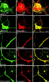
Double immunofluorescent staining of fully polarized hippocampal neurons derived from PS1wt transgenic embryos for PS1 (red in all pictures) and rSEC61αp (a–c), calnexin (d–f), BAP31 (g–i), SEC23 (j–l), and ERGIC-53/p58 (m–o). Fields c, f, i, l, and o represent color overlays. mAb 5.2 was used to label human PS1wt and detection was done with CY3 anti-mouse secondary antibodies. All other marker proteins were counterstained using CY2-conjugated anti–rabbit secondary antibodies. Notice the relative weak colocalization of PS1 with the RER marker rSEC61αp (a–c, arrowheads) mainly in the perinuclear region (a-c, arrow). PS1 colocalizes much better with other ER marker proteins such as calnexin (d–f), BAP31 (g–i), and SEC23 (j–l), or with ERGIC-53/p58, a resident protein of the IC. The pictures in (j–o) give a detailed area of the fully polarized neuron as overviewed in the insets. Bars: (a-i) 20 μm; (j-o) 10 μm.
Figure 3
Figure 3
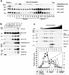
Analysis of PS1 in enriched fractions of the IC isolated from cortices of 5–7-d-old mice (adapted from Schweizer et al. 1991). (a) A postnuclear supernatant (PNS) was mixed with Percoll to give a final density of 1.129 g/ml and centrifuged (at 37,000 g max for 41 min). Twenty-two fractions of 1.5 ml were collected and analyzed by SDS-PAGE and Western blotting (equal amount of fraction volume). PS1 immunoreactivity was distributed bimodally with most of the immunoreactivity recovered in fractions 15–19. (b) Fractions 5–14 were pooled (Pool I), adjusted to 30% (wt/wt) Nycodenz, and overlaid with 27% and 18.5% (wt/wt) Nycodenz. After isopycnic centrifugation (85,800 g max for 20 h), three interfaces were collected (F1, F2, and F3). The 18.5–27% interface F3 is highly enriched in marker proteins for the intermediate compartment (IC), such as p58/ERGIC-53. Comparable enrichments were found for both PS1-NTF and -CTF. Equal amounts of protein were analyzed by SDS-PAGE and Western blotting. For PS1-NTF, an additional long exposure of the same blot is shown to reveal the full-length PS1. Notice that the F3 fraction is still contaminated with membranes derived from the RER as demonstrated by two specific marker proteins, rSEC61αp and TRAPα. (c) The interface fraction F3 was diluted twofold with buffer C (Schweizer et al. 1991) and loaded on a continuous sucrose gradient (0.8–1.6 M sucrose in buffer C). After isopycnic gradient centrifugation (SW41 rotor, at 250,000 g max for 4 h), 12 fractions were collected from the top and analyzed by Western blotting. Calnexin, ERGIC-53/p58 and PS1 fragments all display a bimodal distribution, whereas TRAPα and rSEC61αp are only distributed in the second half of the sucrose gradient. The bottom panel shows the distribution of the different marker proteins expressed per milligram of protein in each fraction (arbitrary units/mg protein). It should be noted that at low sucrose densities (first half of the gradient), both PS1 fragments clearly coenrich with the first pool of ERGIC-53/p58–positive membranes, whereas no clear enrichment is observed for calnexin. The data represent one out of two independent experiments.
Figure 4
Figure 4
Analysis of PS1 immunoreactivity in the Golgi apparatus by cell fractionation (A) and confocal microscopy (B). (A) Fractions 15–19 of the Percoll gradient (see Fig. 3 a) were pooled (Pool II), adjusted to 25% (wt/wt) Nycodenz, and overlaid with 21, 14, and 10% (wt/wt) Nycodenz. After isopycnic centrifugation (SW41 rotor; at 85,800 g max for 20 h), three interfaces were collected (IF1, IF2, and IF3, not shown). Interface IF3, containing most of the PS1 immunoreactivity (not shown) was diluted twofold with buffer C (Schweizer et al. 1991) and further fractionated by continuous sucrose density gradient centrifugation (0.3–1.7 M sucrose, at 250,000 g max for 2 h). Fractions were collected from the bottom and an equal volume of each fraction was analyzed. The peak immunoreactivity for PS1-NTF and -CTF was slightly shifted to less dense regions of the gradient, thereby codistributing with BAP31, RAP, and immature TGN38. Mature TGN38 immunoreactivity was mainly recovered in the bottom fraction of the gradient. GM130, a Golgi marker, is distributed in the high density region of the gradient where no peak of PS1 immunoreactivity was observed. (B) PS1 does not distribute extensively into the Golgi apparatus. Fully polarized neurons isolated from control, nontransgene embryos were double stained for PS1 (pAb B17.2; red) and p58/Golgi (green). Detection was performed with CY3 anti-rabbit and CY2 anti-mouse secondary antibodies, respectively. In contrast to PS1 labeling (b), Golgi staining (a) is restricted to the soma and proximal part of the dendrites as can be seen in the overview of a neuron (a–c, arrow). (d–f) Enlarged area of the proximal dendrite in the same neuron stained for PS1 (red) and p58/Golgi (green). The asterisk indicates regions in the cell body with a relative low level of colocalization. Arrowheads point to areas of codistribution. (g–i) Section in the vertical plane of the neuron at the position in the proximal dendrite indicated by the arrow in (d–f). Only a partial overlap is observed and in many cases, PS1 immunoreactivity is very closely juxtaposed to p58/Golgi staining (arrowheads in g–i). c, f, and i are the merged signals. PS1 and Golgi stainings are mainly not overlapping. Bars, 10 μm.
Figure 4
Figure 4
Analysis of PS1 immunoreactivity in the Golgi apparatus by cell fractionation (A) and confocal microscopy (B). (A) Fractions 15–19 of the Percoll gradient (see Fig. 3 a) were pooled (Pool II), adjusted to 25% (wt/wt) Nycodenz, and overlaid with 21, 14, and 10% (wt/wt) Nycodenz. After isopycnic centrifugation (SW41 rotor; at 85,800 g max for 20 h), three interfaces were collected (IF1, IF2, and IF3, not shown). Interface IF3, containing most of the PS1 immunoreactivity (not shown) was diluted twofold with buffer C (Schweizer et al. 1991) and further fractionated by continuous sucrose density gradient centrifugation (0.3–1.7 M sucrose, at 250,000 g max for 2 h). Fractions were collected from the bottom and an equal volume of each fraction was analyzed. The peak immunoreactivity for PS1-NTF and -CTF was slightly shifted to less dense regions of the gradient, thereby codistributing with BAP31, RAP, and immature TGN38. Mature TGN38 immunoreactivity was mainly recovered in the bottom fraction of the gradient. GM130, a Golgi marker, is distributed in the high density region of the gradient where no peak of PS1 immunoreactivity was observed. (B) PS1 does not distribute extensively into the Golgi apparatus. Fully polarized neurons isolated from control, nontransgene embryos were double stained for PS1 (pAb B17.2; red) and p58/Golgi (green). Detection was performed with CY3 anti-rabbit and CY2 anti-mouse secondary antibodies, respectively. In contrast to PS1 labeling (b), Golgi staining (a) is restricted to the soma and proximal part of the dendrites as can be seen in the overview of a neuron (a–c, arrow). (d–f) Enlarged area of the proximal dendrite in the same neuron stained for PS1 (red) and p58/Golgi (green). The asterisk indicates regions in the cell body with a relative low level of colocalization. Arrowheads point to areas of codistribution. (g–i) Section in the vertical plane of the neuron at the position in the proximal dendrite indicated by the arrow in (d–f). Only a partial overlap is observed and in many cases, PS1 immunoreactivity is very closely juxtaposed to p58/Golgi staining (arrowheads in g–i). c, f, and i are the merged signals. PS1 and Golgi stainings are mainly not overlapping. Bars, 10 μm.
Figure 5
Figure 5
Analysis of the distribution of APP holoprotein and carboxy-terminal fragments in ER- and IC-enriched fractions isolated from cortices of 5–7-d-old mice using the same fractionation protocol as described in Fig. 3, a and b, and Fig. 4 (A). (a) Distribution of APP after Percoll gradient centrifugation of a postnuclear fraction. Like for PS1, both APP and APP stubs display a bimodal distribution with the major amount of immunoreactivity in fractions 15–20 of the gradient. (b) APP carboxy-terminal fragments enrich in the interface F3. (c) APP fragments could be further enriched after Nycodenz and sucrose gradient centrifugation of Pool II from the Percoll gradient (a). The distribution of APP carboxy-terminal fragments is identical to that of PS1-NTF and -CTF (Fig. 4 A).
Figure 6
Figure 6
Purification of lysosomes from cortices of 5–7-d-old mice. Highly purified lysosomes were obtained by a combination of low speed centrifugation in 0.25 M KCl and velocity gradient centrifugation in Percoll according to the method of Maguire and Luzio 1985. The final lysosomal fraction (LYS) is clearly enriched in cathepsin D. Hardly any PS1-NTF or -CTF immunoreactivity was detected in this fraction.
Figure 7
Figure 7
PS1 is not localized in recycling endosomes and synaptic vesicles of hippocampal neurons derived from control, nontransgene embryos. (a–f) Within the somatodendritic area of 14-d-old hippocampal neurons, PS1 (pAb B17.2; red) is not colocalized with the transferrin receptor (green). A detailed area of distal parts of the dendritic arbor (a–c) and a growth cone (d–f) are shown. (d–i) Double immunofluorescent staining of hippocampal neurons for PS1 (pAb B17.2; red) and the synaptic vesicle marker synaptobrevin II (mAb 69.1, green). Axonal, presynaptic boutons are marked by synaptobrevin II immunoreactive punctae and are devoid of PS1 immunoreactivity, most clearly seen in the enlarged area displaying an axon running parallel with a dendrite while making several synaptic contacts (g–i). PS1 is distributed into the postsynaptic areas. PS1 was stained with CY3 anti-rabbit, the transferrin receptor and synaptobrevin II with CY2 anti-mouse secondary antibodies. Bars, 10 μm.
Figure 8
Figure 8
Processing of human wild-type APP (APPwt), the London mutant (APP-Lon), and the APP sorting mutants, APPΔCT, APP/KK, and APP/LDLR in SFV-infected hippocampal neurons. Immunoprecipitation of total βA4 from the culture supernatant (MEDIUM) and carboxy-terminal β-stubs and total βA4 from the cell lysates (CELL) of metabolically labeled hippocampal neurons using antibody B7/7 that recognizes an epitope on the amino terminus of the βA4 sequence. Immunoprecipitates were electrophoresed on 10–20% Tris-Tricine gels and analyzed by phosphorimaging. Lower amounts of secreted βA4 are recovered in the supernatants of cultures infected with APPΔCT and APP/KK as compared with the other virus constructs. Notice the higher expression of APPΔCT in the cell extracts. As expected, infection with the SFV-APPΔCT construct resulted in a shorter carboxy-terminal β-stub. The contribution of carboxyl-terminal fragments and βA4 from endogenous APP processing is neglectable as it is below detection level in untransfected cultures (No Virus). Antibody B7/7 used for the βA4 immunoprecipitation experiments reacts also with low affinity with APP-ectodomain, recovered in low quantities from the conditioned media (APPs).
Figure 9
Figure 9
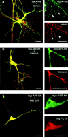
Newly synthesized APP wild-type, but not APP/KK, is sorted to axons in fully polarized hippocampal neurons. (a) Double immunofluorescent staining for APPwt (green) and calnexin (red). Newly expressed APP was detected using a myc antibody. Infection with the SFV-APPwt clearly resulted in axonal localization of the expressed protein. (b) Newly expressed APP/KK was entirely retained in the somatodendritic ER as demonstrated by the virtual complete colocalization with calnexin. APP and calnexin labeling were detected with CY2 anti-mouse and lissamine rhodamine anti-rabbit secondary antibodies, respectively. Asterisk marks a nontransfected neuron. (c) Endogenous PS1 resides in membrane-bound compartments involved in the trafficking and retrieval of dilysine-tagged proteins. The newly expressed APP/KK (green) colocalizes with endogenous PS1 (red) in polarized hippocampal neurons. The colocalization was most obvious in the somatic area (inset). Detection of primary antibodies was with CY2 anti-mouse (APP) and CY3 anti-rabbit (PS1-CTF) secondary antibodies. Bars, 20 μm.

Similar articles

Cited by

References

    1. Annaert W.G., Becker B., Kistner U., Reth M., Jahn R. Export of cellubrevin from the endoplasmic reticulum is controlled by BAP31. J. Cell Biol. 1997;139:1397–1410. - PMC - PubMed
    1. Arar C., Carpentier V., Le Caer J.P., Monsigny M., Legrand A., Roche A.C. ERGIC-53, a membrane protein of the endoplasmic reticulum-Golgi intermediate compartment, is identical to MR60, an intracellular mannose-specific lectin of myelomonocytic cells. J. Biol. Chem. 1995;270:3551–3553. - PubMed
    1. Aridor M., Weissman J., Bannykh S., Nuoffer C., Balch W.E. Cargo selection by the COPII budding machinery during export from the ER. J. Cell Biol. 1998;141:61–70. - PMC - PubMed
    1. Bannykh S.I., Balch W.E. Membrane dynamics at the endoplasmic reticulum–Golgi interface. J. Cell Biol. 1997;138:1–4. - PMC - PubMed
    1. Bannykh S.I., Balch W.E. Selective transport of cargo between the endoplasmic reticulum and Golgi compartments. Histochem. Cell Biol. 1998;109:463–475. - PubMed

Publication types