

Lentiviral delivery of HIV-1 Vpr protein induces apoptosis in transformed cells
- PMID: 10518572
- PMCID: PMC18408
- DOI: 10.1073/pnas.96.21.12039
Lentiviral delivery of HIV-1 Vpr protein induces apoptosis in transformed cells
Abstract
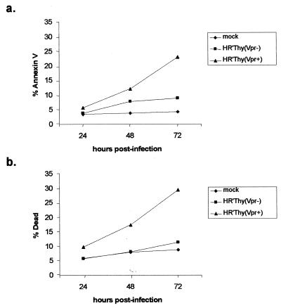
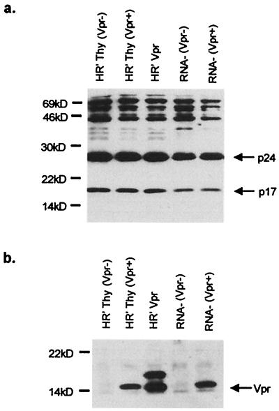
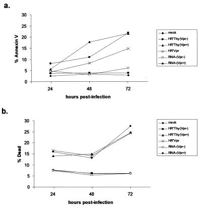
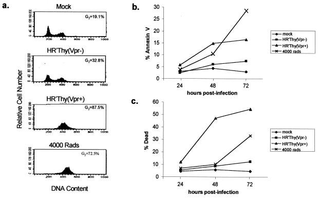
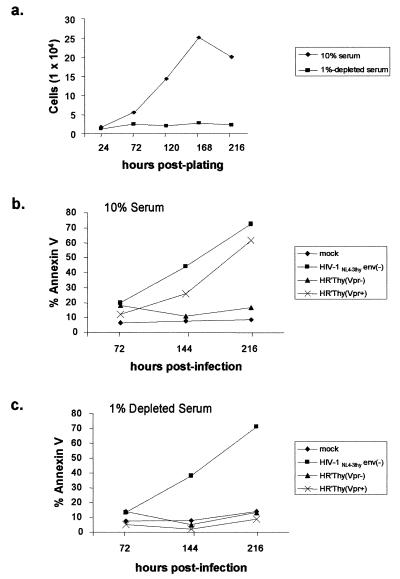
Most current anticancer therapies act by inducing tumor cell stasis followed by apoptosis. HIV-1 Vpr effectively induces apoptosis of T cells after arrest of cells at a G(2)/M checkpoint. Here, we investigated whether this property of Vpr could be exploited for use as a potential anticancer agent. As a potentially safer alternative to transfer of genes encoding Vpr, we developed a method to efficiently introduce Vpr protein directly into cells. Vpr packaged into HIV-1 virions lacking a genome induced efficient cell cycle arrest and apoptosis. Introduction of Vpr into tumor cell lines of various tissue origin, including those bearing predisposing mutations in p53, XPA, and hMLH1, induced cell cycle arrest and apoptosis with high efficiency. Significantly, apoptosis mediated by virion-associated Vpr was more effective on rapidly dividing cells compared with slow-growing cells, thus, in concept, providing a potential differential effect between some types of tumor cells and surrounding normal cells. This model system provides a rationale and proof of concept for the development of potential cancer therapeutic agents based on the growth-arresting and apoptotic properties of Vpr.
Figures
Similar articles
-
Roles of p53 and caspases in the induction of cell cycle arrest and apoptosis by HIV-1 vpr.Exp Cell Res. 1999 Aug 25;251(1):156-65. doi: 10.1006/excr.1999.4568. Exp Cell Res. 1999. PMID: 10438581
-
Comparison of cell cycle arrest, transactivation, and apoptosis induced by the simian immunodeficiency virus SIVagm and human immunodeficiency virus type 1 vpr genes.J Virol. 2001 Apr;75(8):3791-801. doi: 10.1128/JVI.75.8.3791-3801.2001. J Virol. 2001. PMID: 11264368 Free PMC article.
-
Adenovirus encoding HIV-1 Vpr activates caspase 9 and induces apoptotic cell death in both p53 positive and negative human tumor cell lines.Oncogene. 2002 Jul 11;21(30):4613-25. doi: 10.1038/sj.onc.1205549. Oncogene. 2002. PMID: 12096338
-
Partner molecules of accessory protein Vpr of the human immunodeficiency virus type 1.DNA Cell Biol. 2004 Apr;23(4):193-205. doi: 10.1089/104454904773819789. DNA Cell Biol. 2004. PMID: 15142377 Review.
-
Effect of HIV-1 Vpr on cell cycle regulators.DNA Cell Biol. 2004 Apr;23(4):249-60. doi: 10.1089/104454904773819833. DNA Cell Biol. 2004. PMID: 15142382 Review.
Cited by
-
Protein kinase A phosphorylation activates Vpr-induced cell cycle arrest during human immunodeficiency virus type 1 infection.J Virol. 2010 Jul;84(13):6410-24. doi: 10.1128/JVI.02273-09. Epub 2010 Apr 14. J Virol. 2010. PMID: 20392842 Free PMC article.
-
HIV-1 Vpr-induced apoptosis is cell cycle dependent and requires Bax but not ANT.PLoS Pathog. 2006 Dec;2(12):e127. doi: 10.1371/journal.ppat.0020127. PLoS Pathog. 2006. PMID: 17140287 Free PMC article.
-
Depletion of Wee-1 kinase is necessary for both human immunodeficiency virus type 1 Vpr- and gamma irradiation-induced apoptosis.J Virol. 2003 Feb;77(3):2063-70. doi: 10.1128/jvi.77.3.2063-2070.2003. J Virol. 2003. PMID: 12525641 Free PMC article.
-
HIV-1 Vpr-a still "enigmatic multitasker".Front Microbiol. 2014 Mar 31;5:127. doi: 10.3389/fmicb.2014.00127. eCollection 2014. Front Microbiol. 2014. PMID: 24744753 Free PMC article. Review.
-
Human T-cell leukemia virus type 1 tax oncoprotein suppression of multilineage hematopoiesis of CD34+ cells in vitro.J Virol. 2003 Nov;77(22):12152-64. doi: 10.1128/jvi.77.22.12152-12164.2003. J Virol. 2003. PMID: 14581552 Free PMC article.
References
Publication types
MeSH terms
Substances
Grants and funding
LinkOut - more resources
Full Text Sources
Other Literature Sources
Research Materials
Miscellaneous
