. 1999 Oct;19(10):6632-41.
doi: 10.1128/MCB.19.10.6632.

RBP1 recruits both histone deacetylase-dependent and -independent repression activities to retinoblastoma family proteins

Affiliations

RBP1 recruits both histone deacetylase-dependent and -independent repression activities to retinoblastoma family proteins

A Lai et al. Mol Cell Biol. 1999 Oct.

Abstract

Retinoblastoma (RB) tumor suppressor family proteins block cell proliferation in part by repressing certain E2F-specific promoters. Both histone deacetylase (HDAC)-dependent and -independent repression activities are associated with the RB "pocket." The mechanism by which these two repression functions occupy the pocket is unknown. A known RB-binding protein, RBP1, was previously found by our group to be an active corepressor which, if overexpressed, represses E2F-mediated transcription via its association with the pocket. We show here that RBP1 contains two repression domains, one of which binds all three known HDACs and represses them in an HDAC-dependent manner while the other domain functions independently of the HDACs. Thus, RB family members repress transcription by recruiting RBP1 to the pocket. RBP1, in turn, serves as a bridging molecule to recruit HDACs and, in addition, provides a second HDAC-independent repression function.


Figures

FIG. 1
FIG. 1
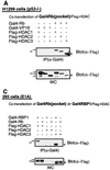
Endogenous interactions of pRB (A) and p107 (B) with HDACs. (A) Immunoprecipitations were done in lysates from either H1299 or 293T cells. Plates (100 mm) of both cell types were lysed in low-stringency buffer. Cell extracts were incubated with 1 μg of the indicated antisera and 30 μl of a 50% slurry of protein G-Sepharose for at least 12 h. Immunoprecipitated complexes were washed six times with the 150 mM low-stringency buffer and eluted by boiling with 2× sample buffer. Eluted proteins were subjected to SDS-PAGE with a 6% polyacrylamide gel. The presence of pRB was detected with monoclonal antibody (G3-245) against pRB (PharMingen). (B) Binding studies similar to those described for panel A were done, but coimmunoprecipitation of p107 was detected with a rabbit polyclonal antibody (C-18) against p107 (α-107) (Santa Cruz).
FIG. 2
FIG. 2
In vivo interaction of the small pocket of pRB with HDACs. (A) cDNAs encoding the Gal4DBD fused with the pocket of pRB were overexpressed with those encoding Flag-tagged HDAC1, HDAC2, or HDAC3 in H1299 cells. RK5C1 (anti-Gal4DBD) antibody (Santa Cruz) was used to immunoprecipitate Gal4-tagged overexpressed proteins, and the coimmunoprecipitation of HDACs was detected by Western blotting with anti-Flag M2 monoclonal antibody (Sigma). WC, whole-cell extracts; IP, anti-Gal4-immunoprecipitated proteins. Binding studies similar to those described for panel A were performed with 293T (B) and 293 (C) cells. (D) A modified binding experiment similar to that described for panel A was done with H1299 cells by incubating cell extracts with increasing amounts (0 to 20 μg) of E1A protein (the first exon of the gene containing the LXCXE motif) fused to GST (GST-E1A) and synthesized in vitro in bacteria (6). The positions of migration of Flag-tagged HDAC1 (open arrow), HDAC2 (closed arrow), and HDAC3 (arrowhead) are indicated.
FIG. 2
FIG. 2
In vivo interaction of the small pocket of pRB with HDACs. (A) cDNAs encoding the Gal4DBD fused with the pocket of pRB were overexpressed with those encoding Flag-tagged HDAC1, HDAC2, or HDAC3 in H1299 cells. RK5C1 (anti-Gal4DBD) antibody (Santa Cruz) was used to immunoprecipitate Gal4-tagged overexpressed proteins, and the coimmunoprecipitation of HDACs was detected by Western blotting with anti-Flag M2 monoclonal antibody (Sigma). WC, whole-cell extracts; IP, anti-Gal4-immunoprecipitated proteins. Binding studies similar to those described for panel A were performed with 293T (B) and 293 (C) cells. (D) A modified binding experiment similar to that described for panel A was done with H1299 cells by incubating cell extracts with increasing amounts (0 to 20 μg) of E1A protein (the first exon of the gene containing the LXCXE motif) fused to GST (GST-E1A) and synthesized in vitro in bacteria (6). The positions of migration of Flag-tagged HDAC1 (open arrow), HDAC2 (closed arrow), and HDAC3 (arrowhead) are indicated.
FIG. 3
FIG. 3
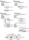
In vivo interaction of RBP1 with HDACs. Binding studies similar to those described for Fig. 2A and B were performed with H1299 cells (A) and 293T cells (B) by using Gal4-RBP1 instead of Gal4-pRB. (C) Binding studies similar to those described for panel B were performed in 293T cells transfected with the RBP1-HA construct (24) and the Gal4-HDAC1, Gal4-HDAC2, and Gal4-HDAC3 constructs (42, 43). Molecular mass was determined with Rainbow Color Marker RPN-756 (Amersham Life Science). (D) A binding study similar to that described for panels B and C was done with the Gal4-RBP1dl-LXCXE and RBP1dl-LXCXE-HA mutants, which lack the LXCXE pocket-binding motif (24). The positions of migration of Flag-tagged HDAC1 (open arrows), HDAC2 (closed arrow), and HDAC3 (arrowhead) are indicated. (E) Coimmunoprecipitation studies similar to those for Fig. 1 were done with both H1299 and 293T cells. In addition to the antisera described, antibodies against pRB (G3-245), p130 (C-20 polyclonal) (Santa Cruz), p107 (C-18), and the HA epitope (HA.11) were used. Coimmunoprecipitation with RBP1 was detected with a monoclonal antibody raised specifically against RBP1 (LY32).
FIG. 4
FIG. 4
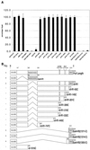
Mapping of transcriptional repression domains in RBP1 and effect of TSA on RBP1 repression activity. (A) Repression by RBP1 mutants. CAT assays were performed with CHO cells, as described previously (24), with G5TKCAT as the reporter. (B) Illustration of the Gal4DBD-RBP1 mutants. The name of each mutant is provided, as well as the amino acid residues affected by deletions. A summary of repression results (Rep.) obtained in experiments described for panel A is shown at the left. NLS, nuclear localization signal. (C) Effect of TSA on repression. Repression assays were carried out as for panel A, except that some cells were incubated with 330 nM TSA for 24 h prior to harvesting and the G5MLPCAT reporter was assayed instead of the G5TKCAT reporter. (D) An experiment similar to that described for panel C was performed with a reporter construct consisting of the E2F-1 promoter linked to a cDNA encoding luciferase, as described previously (24), and either RBP1, RBP1dl-LXCXE, the pocket of pRB, or an inactive pRB pocket mutant, mRB(C706F) mutant (22). All data are the averages (± standard errors) of at least six individual experiments.
FIG. 4
FIG. 4
Mapping of transcriptional repression domains in RBP1 and effect of TSA on RBP1 repression activity. (A) Repression by RBP1 mutants. CAT assays were performed with CHO cells, as described previously (24), with G5TKCAT as the reporter. (B) Illustration of the Gal4DBD-RBP1 mutants. The name of each mutant is provided, as well as the amino acid residues affected by deletions. A summary of repression results (Rep.) obtained in experiments described for panel A is shown at the left. NLS, nuclear localization signal. (C) Effect of TSA on repression. Repression assays were carried out as for panel A, except that some cells were incubated with 330 nM TSA for 24 h prior to harvesting and the G5MLPCAT reporter was assayed instead of the G5TKCAT reporter. (D) An experiment similar to that described for panel C was performed with a reporter construct consisting of the E2F-1 promoter linked to a cDNA encoding luciferase, as described previously (24), and either RBP1, RBP1dl-LXCXE, the pocket of pRB, or an inactive pRB pocket mutant, mRB(C706F) mutant (22). All data are the averages (± standard errors) of at least six individual experiments.
FIG. 5
FIG. 5
Mapping of specific binding of HDAC3 to RBP1. Binding studies similar to those described for Fig. 3B were done with 293T cells by using either Gal4-R1, Gal4-R2(1314-C), Gal4-R2(1263-C), Gal4 alone, Gal4-RBP1 (wild type [WT]), Gal4-ARID, Gal4-dlR1, or Gal4-dl-93C(dl-R2).
FIG. 6
FIG. 6
Model of repression of E2F-dependent promoters by RB family members and RBP1.

Similar articles

Cited by

References

    1. Adnane J, Shao Z, Robbins P D. The retinoblastoma susceptibility gene product represses transcription when directly bound to the promoter. J Biol Chem. 1995;270:8837–8843. - PubMed
    1. Brehm A, Miska E A, McCance D J, Reid J L, Bannister A J, Kouzarides T. Retinoblastoma protein recruits histone deacetylase to repress transcription. Nature. 1998;391:597–601. - PubMed
    1. Bremner R, Cohen B L, Sopta M, Hamel P A, Ingles C J, Gallie B L, Phillips R A. Direct transcriptional repression by pRB and its reversal by specific cyclins. Mol Cell Biol. 1995;15:3256–3265. - PMC - PubMed
    1. Buyse I M, Shao G, Huang S. The retinoblastoma protein binds to RIZ, a zinc-finger protein that shares an epitope with the adenovirus E1A protein. Proc Natl Acad Sci USA. 1995;92:4467–4471. - PMC - PubMed
    1. Chow K N B, Dean D C. Domains A and B in the Rb pocket interact to form a transcriptional repressor motif. Mol Cell Biol. 1996;16:4862–4868. - PMC - PubMed

Publication types

LinkOut - more resources