. 1999 Sep;10(9):2829-45.
doi: 10.1091/mbc.10.9.2829.

A novel Ras-interacting protein required for chemotaxis and cyclic adenosine monophosphate signal relay in Dictyostelium

Affiliations
Free PMC article

A novel Ras-interacting protein required for chemotaxis and cyclic adenosine monophosphate signal relay in Dictyostelium

S Lee et al. Mol Biol Cell. 1999 Sep.
Free PMC article

Abstract

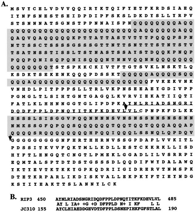
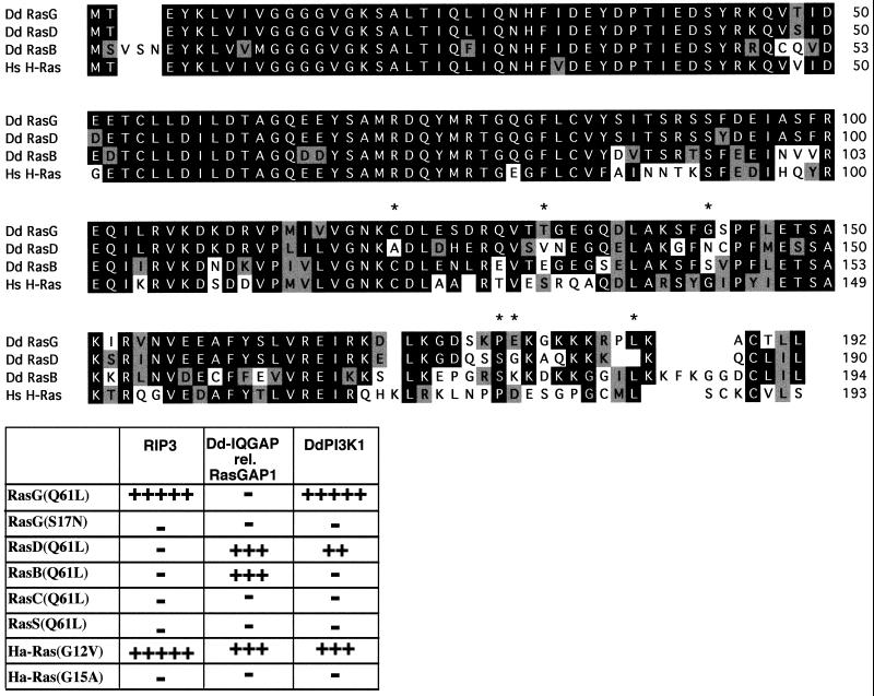
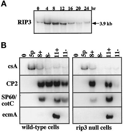
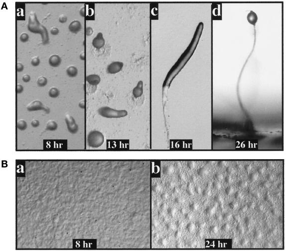
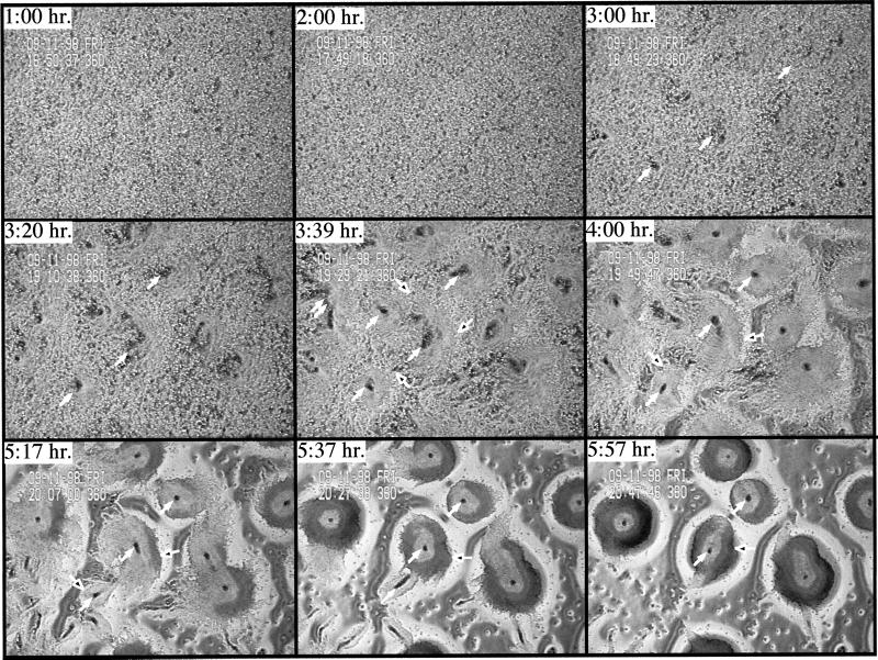
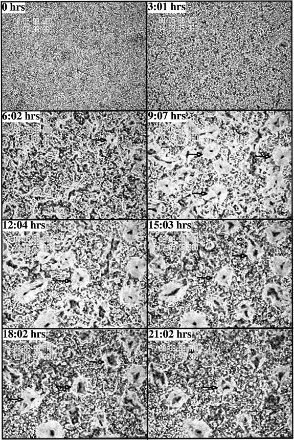
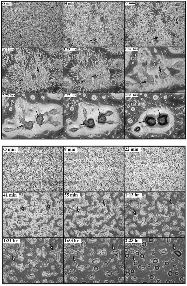
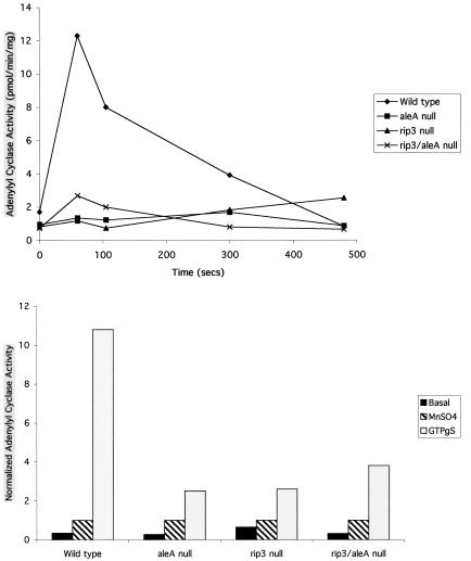
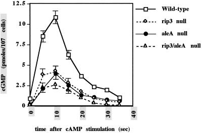
We have identified a novel Ras-interacting protein from Dictyostelium, RIP3, whose function is required for both chemotaxis and the synthesis and relay of the cyclic AMP (cAMP) chemoattractant signal. rip3 null cells are unable to aggregate and lack receptor activation of adenylyl cyclase but are able, in response to cAMP, to induce aggregation-stage, postaggregative, and cell-type-specific gene expression in suspension culture. In addition, rip3 null cells are unable to properly polarize in a cAMP gradient and chemotaxis is highly impaired. We demonstrate that cAMP stimulation of guanylyl cyclase, which is required for chemotaxis, is reduced approximately 60% in rip3 null cells. This reduced activation of guanylyl cyclase may account, in part, for the defect in chemotaxis. When cells are pulsed with cAMP for 5 h to mimic the endogenous cAMP oscillations that occur in wild-type strains, the cells will form aggregates, most of which, however, arrest at the mound stage. Unlike the response seen in wild-type strains, the rip3 null cell aggregates that form under these experimental conditions are very small, which is probably due to the rip3 null cell chemotaxis defect. Many of the phenotypes of the rip3 null cell, including the inability to activate adenylyl cyclase in response to cAMP and defects in chemotaxis, are very similar to those of strains carrying a disruption of the gene encoding the putative Ras exchange factor AleA. We demonstrate that aleA null cells also exhibit a defect in cAMP-mediated activation of guanylyl cyclase similar to that of rip3 null cells. A double-knockout mutant (rip3/aleA null cells) exhibits a further reduction in receptor activation of guanylyl cyclase, and these cells display almost no cell polarization or movement in cAMP gradients. As RIP3 preferentially interacts with an activated form of the Dictyostelium Ras protein RasG, which itself is important for cell movement, we propose that RIP3 and AleA are components of a Ras-regulated pathway involved in integrating chemotaxis and signal relay pathways that are essential for aggregation.


Figures

Figure 1
Figure 1
Sequence of RIP3. (A) The derived amino acid sequence of RIP3. The shaded areas contain a repetitive amino acid sequence that is often found in Dictyostelium ORFs and that is not thought to have an important function in the protein. RIP3 has a higher fraction of this sequence than other identified proteins. The domain that is homologous to the mammalian protein cloned, JC310, which was identified in a screen for mammalian genes that inhibit activated Ras function in yeast, is underlined. The two arrows mark the beginning of the ORFs of two different two-hybrid clones that were identified in our screen using activated mammalian Ha-Ras as bait. (B) Comparison of the amino acid homology between RIP3 and mammalian ORF JC310.
Figure 2
Figure 2
Interaction of Dictyostelium RIP3 with different Ras proteins. (A) Amino acid sequence comparison of Dictyostelium RasG, RasD, and RasB and human Ha-Ras. The asterisks indicate positions of conservation between RasG and Ha-Ras but not RasD. (B) The carboxyl-terminal region of RIP3 that was identified and cloned in two-hybrid screens using mammalian Ha-RasG12V as bait was used in two-hybrid assays to examine interaction of this domain with the activated form of the five identified Dictyostelium Ras proteins (RasG, RasD, RasB, RasC, and RasS). In addition, the dominant negative or nonactivatable form of RasG (RasGS17N) and interaction with Ha-RasG12V and Ha-RasG15A is shown. As controls, the interactions of the previously identified Dictyostelium IQGAP and the related gene RasGAPa as well as the Ras-interacting domain of Dictyostelium P110-related PI3 kinase DdPI3K1 are shown. The level of two-hybrid interactions was quantitated by the level of β-galactosidase production and the intensity of blue staining of yeast colonies.
Figure 3
Figure 3
Developmental kinetics of RIP3 gene expression and the requirement of RIP3 for expression of aggregation and postaggregative genes. (A) RNA blot analysis of the expression pattern of RIP3 using RNA isolated at different stages of Dictyostelium development. Mound formation occurs at ∼8 h and culmination initiates at ∼18–20 h. (B) Expression of aggregation-stage, postaggregative, and cell-type-specific genes in wild-type and rip3 null cells in suspension culture in response to cAMP. Cells were pulsed for 5 h with 30 nM cAMP (5p). Cells were given cAMP under slow-shake suspension conditions, which allow cell-cell interactions (120 rpm), to 300 μM and were supplemented to 300 μM every 2 h. 8+ and 11+ represent time points (hours) after the initiation of the experiment. The plus sign represents the addition of exogenous cAMP. Minus cultures (8− and 11−) did not receive exogenous cAMP. Cultures were split after 5 h of cAMP pulsing. csA (Contact Sites A) is a marker for aggregation-stage gene expression; CP2 is a marker for postaggregative gene expression. SP60/CotC is a prespore-specific gene; ecmA is a prestalk-specific gene.
Figure 4
Figure 4
Developmental phenotypes of wild-type and rip3 null cells. (A) The developmental phenotypes of wild-type KAx-3 cells. Mound formation is visible at 8 h. At 13 h, most of the aggregates have formed a tip, and at 16 h, the cells have formed a migrating slug or pseudoplasmodium. Cells have culminated by 24–26 h. (B) Developmental phenotype of rip3 null cells. At 8 h, the cells exhibit a small amount of rippling. At 24 h, very loose cellular associations are observed. Half or less of the cells are associated with the aggregates. Most cells show no sign of participation in aggregate formation.
Figure 5
Figure 5
Phase contrast video microscopic analysis of aggregate formation in wild-type and rip3 null cells. (A) Phase contrast video microscopy of wild-type cells is depicted as described previously. Briefly, cells are plated as a monolayer on nonnutrient agar and examined by phase contrast video microscopy using a 4× objective. The solid white arrows point to some of the aggregation centers. The open white arrows point to the outer regions of individual aggregation domains. Only a few of these are marked. (B) rip3 null cells. A similar analysis was performed on rip3 null cells.
Figure 5
Figure 5
Phase contrast video microscopic analysis of aggregate formation in wild-type and rip3 null cells. (A) Phase contrast video microscopy of wild-type cells is depicted as described previously. Briefly, cells are plated as a monolayer on nonnutrient agar and examined by phase contrast video microscopy using a 4× objective. The solid white arrows point to some of the aggregation centers. The open white arrows point to the outer regions of individual aggregation domains. Only a few of these are marked. (B) rip3 null cells. A similar analysis was performed on rip3 null cells.
Figure 6
Figure 6
Phase contrast video microscopic analysis of wild-type and rip3 null cells aggregating after being pulsed for 5 h with cAMP. Wild-type (A) or rip3 null (B) cells were pulsed for 5 h with 30 nM cAMP and plated on nonnutrient agar as described in Figure 5.
Figure 7
Figure 7
Adenylyl cyclase activation in wild-type and mutant cell lines. (A) Receptor-mediated stimulation of adenylyl cyclase activity. Differentiated cells were stimulated with 10 μM cAMP, lysed at specific time points, and immediately assayed as described in MATERIALS AND METHODS. (B) GTPγS-mediated stimulation of adenylyl cyclase activity. Differentiated cells were lysed with or without GTPγS, or in the presence MnSO4 as a measure of unregulated enzyme activity, incubated on ice for 4 min, and assayed as described in MATERIALS AND METHODS. The results are expressed as a ratio of the adenylyl cyclase activity to MnSO4 activity. The absolute MnSO4 activities are 4.0, 3.9, 2.5, and 2.5 pmol·min−1·mg−1 for wild-type, aleA null, rip3 null, and rip3/aleA null cells, respectively. The results presented are representative of at least two independent experiments.
Figure 8
Figure 8
cAMP stimulation of guanylyl cyclase activity in wild-type, rip3 null, aleA null, and rip3/aleA null strains. Cells were pulsed for 5 h with 30 nM cAMP and stimulated with cAMP, and measurements were taken for the quantitation of cGMP as described in MATERIALS AND METHODS. Unregulated activity of the enzyme was measured in the presence of 5 mM MnSO4. Each strain was analyzed a minimum of three times in association with wild-type cells. The results of representative experiments are shown. The differences between rip3 and aleA null cells, wild-type cells and rip3/aleA null cells, and rip3 and aleA null strains are reproducible.
Figure 9
Figure 9
Chemotaxis of wild-type, rip3 null, and rip3/aleA null strains. Cells were pulsed for 5 h with 30 nM cAMP, plated on coverslips as described previously, and examined by differential interference contrast microscopy. The micropipet shown contains 150 μM cAMP. Cells chemotax toward the cAMP gradient produced by diffusion of the cAMP from the micropipet. Insets in A and B depict the shapes of wild-type and rip3 null cells, respectively. (A) Analysis of wild-type cells was done with a 20× objective, whereas that of rip3/aleA null cells was done with a 40× objective. (A) Wild-type cells. (B) rip3 null cells. (C) rip3/aleA null cells.
Figure 9
Figure 9
Chemotaxis of wild-type, rip3 null, and rip3/aleA null strains. Cells were pulsed for 5 h with 30 nM cAMP, plated on coverslips as described previously, and examined by differential interference contrast microscopy. The micropipet shown contains 150 μM cAMP. Cells chemotax toward the cAMP gradient produced by diffusion of the cAMP from the micropipet. Insets in A and B depict the shapes of wild-type and rip3 null cells, respectively. (A) Analysis of wild-type cells was done with a 20× objective, whereas that of rip3/aleA null cells was done with a 40× objective. (A) Wild-type cells. (B) rip3 null cells. (C) rip3/aleA null cells.

Similar articles

Cited by

References

    1. Aubry L, Maeda M, Insall R, Devreotes PN, Firtel RA. The Dictyostelium mitogen-activated protein kinase ERK2 is regulated by Ras and cAMP-dependent protein kinase (PKA) and mediates PKA function. J Biol Chem. 1997;272:3883–3886. - PubMed
    1. Burki E, Anjard C, Scholder JC, Reymond CD. Isolation of two genes encoding putative protein kinases regulated during Dictyostelium discoideum development. Gene. 1991;102:57–65. - PubMed
    1. Chen MY, Insall RH, Devreotes PN. Signaling through chemoattractant receptors in Dictyostelium. Trends Genet. 1996;12:52–57. - PubMed
    1. Chen MY, Long Y, Devreotes PN. A novel cytosolic regulator, pianissimo, is required for chemoattractant receptor and G protein-mediated activation of the 12 transmembrane domain adenylyl cyclase in Dictyostelium. Genes Dev. 1997;11:3218–3231. - PMC - PubMed
    1. Chung CY, Firtel RA. Dictyostelium: a model experimental system for elucidating the pathways and mechanisms controlling chemotaxis. In: Conn PM, Means A, editors. Molecular Regulation. Totowa, NJ: Humana Press; 1999. (in press).

Publication types

MeSH terms

Associated data

LinkOut - more resources