

CoREST: a functional corepressor required for regulation of neural-specific gene expression
- PMID: 10449787
- PMCID: PMC22303
- DOI: 10.1073/pnas.96.17.9873
CoREST: a functional corepressor required for regulation of neural-specific gene expression
Abstract
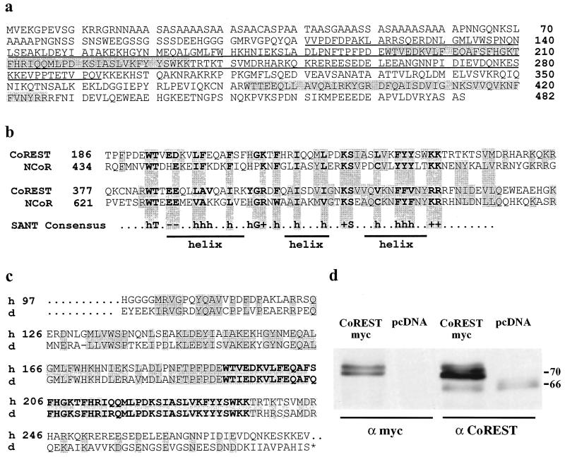
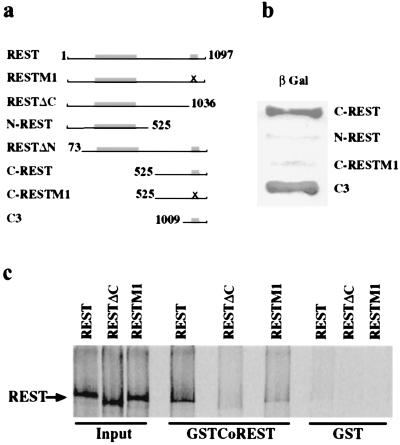
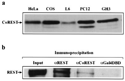
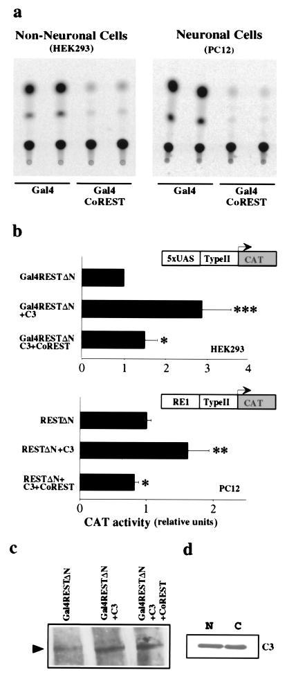
Several genes encoding proteins critical to the neuronal phenotype, such as the brain type II sodium channel gene, are expressed to high levels only in neurons. This cell specificity is due, in part, to long-term repression in nonneural cells mediated by the repressor protein REST/NRSF (RE1 silencing transcription factor/neural-restrictive silencing factor). We show here that CoREST, a newly identified human protein, functions as a corepressor for REST. A single zinc finger motif in REST is required for CoREST interaction. Mutations of the motif that disrupt binding also abrogate repression. When fused to a Gal4 DNA-binding domain, CoREST functions as a repressor. CoREST is present in cell lines that express REST, and the proteins are found in the same immunocomplex. CoREST contains two SANT (SW13/ADA2/NCoR/TFIIIB B) domains, a structural feature of the nuclear receptor and silencing mediator for retinoid and thyroid human receptors (SMRT)-extended corepressors that mediate inducible repression by steroid hormone receptors. Together, REST and CoREST mediate repression of the type II sodium channel promoter in nonneural cells, and the REST/CoREST complex may mediate long-term repression essential to maintenance of cell identity.
Figures
Similar articles
-
Differential deployment of REST and CoREST promotes glial subtype specification and oligodendrocyte lineage maturation.PLoS One. 2009 Nov 3;4(11):e7665. doi: 10.1371/journal.pone.0007665. PLoS One. 2009. PMID: 19888342 Free PMC article.
-
Corepressor for element-1-silencing transcription factor preferentially mediates gene networks underlying neural stem cell fate decisions.Proc Natl Acad Sci U S A. 2010 Sep 21;107(38):16685-90. doi: 10.1073/pnas.0906917107. Epub 2010 Sep 7. Proc Natl Acad Sci U S A. 2010. PMID: 20823235 Free PMC article.
-
REST and CoREST modulate neuronal subtype specification, maturation and maintenance.PLoS One. 2009 Dec 7;4(12):e7936. doi: 10.1371/journal.pone.0007936. PLoS One. 2009. PMID: 19997604 Free PMC article.
-
CoREST-like complexes regulate chromatin modification and neuronal gene expression.J Mol Neurosci. 2006;29(3):227-39. doi: 10.1385/JMN:29:3:227. J Mol Neurosci. 2006. PMID: 17085781 Review.
-
More than a Corepressor: The Role of CoREST Proteins in Neurodevelopment.eNeuro. 2020 Mar 10;7(2):ENEURO.0337-19.2020. doi: 10.1523/ENEURO.0337-19.2020. Print 2020 Mar/Apr. eNeuro. 2020. PMID: 32075869 Free PMC article. Review.
Cited by
-
LSD1 inhibition induces differentiation and cell death in Merkel cell carcinoma.EMBO Mol Med. 2020 Nov 6;12(11):e12525. doi: 10.15252/emmm.202012525. Epub 2020 Oct 7. EMBO Mol Med. 2020. PMID: 33026191 Free PMC article.
-
Inferring transcriptional and microRNA-mediated regulatory programs in glioblastoma.Mol Syst Biol. 2012;8:605. doi: 10.1038/msb.2012.37. Mol Syst Biol. 2012. PMID: 22929615 Free PMC article.
-
TSPYL2 is an essential component of the REST/NRSF transcriptional complex for TGFβ signaling activation.Cell Death Differ. 2015 Aug;22(8):1353-62. doi: 10.1038/cdd.2014.226. Epub 2015 Jan 23. Cell Death Differ. 2015. PMID: 25613376 Free PMC article.
-
The CoREST repressor complex mediates phenotype switching and therapy resistance in melanoma.J Clin Invest. 2024 Feb 1;134(6):e171063. doi: 10.1172/JCI171063. J Clin Invest. 2024. PMID: 38300709 Free PMC article.
-
REST and RCOR genes display distinct expression profiles in neurons and astrocytes using 2D and 3D human pluripotent stem cell models.Heliyon. 2024 Jun 10;10(12):e32680. doi: 10.1016/j.heliyon.2024.e32680. eCollection 2024 Jun 30. Heliyon. 2024. PMID: 38975076 Free PMC article.
References
-
- Chong J A, Tapia-Ramírez J, Kim S, Toledo-Aral J J, Zheng Y, Boutros M C, Altshuller Y M, Frohman M A, Kraner S D, Mandel G. Cell. 1995;80:949–957. - PubMed
-
- Schoenherr C J, Anderson D J. Science. 1995;267:1360–1363. - PubMed
-
- Chen Z F, Paquette A J, Anderson D J. Nat Genet. 1998;20:136–142. - PubMed
-
- Goodman R H, Mandel G. Curr Opin Neurobiol. 1998;8:413–417. - PubMed
Publication types
MeSH terms
Substances
Associated data
- Actions
Grants and funding
LinkOut - more resources
Full Text Sources
Other Literature Sources
Molecular Biology Databases
