. 1999 Aug 17;96(17):9815-20.
doi: 10.1073/pnas.96.17.9815.

Vascular endothelial-cadherin is an important determinant of microvascular integrity in vivo

Affiliations

Vascular endothelial-cadherin is an important determinant of microvascular integrity in vivo

M Corada et al. Proc Natl Acad Sci U S A. .

Abstract

In the present paper, we characterize an antibody, mAb BV13, directed to mouse vascular endothelial (VE)-cadherin, a major adhesive protein of interendothelial adherens junctions. When added to cultured endothelial cells, BV13 induces a redistribution of VE-cadherin from intercellular junctions. VE-cadherin redistribution did not change the localization of platelet endothelial cell adhesion molecule or tight junction markers such as zonula occludens 1, cingulin, and junctional adhesion molecule. Intravenous administration of mAb BV13 induced a concentration- and time-dependent increase in vascular permeability in heart and lungs. By electron microscopy, interstitial edema and accumulation of mixed types of inflammatory cells in heart and lungs were observed. Injection of (rhodamine-labeled) Ricinus communis I lectin showed focal spots of exposed basement membrane in the alveolar capillaries and in some larger pulmonary vessels. These data indicate that VE-cadherin is required for vascular integrity and normal organ functions.


Figures
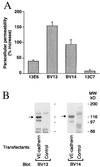
Figure 1
Figure 1
Characterization of monoclonal antibodies to VE-cadherin. (A) mAbs BV13 and BV14 increase paracellular permeability in comparison to other anti-VE-cadherin mAbs (13E6 and 13C7). Data are means ± SEM of three different experiments and are expressed as percentage increase in comparison to the values obtained with an irrelevant mAb (26.1 ± 2.4 was the fluorescence unit value of the irrelevant mAb at the same time). (B) mAb BV13 and BV14 specifically recognize a protein of the molecular weight of VE-cadherin. VE-cadherin and control Chinese hamster ovary transfectants were analyzed by Western blot using BV13 and BV14.
Figure 2
Figure 2
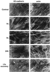
Effect of BV13 on VE-cadherin organization in endothelial cells. BV13 (50 μg/ml) was added to cultured endothelial monolayers (1G11). VE-cadherin staining was strongly reduced at junctions within 1 hour, and this effect lasted up to 24 hours of incubation with the mAb. When BV13 was removed after 7 hours of incubation and the cells were cultured for additional 17 hours, a partial recovery of VE-cadherin at junctions was detected (white arrowheads). Actin staining shows that reduction of VE-cadherin staining from junctions was not accompanied by cell retraction. Comparable results were obtained when, after fixation of the cells, VE-cadherin was detected by using either mAb BV14 or a VE-cadherin rabbit polyclonal antiserum (data not shown). (Bar = 20 μm.)
Figure 3
Figure 3
Effect of BV13 on VE-cadherin internalization. Endothelial cells (1G11) were incubated with 50 μg/ml of BV13 for 1 hour and 7 hours (1 h and 7 h lanes) or nonimmune IgG for 7 hours (Control lane). Before extraction, endothelial cells were surface-labeled with biotin by using sulfo-nitrohydroxysuccinimido-biotin (16).
Figure 4
Figure 4
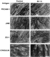
Effect of BV13 on the organization of other junctional components in the endothelium. Endothelial cell monolayers were incubated with BV13 (50 μg/ml) for 7 hours. Cells then were fixed, and the distribution of junctional proteins was analyzed by immunofluorescence microscopy. PECAM-1, JAM, ZO-1, and cingulin distribution was not significantly affected by addition of BV13. (Bar = 20 μm.)
Figure 5
Figure 5
Effect of BV13 administration on vascular permeability in vivo. (A) BV13 (100 μg/mouse) induced a significant and time-dependent increase in Evans blue accumulation in heart (white columns) and lungs (striped columns). (B) BV13 increased vascular permeability in a concentration-dependent fashion. Different doses—10 μg/mouse (white columns), 50 μg/mouse (striped columns), or 100 μg/mouse (black columns) of BV13—were administered, and, after 7 hours, Evans blue extravasation was evaluated. (C) BV13 or BV14, 25 μg/mouse (gray columns) and 50 μg/mouse (stripped columns), was administered, and, after 7 hours, Evans blue leakage was measured. For the animals treated with BV13 and BV14, data are expressed as percentage increase in Evans blue content of the different organs in comparison to mice treated with the same concentration of the control mAb MEC 7.46 for the same time. Data are means ± SEM of at least five experiments, each performed in quadruplicates.
Figure 6
Figure 6
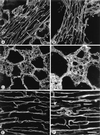
Endothelial cell junctions stained by i.v. injection of VE-cadherin antibody. Blood vessels were labeled by i.v. injection of BV13 and ex vivo incubation in Cy3-labeled secondary antibody. Images of vessels in thick tissue sections were obtained by fluorescence confocal microscopy. Shown are intrapulmonary segments of pulmonary arterioles at 20 minutes (A) or 2 hours (B) after i.v. injection of BV13. In the large arterioles and venules, antibody bound continuously along endothelial cell–cell junctions at 20 minutes and 2 hours (arrows in A and B). In small pulmonary vessels near arterioles, antibody also bound in sharp, continuous lines along endothelial cell junctions at 20 minutes (arrowheads in A), but, in contrast, binding was discontinuous and diffuse at 2 hours (arrowheads in B). Similarly, in alveolar capillaries, antibody bound in sharp, continuous lines at 20 minutes (arrow in C), but binding was discontinuous and diffuse at 2 hours (arrow in D). In capillaries of cardiac muscle, antibody bound in sharp, continuous lines at 20 minutes (arrow in E) but was discontinuous and diffuse at 2 hours (arrow in F). (Bar = 25 μm.)
Figure 7
Figure 7
Complement requirements for BV13 effects on vascular permeability. BV13 was able to increase heart (white columns) and lung (striped columns) permeability in C5 complement-deficient animals (A). Mice DBA/2J (C5 deficient) and their matched controls C57BL/6N (Control) were injected with BV13 or the control mAb MEC 7.46 (100 μg/mouse), and permeability was evaluated after 7 hours. (B) Mice depleted of C3 complement. Animals were treated with either cobra venom factor as described in Materials and Methods to deplete C3 complement element (C3-depleted) or saline (Control). The mice then were injected i.v. with BV13 or MEC 7.46 (100 μg/mouse), and, after 7 hours, permeability in heart and lungs was measured. (C) Fab fragments of BV13 (200 μg/mouse) or BV13 whole mAb (100 μg/mouse) were injected i.v., and the organs were extracted after 2 hours. Data are expressed as percentage increase in Evans blue content of the different organs and are means ± SEM of three experiments, each performed in triplicates.
Figure 8
Figure 8
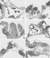
Histological analysis of the effects of BV13 on heart and lungs. Morphological features of effects of BV13 on heart (AC) and lungs (DF). In the hearts of control animals, the characteristics of normal myocardium are evident (A). At 7 (B) and 9 hours (C) after BV13, there are interstitial edema and hemorrhage (open arrows) and interstitial accumulations of neutrophils (black arrows) and mononuclear cells (white arrows). The features of normal lung, including intact alveolar walls and venules, are apparent in control animals (D) whereas 9 hours after infusion of BV13, endothelial cell bleb formation is evident (open arrowheads) (E), together with intravascular neutrophils (black arrows), bleb formation involving small capillaries and epithelial cell (black arrowheads), and intravascular platelet aggregates (F). (Toluidine blue stain; 100×.)
Figure 9
Figure 9
Electron micrographs of lung samples 0 (A), 1 (B and C) 7 (D), and 9 hours (E and F) after BV13 infusion. In A, normal vascular endothelial cell morphology is present. In B, some neutrophils are evident (open arrows) in lung capillaries. C shows platelet (P) aggregates present in venules. In D, endothelial cells present blebbing (black arrows). In E and F, multiple gaps and breaks are evident (black arrows), and platelets aggregates are present (P). Neutrophils (open arrows) appear to be adhering to denuded basement membrane and passing through the gaps in vascular wall (E). Interstitial cell debris are present in the vascular lumen (F, double-headed arrow), where the endothelial cell barrier has been lost. Sections were stained with uranyl acetate and lead citrate. (×4,800.)
Figure 10
Figure 10
Confocal micrographs of pulmonary vessels stained by injection of rhodamine-labeled Ricin lectin. (A) Alveolar capillaries in mouse given rat IgG. Luminal surface of endothelial cells stains uniformly faint whereas adherent leukocytes stain more brightly (arrowhead). (B) Alveolar capillaries in mouse given BV13 (50 μg, 4 hours). Bright focal patches of staining (arrows) indicate exposed basement membrane. (Bar = 25 μm.)

Similar articles

Cited by

References

    1. van Hinsbergh W M. Arterioscler Thromb Vasc Biol. 1997;17:1018–1023. - PubMed
    1. Lum H, Malik A B. Am J Physiol. 1994;267:L223–L241. - PubMed
    1. Anderson R G. Proc Natl Acad Sci USA. 1993;90:10909–10913. - PMC - PubMed
    1. Schnitzer J E. Cardiovasc Med. 1993;3:124–130. - PubMed
    1. Dvorak A M, Kohn S, Morgan E S, Fox P, Nagy J A, Dvorak H F. J Leukocyte Biol. 1996;59:100–115. - PubMed

Publication types

LinkOut - more resources