. 1999 Aug 9;146(3):573-84.
doi: 10.1083/jcb.146.3.573.

Cdc25B and Cdc25C differ markedly in their properties as initiators of mitosis

Affiliations

Cdc25B and Cdc25C differ markedly in their properties as initiators of mitosis

C Karlsson et al. J Cell Biol. .

Abstract

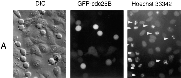
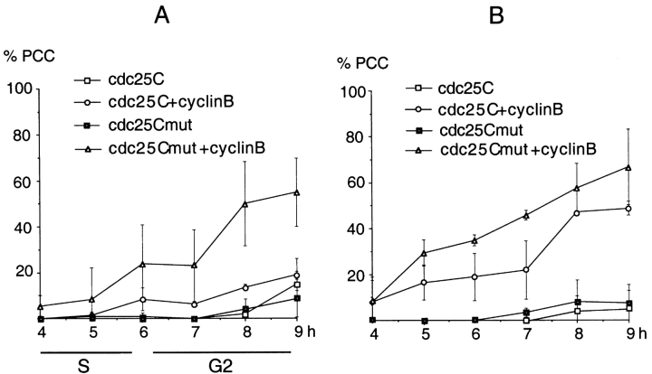
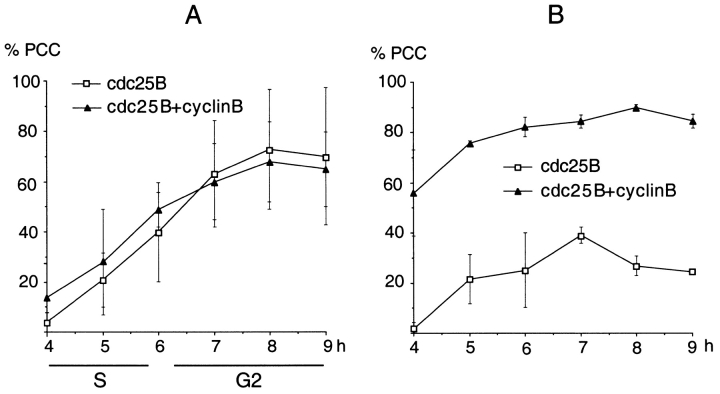
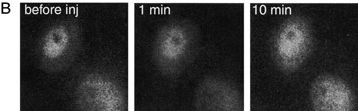
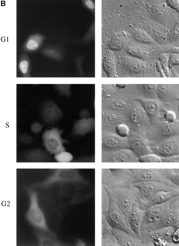
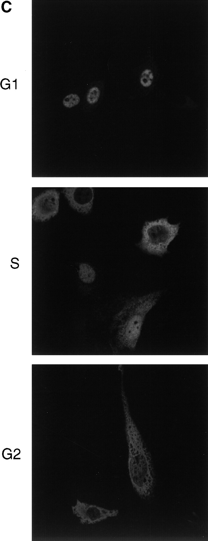
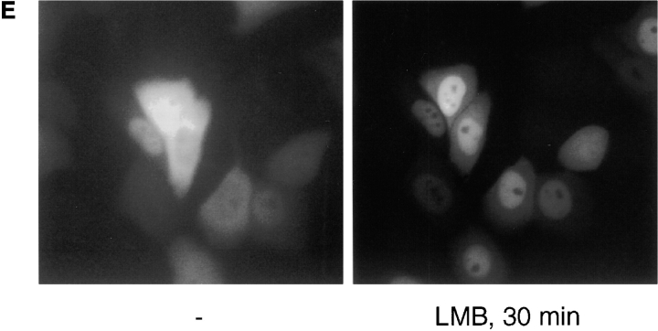
We have used time-lapse fluorescence microscopy to study the properties of the Cdc25B and Cdc25C phosphatases that have both been implicated as initiators of mitosis in human cells. To differentiate between the functions of the two proteins, we have microinjected expression constructs encoding Cdc25B or Cdc25C or their GFP-chimeras into synchronized tissue culture cells. This assay allows us to express the proteins at defined points in the cell cycle. We have followed the microinjected cells by time-lapse microscopy, in the presence or absence of DNA synthesis inhibitors, and assayed whether they enter mitosis prematurely or at the correct time. We find that overexpressing Cdc25B alone rapidly causes S phase and G2 phase cells to enter mitosis, whether or not DNA replication is complete, whereas overexpressing Cdc25C does not cause premature mitosis. Overexpressing Cdc25C together with cyclin B1 does shorten the G2 phase and can override the unreplicated DNA checkpoint, but much less efficiently than overexpressing Cdc25B. These results suggest that Cdc25B and Cdc25C do not respond identically to the same cell cycle checkpoints. This difference may be related to the differential localization of the proteins; Cdc25C is nuclear throughout interphase, whereas Cdc25B is nuclear in the G1 phase and cytoplasmic in the S and G2 phases. We have found that the change in subcellular localization of Cdc25B is due to nuclear export and that this is dependent on cyclin B1. Our data suggest that although both Cdc25B and Cdc25C can promote mitosis, they are likely to have distinct roles in the controlling the initiation of mitosis.


Figures

Figure 1
Figure 1
Overexpressing cdc25B induces premature mitosis and not apoptosis. (A) HeLa cells in S phase were microinjected with GFP-cdc25B3 cDNA at a concentration of 0.1μg/μl and cells were followed by time-lapse DIC microscopy. After 5 h cells were analyzed. Arrows indicate GFP-cdc25B–expressing cells. DIC (left), GFP fluorescence (center), and Hoechst 33342 staining (right). (B) GFP fluorescence (left) and β-tubulin immunofluorescence (right). (C) GFP fluorescence (left) and MPM2 immunofluorescence (right).
Figure 1
Figure 1
Overexpressing cdc25B induces premature mitosis and not apoptosis. (A) HeLa cells in S phase were microinjected with GFP-cdc25B3 cDNA at a concentration of 0.1μg/μl and cells were followed by time-lapse DIC microscopy. After 5 h cells were analyzed. Arrows indicate GFP-cdc25B–expressing cells. DIC (left), GFP fluorescence (center), and Hoechst 33342 staining (right). (B) GFP fluorescence (left) and β-tubulin immunofluorescence (right). (C) GFP fluorescence (left) and MPM2 immunofluorescence (right).
Figure 3
Figure 3
Frequency of PCC induced by overexpressing cdc25C and cdc25C(S216G) in the S phase. Constructs encoding Cdc25C or a Cdc25C(S216G) mutant were microinjected with or without cyclin B1 into S phase HeLa cells in the absence (A) or presence (B) of 2.5 mM hydroxyurea and followed by DIC microscopy as described in Fig. 2. (A) Frequency of PCC in cells after release from an aphidicolin block. (B) Frequency of PCC in cells after release from an aphidicolin block in the presence of hydroxyurea. Data are the mean of three different experiments.
Figure 2
Figure 2
Frequency of PCC induced by overexpressing cdc25B in the S phase. Constructs encoding Cdc25B were microinjected with or without constructs encoding cyclin B1 into S phase HeLa cells, ∼1 h after release from a thymidine/aphidicolin block in the absence (A) or presence (B) of 2.5 mM hydroxyurea. One of the constructs was expressed as a GFP-chimera to detect injected cells. (Similar results were obtained whether Cdc25B or cyclin B1 was linked to GFP.) Cells were followed by DIC microscopy and cells that rounded up were counted at the indicated time points after release. At the end of the experiment, Hoechst 33342 was added to detect abnormally condensed chromatin and the cells expressing the GFP-chimera were counted and the frequency of PCC was estimated. (A) Frequency of PCC in cells overexpressing cdc25B+/− cyclin B1. (B) Frequency of PCC in cells overexpressing cdc25B+/− cyclin B1 in the presence of hydroxyurea. All constructs were injected at a concentration of 0.02 μg/μl. For cells in the absence of hydroxyurea, the duration of S and G2 phases in the control cells are indicated. Data are the mean of three different experiments.
Figure 4
Figure 4
Overexpression of cdc25B together with cyclin B1 can force G1 cells into premature mitosis. Cells in the G1 phase were recognized by virtue of being attached after cytokinesis. Cells were injected with constructs expressing GFP-cdc25B or GFP-Cdc25C with or without constructs expressing a nondestructible cyclin B1 (R42A) mutant. The frequency of PCC was determined 6 h after microinjection as described in Fig. 2. Data are representative of three different experiments.
Figure 5
Figure 5
Wee1 can rescue cells overexpressing cdc25B from PCC. Cells were microinjected with expression constructs encoding cdc25B with or without constructs expressing Wee1 at the indicated concentrations (in μg/μl). 5 h later, Hoechst 33342 was added and the frequency of PCC was determined as described in Fig. 2. Data are the mean of four different experiments.
Figure 7
Figure 7
GFP-cdc25B is exported from the nucleus after injection of Cyclin B1/CDK1K33R and can bind to cyclin B1 in vitro. (A) HeLa cells were synchronized in the S phase and microinjected with a GFP-cdc25B expression construct immediately after release from an aphidicolin block. 3 h later, ∼40 pg of cyclin B1/CDK1K33R expressed in, and purified from, baculovirus was microinjected into the nucleus and GFP images were taken at the indicated time points. 9 min after the injection of cyclinB1/CDK1K33R, 20 nM LMB was added to the cells and a final image captured 10 min later. (B) HeLa cells were synchronized in the S phase and microinjected with a YFP-cdc25B expression construct 3 h after mitosis. 3 h later, ∼40 pg of a nuclear export–defective cyclin B1 (F146A)-GFP was microinjected into the nucleus and YFP images were taken at the indicated time points using a filter set that distinguishes between GFP and YFP (Hagting et al. 1999). (C) Physical interaction between cdc25B and cyclin B1. (Left) 35S-Labeled, in vitro translated cdc25B2 and cdc25B3 and Cdc25C were incubated with GST alone or GST-cyclin B1. (Right) 35S-Labeled in vitro translated cyclin B1 was incubated with GST alone, or GST-Cdc25B2, or GST-Cdc25B3. The complexes were bound to glutathione-Sepharose, washed extensively, separated by SDS-PAGE, and exposed to film.
Figure 7
Figure 7
GFP-cdc25B is exported from the nucleus after injection of Cyclin B1/CDK1K33R and can bind to cyclin B1 in vitro. (A) HeLa cells were synchronized in the S phase and microinjected with a GFP-cdc25B expression construct immediately after release from an aphidicolin block. 3 h later, ∼40 pg of cyclin B1/CDK1K33R expressed in, and purified from, baculovirus was microinjected into the nucleus and GFP images were taken at the indicated time points. 9 min after the injection of cyclinB1/CDK1K33R, 20 nM LMB was added to the cells and a final image captured 10 min later. (B) HeLa cells were synchronized in the S phase and microinjected with a YFP-cdc25B expression construct 3 h after mitosis. 3 h later, ∼40 pg of a nuclear export–defective cyclin B1 (F146A)-GFP was microinjected into the nucleus and YFP images were taken at the indicated time points using a filter set that distinguishes between GFP and YFP (Hagting et al. 1999). (C) Physical interaction between cdc25B and cyclin B1. (Left) 35S-Labeled, in vitro translated cdc25B2 and cdc25B3 and Cdc25C were incubated with GST alone or GST-cyclin B1. (Right) 35S-Labeled in vitro translated cyclin B1 was incubated with GST alone, or GST-Cdc25B2, or GST-Cdc25B3. The complexes were bound to glutathione-Sepharose, washed extensively, separated by SDS-PAGE, and exposed to film.
Figure 6
Figure 6
The localization of Cdc25B and GFP-cdc25B in HeLa cells depends on nuclear export. (A) Localization of GFP-cdc25C after microinjection of expression constructs into cells synchronized in G1, S, and G2 phases. GFP fluorescence (left) and DIC image (right). (B) Localization of GFP-cdc25B after microinjection of expression constructs into cells synchronized in G1, S, and G2 phases. GFP fluorescence (left) and DIC image (right). (C) Localization by immunofluorescence of a myc-tagged cdc25B construct in synchronized G1, S, and G2 HeLa cells. (D) Time-lapse sequence of HeLa cells expressing GFP-cdc25B in the S phase. Cells where GFP-cdc25B gradually becomes less nuclear (open arrows). Cells where GFP-cdc25B is completely exported to the cytoplasm (closed arrows). Images were captured at 10-min intervals. (E) Localization of GFP-cdc25B in cells before (left) and 30 min after (right) the addition of 20 nM LMB.
Figure 6
Figure 6
The localization of Cdc25B and GFP-cdc25B in HeLa cells depends on nuclear export. (A) Localization of GFP-cdc25C after microinjection of expression constructs into cells synchronized in G1, S, and G2 phases. GFP fluorescence (left) and DIC image (right). (B) Localization of GFP-cdc25B after microinjection of expression constructs into cells synchronized in G1, S, and G2 phases. GFP fluorescence (left) and DIC image (right). (C) Localization by immunofluorescence of a myc-tagged cdc25B construct in synchronized G1, S, and G2 HeLa cells. (D) Time-lapse sequence of HeLa cells expressing GFP-cdc25B in the S phase. Cells where GFP-cdc25B gradually becomes less nuclear (open arrows). Cells where GFP-cdc25B is completely exported to the cytoplasm (closed arrows). Images were captured at 10-min intervals. (E) Localization of GFP-cdc25B in cells before (left) and 30 min after (right) the addition of 20 nM LMB.

Similar articles

Cited by

References

    1. Amon A., Surana U., Muroff I., Nasmyth K. Regulation of p34CDC28 tyrosine phosphorylation is not required for entry into mitosis in S. cerevisiae . Nature. 1992;355:368–371. - PubMed
    1. Amon A., Irniger S., Nasmyth K. Closing the cell cycle circle in yeastG2 cyclin proteolysis initiated at mitosis persists until the activation of G1 cyclins in the next cycle. Cell. 1994;77:1037–1050. - PubMed
    1. Atherton Fessler S., Parker L.L., Geahlen R.L., Piwnica-Worms H. Mechanisms of p34cdc2 regulation. Mol. Cell Biol. 1993;13:1675–1685. - PMC - PubMed
    1. Baldin V., Cans C., Knibiehler M., Ducommun B. Phosphorylation of human CDC25B phosphatase by CDK1-cyclin A triggers its proteasome-dependent degradation J. Biol. Chem 272 1997. 32731 32734a - PubMed
    1. Baldin V., Cans C., Superti Furga G., Ducommun B. Alternative splicing of the human CDC25B tyrosine phosphatase. Possible implications for growth control? Oncogene 14 1997. 2485 2495b - PubMed

Publication types

MeSH terms