. 1999 Sep;73(9):7607-18.
doi: 10.1128/JVI.73.9.7607-7618.1999.

Targeted recombination demonstrates that the spike gene of transmissible gastroenteritis coronavirus is a determinant of its enteric tropism and virulence

Affiliations
Free PMC article

Targeted recombination demonstrates that the spike gene of transmissible gastroenteritis coronavirus is a determinant of its enteric tropism and virulence

C M Sánchez et al. J Virol. 1999 Sep.
Free PMC article

Abstract

Targeted recombination within the S (spike) gene of transmissible gastroenteritis coronavirus (TGEV) was promoted by passage of helper respiratory virus isolates in cells transfected with a TGEV-derived defective minigenome carrying the S gene from an enteric isolate. The minigenome was efficiently replicated in trans and packaged by the helper virus, leading to the formation of true recombinant and pseudorecombinant viruses containing the S proteins of both enteric and respiratory TGEV strains in their envelopes. The recombinants acquired an enteric tropism, and their analysis showed that they were generated by homologous recombination that implied a double crossover in the S gene resulting in replacement of most of the respiratory, attenuated strain S gene (nucleotides 96 to 3700) by the S gene of the enteric, virulent isolate. The recombinant virus was virulent and rapidly evolved in swine testis cells by the introduction of point mutations and in-phase codon deletions in a domain of the S gene (nucleotides 217 to 665) previously implicated in the tropism of TGEV. The helper virus, with an original respiratory tropism, was also found in the enteric tract, probably because pseudorecombinant viruses carrying the spike proteins from the respiratory strain and the enteric virus in their envelopes were formed. These results demonstrated that a change in the tropism and virulence of TGEV can be engineered by sequence changes in the S gene.


Figures

FIG. 1
FIG. 1
Nomenclature and relationship among the TGEVs used in this work. All the TGEVs used were derived from an original isolate of the Purdue virus (24, 34) that has been named PUR46 in reference to the place and year that it was reported for the first time. The PUR46-SW11 (SW11) strain was obtained from PUR46 by 11 passages in swine intestine; this virus was kindly provided as a 20% suspension of small intestine cells by M. Pensaert (24, 34). The PUR46-SW11-ST2 (ST2) virus was obtained from SW11 after two passages in ST cells (40). The PUR46-SW11-ST2-C8 (C8) and PUR46-SW11-ST2-C11 (C11) isolates were plaque-purified isolates derived from the PUR46-SW11-ST1 (ST1) at CNB, Madrid, Spain. The PUR46-ST115 was independently derived from the PUR46-SW11 (or a similar passage number in swine) after 115 passages in ST cells, by L. Saif at the OARDC (Ohio State University) (50). From this strain we obtained the PUR46-MAD-ST120 by five additional passages on ST cells including three cloning steps by plaque purification (54). The PTV virus was derived from a Purdue type strain by sequential passage in gnotobiotic pigs by the pulmonary route, pig lung cell cultures, and diploid ST cell cultures. During this time the virus was exposed to an acidic (pH 3) environment and trypsin (66).
FIG. 2
FIG. 2
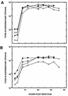
Growth kinetics of isolates C8 and C11 in ST cells. The growth of isolates C8 (A) and C11 (B) in ST cells is shown. Virus replication in ST cells that just reached confluence (○, □) or were grown for 1 more day following confluence (●, ■) after infection at an MOI of 1 (○, ●) or 10 (□, ■) is shown. Aliquots (0.2 ml) were collected at the indicated times and titrated on ST cells. Results of a representative experiment among three that gave similar results are shown.
FIG. 3
FIG. 3
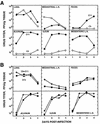
Growth kinetics of isolates C8 and C11 in swine. Two- to three-day-old non-colostrum-deprived NIH miniswine were used to study the growth kinetics of isolate C8 (○) or C11 (●) alone (A) or the growth kinetics of a mixture of isolates C8 and C11 (70% C8 plus 30% C11) (■) or a virulent TGEV isolate (ST2) (⧫) that includes both isolates (B). Groups of four minipigs were oronasally (2 × 10⁸ PFU/pig) and intragastrically (3 × 10⁸ PFU/pig) inoculated. Virus titers at the indicated number of days postinfection were determined in the indicated tissue extracts. L.N., lymph nodes. The whole organs were homogenized in order to obtain representative samples. Results of a representative experiment among three that gave similar results are shown in panels A and B.
FIG. 4
FIG. 4
Pathogenesis of isolates C8 and C11 in minipigs. Groups of eight minipigs were inoculated with the indicated TGEV isolates (isolates C8 and C11, a mixture of C8 and C11 [70% C8 plus 30% C11], or the virulent uncloned isolate ST2) as described in the legend for Fig. 3. The number of piglets without enteritis (■) or surviving (●) is shown at different days postinfection. Results of a representative experiment of two that gave similar results are shown.
FIG. 5
FIG. 5
Sequence comparison of isolates C8 and C11. The nucleotide substitutions in isolate C8 in relationship to isolate C11 are shown for all genes except ORF 1. The top bar indicates the different viral genes, and the numbers above the second bar indicate the positions of the substituted nucleotides, with nt 1 of each gene considered to be the A of the initiation codon. Letters within the bars indicate the corresponding nucleotides at the indicated positions. Letters below the bars indicate the amino acid substitutions encoded by the nucleotides around the indicated position. Δ6 nt, deletion of six nucleotides. The arrow indicates the position of the S gene stop codon.
FIG. 6
FIG. 6
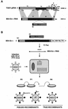
Diagram of the pseudorecombinant and true recombinant virus isolation protocol. (A) Structure of the M54 minigenome indicating the sequence position where the S gene from the enteric C11 isolate was cloned. Letters and numbers above the top bar indicate the TGEV ORFs. Numbers below this bar indicate the nucleotide sequences of the helper virus incorporated into the M54 minigenome. Numbers above the second bar indicate the four sequence domains that were linked during the generation of the minigenome. Numbers to the right of the bars indicate sizes of the genomes in nucleotides. gRNA, genomic RNA. IG, intergenic sequences preceding the S gene of the C11 isolate, which is identical to that of the PUR46-MAD strain of TGEV. An, poly(A). (B) Generation of recombinants. The S gene from an enteric TGEV (isolate C11) was cloned into minigenome M54, generating the minigenome M54-Sc11 with the structure diagrammatically shown in panel A. This minigenome has been cloned after the T7 promoter (T7) (black box) and preceding the hepatitis delta virus ribozyme (HDV Rz) sequences and the T7 terminator sequences (TΦ). ST cells were infected with C8 or PTV viruses. At 4 to 6 h p.i., cells were electroporated with in vitro-transcribed RNA and the supernatants from these cultures were passaged by using ST cells. The potential pseudorecombinants with the S protein from the respiratory helper viruses (light circles) or from the enteric C11 isolate (dark circles) containing either the genome of the helper virus (large bar) or the minigenome with the S gene of the C11 isolate are diagrammatically represented (bottom left). True recombinants with a chimeric S protein generated by two crossovers between the S gene from the helper virus and the S gene from isolate C11 are also indicated (bottom right).
FIG. 7
FIG. 7
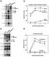
Northern blot analysis and growth kinetics of the potential recombinants in the enteric tract. The characterization of the viruses obtained after minigenome M54-Sc11 rescue using as helper virus isolate C8 (A and B) or PTV (C and D) is shown. The Northern blot analysis of the viral RNAs obtained after infecting ST cells for eight passages with the indicated helper virus (either C8 or PTV) alone or with the helper virus plus the minigenome M54 or plus minigenome M54-Sc11 is shown (A and C). Numbers and arrows to the left of boxes A and C indicate the positions of the helper virus RNAs. Arrows and names to the right of these boxes indicate the expected positions for the minigenomes. UK, unknown RNA. Virus replication in the enteric tract was examined following oronasal and intragastrical inoculation of groups of four newborn piglets. Animals were sacrificed at the indicated days, and the virus content in representative samples of the whole tissue was determined by using the plaque assay on ST cells. Virus content was determined, and the arithmetic mean of the titers of virus recovered from the jejunum, ileum, and intestinal content is shown (panel B). ■, isolate C11; ●, isolate C8 plus minigenome M54-Sc11; ▴, isolate C8 plus minigenome M54. Virus replication using the strain PTV as the helper virus is also shown (panel D). Virus content was measured in the jejunum (●) or in the ileum (■) of piglets infected with strain PTV plus minigenome M54-Sc11; virus content was measured in the jejunum (formula image) or the ileum (░⃞) of piglets infected with the strain PTV plus the minigenome M54.
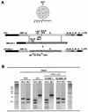
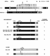
FIG. 8
FIG. 8
Restriction endonuclease analysis of the potential recombinants. (A) A diagram of the procedure followed in the selection of true recombinant and pseudorecombinant viruses growing in the enteric tract is shown. Gut tissue (jejunum), collected from a single pig at 2 d p.i., was homogenized, and lysis plaques were isolated on ST cells. Plaques of two sizes (3- and 1-mm diameter) were observed and cloned twice. Viruses in these plaques were expected to have either the genome of the helper respiratory virus (PTV) or the genome of a true recombinant virus (rPTV/Sc11) formed by a two-crossover event within the S gene (bottom bar). The positions of restriction endonuclease (DraIII and MslI) sites in cDNA, derived by RT-PCR, between nt 487 and 1640 of the S gene are indicated. (B) Prototype restriction endonuclease patterns of cDNAs derived from S genes of the strains PTV and C11 and two isolates (clones 1 and 43) with a restriction endonuclease pattern identical to that of strain C11 or PTV are shown. MWM, molecular weight markers (1-kb DNA ladder; Gibco). Numbers on the left of the MWM lane indicate the sizes of the markers in kilobases.
FIG. 9
FIG. 9
Nucleotide sequences of TGEV recombinants isolated in enteric tissues. Virus was recovered from the enteric tract of a single pig at 2 d p.i. Recombinants with a large plaque size (no. 1, 8, 10, 12, 17, 18, 21, 25, 29, 30, 31, 32, 34, and 41) or a small plaque size (no. 43, 45, and 48) were plaque purified twice and expanded into ST cells, and their RNAs were copied into cDNA and sequenced. The nucleotide differences between the S genes of strains PTV and C11 are indicated (top bars). Numbers above bars indicate the positions of nucleotide substitutions or deletions. The approximate locations of the two crossovers in the S gene are indicated by two sets of crossed lines extending between the two top bars. The asterisk indicates the insertion sequences containing the restriction endonuclease sites SpeI, NotI, PstI, and SalI, derived from the polylinker of plasmid pGEM-T and located after the S gene. In isolates 1 to 41, a C was present at position 2136. This nucleotide (C) was different from that located at the equivalent positions in the S genes of the two parental viruses (T). The presence of codon deletions (▵) and nucleotide and amino acid (aa) substitutions is indicated above the bars. L and S indicate large- and small-plaque phenotypes.

Similar articles

Cited by

References

    1. Bai M, Campisi L, Freimuth P. Vitronectin receptor antibodies inhibit infection of HeLa and A549 cells by adenovirus type 12 but not by adenovirus type 2. J Virol. 1994;68:5925–5932. - PMC - PubMed
    1. Ballesteros M L, Sanchez C M, Enjuanes L. Two amino acid changes at the N-terminus of transmissible gastroenteritis coronavirus spike protein result in the loss of enteric tropism. Virology. 1997;227:378–388. - PMC - PubMed
    1. Benbacer L, Kut E, Besnardeau L, Laude H, Delmas B. Interspecies aminopeptidase-Nchimeras reveal species-specific receptor recognition by canine coronavirus, feline infectious peritonitis virus, and transmissible gastroenteritis virus. J Virol. 1997;71:734–737. - PMC - PubMed
    1. Bergelson J M, Cunningham J A, Droguett G, Kurt-Jones E A, Krithivas A, Hong J S, Horwitz M S, Crowell R L, Finberg R W. Isolation of a common receptor for coxsackie B viruses and adenoviruses 2 and 5. Science. 1997;275:1320–1323. - PubMed
    1. Bergelson J M, Mohanty J G, Crowell R L, St. John N F, Lublin D M, Finberg R W. Coxsackievirus B3 adapted to growth in RD cells binds to decay-accelerating factor (CD55) J Virol. 1995;69:1903–1906. - PMC - PubMed

Publication types

Substances

LinkOut - more resources