. 1999 Sep;73(9):7287-96.
doi: 10.1128/JVI.73.9.7287-7296.1999.

Tyrosine phosphorylation of A17 during vaccinia virus infection: involvement of the H1 phosphatase and the F10 kinase

Affiliations

Tyrosine phosphorylation of A17 during vaccinia virus infection: involvement of the H1 phosphatase and the F10 kinase

M Derrien et al. J Virol. 1999 Sep.

Abstract

Vaccinia virus encodes two protein kinases (B1 and F10) and a dual-specificity phosphatase (VH1), suggesting that phosphorylation and dephosphorylation of substrates on serine/threonine and tyrosine residues are important in regulating diverse aspects of the viral life cycle. Using a recombinant in which expression of the H1 phosphatase can be regulated experimentally (vindH1), we have previously demonstrated that repression of H1 leads to the maturation of noninfectious virions that contain several hyperphosphorylated substrates (K. Liu et al., J. Virol. 69:7823-7834). In this report, we demonstrate that among these is a 25-kDa protein that is phosphorylated on tyrosine residues in H1-deficient virions and can be dephosphorylated by recombinant H1. We demonstrate that the 25-kDa phosphoprotein represents the product of the A17 gene and that A17 is phosphorylated on serine, threonine, and tyrosine residues during infection. Detection of phosphotyrosine within A17 is abrogated when Tyr(203) (but not Tyr(3), Tyr(6), or Tyr(7)) is mutated to phenylalanine, suggesting strongly that this amino acid is the site of tyrosine phosphorylation. Phosphorylation of A17 fails to occur during nonpermissive infections performed with temperature-sensitive mutants defective in the F10 kinase. Our data suggest that this enzyme, which was initially characterized as a serine/threonine kinase, might in fact have dual specificity. This hypothesis is strengthened by the observation that Escherichia coli induced to express F10 contain multiple proteins which are recognized by antiphosphotyrosine antiserum. This study presents the first evidence for phosphotyrosine signaling during vaccinia virus infection and implicates the F10 kinase and the H1 phosphatase as the dual-specificity enzymes that direct this cycle of reversible phosphorylation.


Figures

FIG. 1
FIG. 1
Identification of an encapsidated H1 substrate that is phosphorylated on Tyr residues. (A) A 25-kDa pTyrosine-containing protein is found within the membranes of H1-deficient virions. (Left) Seven independently prepared stocks of wt virions (lanes 1 to 4) and H1-deficient virions (lanes 5 to 7) (approximately 3 μg of each) were subjected to SDS-PAGE and immunoblot analysis. After incubation of the nitrocellulose filters with a polyclonal anti-pTyr serum and an HRP-conjugated secondary antiserum, ECL development allowed the immunoreactive proteins to be visualized on Kodak MR film. (Right) H1-deficient virions (4 μg) were partitioned into membrane and core fractions as described in Materials and Methods. pTyr-containing proteins were detected by immunoblot analysis as described above; only the relevant portion of the filter is shown. (B) The H1 phosphatase can dephosphorylate the 25-kDa protein in vitro. wt or H1-deficient virions (9 μg) were analyzed directly (lanes 1 and 2) or permeabilized with NP-40 plus DTT and then incubated for 1 h at 37°C in the absence (lane 3) or presence (lane 6) of 10 mM sodium orthovanadate or after the addition of 1.5 μg of active or catalytically inert H1 phosphatase (H1 [lane 4] and H1C100S [lane 5], respectively). The samples were then subjected to SDS-PAGE and immunoblot analysis with anti-pTyr serum as described above.
FIG. 2
FIG. 2
The 25-kDa pTyr-containing is the product by the A17 gene. (A) The proteins immunoprecipitated (I.P.) with anti-pTyr and anti-A17 sera comigrate. BSC40 cells were infected with vindH1 (MOI of 10) in the absence of IPTG and metabolically labeled with [35S]methionine. Lysates were subjected to immunoprecipitation with sera directed against pTyr or A17, as shown; after SDS-PAGE, radiolabeled proteins were visualized by fluorography. The electrophoretic profile of protein standards is shown at the left, with molecular masses indicated in kilodaltons; only the relevant portion of the autoradiograph is shown. (B) Detection of the pTyr-containing 25-kDa protein is dependent on expression of the A17 protein. BSC40 cells were left uninfected (lanes 1) or infected (MOI of 10) with wt virus (lanes 2) or recombinants in which expression of A17 (lanes 3) or H1 (lanes 4) is inducible. Duplicate infections were performed in the absence or presence of IPTG, the inducer. Cells were harvested at 12 hpi, and cell lysates were subjected to SDS-PAGE and immunoblot analysis with anti-pTyr serum. Only the relevant portion of the immunoblot is shown. (C) A17 is phosphorylated within infected cells. BSC40 cells were left uninfected or infected (MOI of 2) with wt virus or vindH1 in the absence of IPTG. Cells were radiolabeled with 32PPi from 3 to 17 hpi; cells were then harvested, and lysates were subjected to immunoprecipitation with anti-A17 (lanes 1), anti-pTyr (lanes 2), or anti-I3 (lanes 3). Phosphorylated I3 is indicated with a filled oval; the phosphoproteins immunoprecipitated with anti-A17 and anti-pTyr sera comigrate and are indicated with the filled and open arrows. (D) Immunoprecipitated A17 is recognized by anti-pTyr serum. BSC40 cells were infected with vindH1 in the absence of IPTG (MOI of 10); at 12 hpi, cells were harvested, and lysates were subjected to immunoprecipitation with rabbit polyclonal sera directed against pTyr, A17, or A14. Immunoprecipitates were then resolved on an SDS–15% polyacrylamide gel and transferred electrophoretically to nitrocellulose. The filter was incubated with a murine monoclonal antibody directed against pTyr, and immunoreactive proteins were visualized after ECL development. Lane 1 to 3, proteins precipitated with anti-pTyr, anti-A17, and anti-A14 sera, respectively; lane 4, normal rabbit serum. The arrow at the left indicates the strong immunoreactive signal in lanes 1 and 2. The diamond and open arrow at the right indicate the heavy and light chains of the rabbit immunoglobulin used in the immunoprecipitations; there is cross-species recognition of these proteins by the goat anti-mouse immunoglobulin G used in the immunoblot development.
FIG. 3
FIG. 3
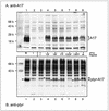
Mutation of Tyr203 to Phe abrogates the phosphorylation of A17 on Tyr residues. BSC40 cells were infected with an inducible A17 recombinant (MOI of 10) in the presence (lanes 1) or absence (lanes 2 to 9) of IPTG. Infected cells were left untransfected (lanes 1 and 2) or transfected with empty plasmid (lane 3) or a plasmid encoding wt A17 (lane 9) or A17 containing the following amino acid substitution: Y3F (lane 4), Y6F (lane 5), Y7F (lane 6), Y203F (lane 7), or Y367F (lane 8). Samples were harvested at 30 hpi, and cell lysates were subjected to SDS-PAGE and immunoblot analysis. (A) Analysis with anti-A17 serum. The A17 species expressed from the genomic (lane 1) or transfected DNA (lanes 4 to 9) are indicated by the brace at the right of panel A. Note that the Y3F protein migrates somewhat more slowly than the other A17 proteins. Prestained protein standards are shown in the leftmost lane, with their MWs indicated. (B) Analysis with anti-pTyr serum. The A17 proteins containing immunoreactive pTyr are indicated by arrowheads; note that the Y3F protein migrates somewhat more slowly than the other pTyr-containing A17 proteins. No immunoreactive pTyr was detected in the A17 protein containing the Y203F substitution.
FIG. 4
FIG. 4
Phosphorylation of A17 is abrogated during nonpermissive infections with ts mutants defective in the F10 kinase. (A) Incorporation of 32PPi into A17 is not observed during nonpermissive infections with ts28 (tsF10). Confluent BSC40 cells were infected (MOI of 2) with wt virus or ts28 (tsF10) and maintained at the permissive (31.5°C) or nonpermissive (39.5°C) temperature. Cultures were metabolically labeled with 32PPi from 3 to 17 hpi, harvested, and subjected to immunoprecipitation with anti-A17 (lanes 1, 4, 7, and 10), anti-pTyr (lanes 2, 5, 8, and 11), or anti-I3 (lanes 3, 6, 9, and 12) serum. Incorporation of 32PPi into I3 (filled oval) was equivalent under all conditions, whereas incorporation of 32PPi into A17 (filled arrow) was lost in cells infected with ts28 at 39.5°C (compare lane 4 with lanes 1, 7, and 10). Since the H1 phosphatase was active during all of these infections, recovery of 32P-labeled A17 with anti-pTyr serum (open arrow) was minimal. (B) A17 does not contain immunoreactive pTyr in cells infected with ts28 under nonpermissive conditions. Cells were left uninfected or infected with wt virus or ts28 (tsF10) under permissive and nonpermissive conditions (MOI of 10). At 12 hpi, cells were harvested and subjected to immunoblot analysis with anti-pTyr serum. All of the immunoreactive pTyr in the A17 protein (open arrow) was lost during nonpermissive infections with ts28 (compare lane 6 with lanes 3 to 5).
FIG. 5
FIG. 5
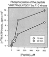
Recombinant F10 kinase phosphorylates a peptide representing the C terminus of the A17 protein. Recombinant F10 kinase (74 ng) was incubated with [γ-32P]ATP and various concentrations (0, 5, 10, 50, 100, or 500 μM) of a peptide substrate whose sequence comprised a basic N-terminal extension followed by 10 amino acids representing residues 194 to 203 of the A17 protein. Reactions were performed for 10 or 30 min, as indicated. To quantitate the incorporation of radiolabel, samples were spotted onto P81 paper, washed extensively, and analyzed by Cerenkov counting as described in Materials and Methods.
FIG. 6
FIG. 6
E. coli cultures induced to express active F10 kinase contain multiple proteins which display immunoreactive pTyr. Mid-log-phase cultures of E. coli BL21(DE3) and BL21(DE3):pET16b-F10 transformants were induced with IPTG as described in Materials and Methods. Cultures were harvested after 48 h of induction at 18°C, and total cell extracts were subjected to SDS-PAGE in duplicate. (A) One portion of the gel was stained with Coomassie blue to visualize total protein content; the two cultures displayed equivalent protein profiles. (B) The proteins in the other portion of the gel were transferred electrophoretically to nitrocellulose and subjected to immunoblot analysis with anti-pTyr serum. No immunoreactive pTyr was detected in the BL21(DE3) culture, whereas the culture expressing enzymatically active F10 kinase contained immunoreactive pTyr on numerous protein species. The arrow in panel A indicates a protein likely to represent the induced F10 species; in panel B, the arrow indicates a protein of similar electrophoretic mobility that is highly reactive with the anti-pTyr serum. In both panels, the electrophoretic profile of protein standards is shown at the left, with molecular masses indicated in kilodaltons.

Similar articles

Cited by

References

    1. Banham A H, Smith G L. Vaccinia virus gene B1R encodes a 34-kDa serine/threonine protein kinase that localizes in cytoplasmic factories and is packaged into virions. Virology. 1992;191:803–812. - PubMed
    1. Beaud G, Sharif A, Topa-Masse A, Leader D P. Ribosomal protein S2/Sa kinase purified from HeLa cells infected with vaccinia virus corresponds to the B1R protein kinase and phosphorylates in vitro the viral ssDNA-binding protein. J Gen Virol. 1994;75:283–293. - PubMed
    1. Betakova T, Wolffe E J, Moss B. Regulation of vaccinia virus morphogenesis: phosphorylation of the A14L and A17L membrane proteins and C-terminal truncation of the A17L protein are dependent on the F10L kinase. J Virol. 1999;73:3534–3543. - PMC - PubMed
    1. Casnellie J E. Assay of protein kinases using peptides with basic residues for phosphocellulose binding. Methods Enzymol. 1991;200:115–121. - PubMed
    1. Condit R C, Motyczka A, Spizz G. Isolation, characterization and physical mapping of temperature sensitive mutants of vaccinia virus. Virology. 1983;128:429–443. - PubMed

Publication types

MeSH terms

LinkOut - more resources