Vascular endothelial growth factor can substitute for macrophage colony-stimulating factor in the support of osteoclastic bone resorption
- PMID: 10432291
- PMCID: PMC2195572
- DOI: 10.1084/jem.190.2.293
Vascular endothelial growth factor can substitute for macrophage colony-stimulating factor in the support of osteoclastic bone resorption
Abstract
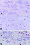
We demonstrated previously that a single injection of recombinant human macrophage colony-stimulating factor (rhM-CSF) is sufficient for osteoclast recruitment and survival in osteopetrotic (op/op) mice with a deficiency in osteoclasts resulting from a mutation in M-CSF gene. In this study, we show that a single injection of recombinant human vascular endothelial growth factor (rhVEGF) can similarly induce osteoclast recruitment in op/op mice. Osteoclasts predominantly expressed VEGF receptor 1 (VEGFR-1), and activity of recombinant human placenta growth factor 1 on osteoclast recruitment was comparable to that of rhVEGF, showing that the VEGF signal is mediated through VEGFR-1. The rhM-CSF-induced osteoclasts died after injections of VEGFR-1/Fc chimeric protein, and its effect was abrogated by concomitant injections of rhM-CSF. Osteoclasts supported by rhM-CSF or endogenous VEGF showed no significant difference in the bone-resorbing activity. op/op mice undergo an age-related resolution of osteopetrosis accompanied by an increase in osteoclast number. Most of the osteoclasts disappeared after injections of anti-VEGF antibody, demonstrating that endogenously produced VEGF is responsible for the appearance of osteoclasts in the mutant mice. In addition, rhVEGF replaced rhM-CSF in the support of in vitro osteoclast differentiation. These results demonstrate that M-CSF and VEGF have overlapping functions in the support of osteoclastic bone resorption.
Figures


Similar articles
-
Transient recruitment of osteoclasts and expression of their function in osteopetrotic (op/op) mice by a single injection of macrophage colony-stimulating factor.J Bone Miner Res. 1993 Jan;8(1):45-50. doi: 10.1002/jbmr.5650080107. J Bone Miner Res. 1993. PMID: 8427048
-
Effects of vascular endothelial growth factor on osteoclast induction during tooth movement in mice.J Dent Res. 2001 Oct;80(10):1880-3. doi: 10.1177/00220345010800100401. J Dent Res. 2001. PMID: 11706945
-
Congenital osteoclast deficiency in osteopetrotic (op/op) mice is cured by injections of macrophage colony-stimulating factor.J Exp Med. 1991 Jan 1;173(1):269-72. doi: 10.1084/jem.173.1.269. J Exp Med. 1991. PMID: 1985123 Free PMC article.
-
Role of CSF-1 in bone and bone marrow development.Mol Reprod Dev. 1997 Jan;46(1):75-83; discussion 83-4. doi: 10.1002/(SICI)1098-2795(199701)46:1<75::AID-MRD12>3.0.CO;2-2. Mol Reprod Dev. 1997. PMID: 8981367 Review.
-
Macrophage differentiation and granulomatous inflammation in osteopetrotic mice (op/op) defective in the production of CSF-1.Mol Reprod Dev. 1997 Jan;46(1):85-91. doi: 10.1002/(SICI)1098-2795(199701)46:1<85::AID-MRD13>3.0.CO;2-2. Mol Reprod Dev. 1997. PMID: 8981368 Review.
Cited by
-
Functional Scaffolds for Bone Tissue Regeneration: A Comprehensive Review of Materials, Methods, and Future Directions.J Funct Biomater. 2024 Sep 25;15(10):280. doi: 10.3390/jfb15100280. J Funct Biomater. 2024. PMID: 39452579 Free PMC article. Review.
-
Compression and hypoxia play independent roles while having combinative effects in the osteoclastogenesis induced by periodontal ligament cells.Angle Orthod. 2016 Jan;86(1):66-73. doi: 10.2319/121414.1. Epub 2015 Apr 6. Angle Orthod. 2016. PMID: 25844508 Free PMC article.
-
Targeting VEGF and Its Receptors for the Treatment of Osteoarthritis and Associated Pain.J Bone Miner Res. 2016 May;31(5):911-24. doi: 10.1002/jbmr.2828. Epub 2016 Apr 8. J Bone Miner Res. 2016. PMID: 27163679 Free PMC article. Review.
-
Normalization of the Immunological Microenvironment and Sustained Minimal Residual Disease Negativity: Do We Need Both for Long-Term Control of Multiple Myeloma?Int J Mol Sci. 2022 Dec 14;23(24):15879. doi: 10.3390/ijms232415879. Int J Mol Sci. 2022. PMID: 36555520 Free PMC article. Review.
-
Osteoclastogenesis in human breast carcinoma.Virchows Arch. 2004 May;444(5):470-2. doi: 10.1007/s00428-004-0989-1. Epub 2004 Mar 11. Virchows Arch. 2004. PMID: 15014987 No abstract available.
References
-
- Marks S.C., Jr., Lane P.W. Osteopetrosis, a new recessive skeletal mutation on chromosome 12 of the mouse. J. Hered. 1976;67:11–18. - PubMed
-
- Marks S.C., Jr. Morphological evidence of reduced bone resorption in osteopetrotic (op) mice. Am. J. Anat. 1982;163:157–167. - PubMed
-
- Yoshida H., Hayashi S., Kunisada T., Ogawa M., Nishikawa S., Okumura H., Sudo T., Shultz L.D., Nishikawa S.-I. The murine mutation osteopetrosis is in the coding region of the macrophage colony stimulating factor gene. Nature. 1990;345:442–444. - PubMed
-
- Felix R., Cecchini M.G., Fleisch H. Macrophage colony-stimulating factor restores in vivo bone resorption in the op/op osteopetrotic mouse. Endocrinology. 1990;127:2592–2594. - PubMed
MeSH terms
Substances
LinkOut - more resources
Full Text Sources
Other Literature Sources
Research Materials
Miscellaneous

