. 1999 Jul 26;146(2):333-44.
doi: 10.1083/jcb.146.2.333.

Sec1p binds to SNARE complexes and concentrates at sites of secretion

Affiliations

Sec1p binds to SNARE complexes and concentrates at sites of secretion

C M Carr et al. J Cell Biol. .

Abstract

Proteins of the Sec1 family have been shown to interact with target-membrane t-SNAREs that are homologous to the neuronal protein syntaxin. We demonstrate that yeast Sec1p coprecipitates not only the syntaxin homologue Ssop, but also the other two exocytic SNAREs (Sec9p and Sncp) in amounts and in proportions characteristic of SNARE complexes in yeast lysates. The interaction between Sec1p and Ssop is limited by the abundance of SNARE complexes present in sec mutants that are defective in either SNARE complex assembly or disassembly. Furthermore, the localization of green fluorescent protein (GFP)-tagged Sec1p coincides with sites of vesicle docking and fusion where SNARE complexes are believed to assemble and function. The proposal that SNARE complexes act as receptors for Sec1p is supported by the mislocalization of GFP-Sec1p in a mutant defective for SNARE complex assembly and by the robust localization of GFP-Sec1p in a mutant that fails to disassemble SNARE complexes. The results presented here place yeast Sec1p at the core of the exocytic fusion machinery, bound to SNARE complexes and localized to sites of secretion.


Figures

Figure 1
Figure 1
MYC-Sec1p binds to all three exocytic SNAREs. (A) Ssop, Sec9p, and Sncp coprecipitate with MYC-Sec1p. 15 A600 units of either untagged (NY1491) or MYC-SEC1 (NY1689) cells were harvested and diluted 10-fold into ice-cold wash buffer (20 mM Tris-Cl, pH 7.5, plus 20 mM NaN3, and 20 mM NaF) to inhibit membrane fusion and to deplete intracellular ATP. Cells were lysed in ice-cold IP buffer (50 mM Hepes, pH 7.4, 150 mM KCl, 1 mM EDTA, 0.5% NP-40 plus protease inhibitors) to solubilize proteins and inhibit ATP hydrolysis. Anti-MYC IPs were probed for coprecipitated SNARE proteins with antibodies to Sec9p, Ssop, and Sncp using standard Western blot analysis. (B) MYC-Sec1p coprecipitates with HA-Sncp. Lysates of untagged (NY1491) and MYC-SEC1 HA-SNC (NY1702) were prepared as in A. To detect coprecipitation of HA-Sncp with MYC-Sec1p, MYC-Sec1p IPs were blotted for HA-Sncp using the monoclonal HA antibody. To detect coprecipitation of MYC-Sec1p with HA-Sncp, HA-Sncp IPs were blotted for MYC-Sec1p using the Sec1p antibody. (C) Nonexocytic SNAREs do not coprecipitate with MYC-Sec1p. IPs from lysates of untagged (NY1491) and MYC-SEC1 (NY1689) strains were divided into four samples and analyzed for coprecipitated SNARE proteins with antibodies to Ssop, Pep12p, Sec22p, and Bos1p. The lysate shown is 2% of the protein used for the IP. (D) Stoichiometry of SNARE proteins bound to MYC-Sec1p. IPs from lysates of untagged (NY1491) and MYC-SEC1 (NY1689) were probed with antibodies to Ssop and Sncp. The ratio of Ssop to Sncp coprecipitated with MYC-Sec1p was compared with twofold serial dilutions of purified, soluble SNARE complexes, in which the ratio of soluble SNAREs, SsopΔTM to SncpΔTM to Sec9p (SNAP-25–like domain), is 1:1:1.
Figure 2
Figure 2
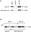
Conditions that alter the abundance of SNARE complexes also alter the Sec1p-Ssop interaction. (A) The interaction of Ssop with either MYC-Sec1p or Sncp is altered in sec mutants. SEC+ (NY1689), sec4-8 (NY1690), and sec18-1 (NY1691) MYC-SEC1 strains were shifted to 37°C for 10 min. The Ssop antibody was used to detect Ssop from the anti-Sncp and the anti-MYC IPs. (B) Sec18p disassembles SNARE complexes and disrupts the Sec1p-Ssop interaction. Lysates of SEC+ (NY1689) and sec18-1 (NY1691) MYC-SEC1 strains were prepared either with ATP (+) or without ATP (−), and the anti-MYC or anti-Sncp IPs were probed for coprecipitated Ssop. When ATP was required, NaN3 and NaF were omitted from the wash buffer, and EDTA was replaced by an ATP-regeneration system (10 μg/ml creatine kinase, 5 mM creatine phosphate, 1 mM ATP, and 1 mM MgCl2) in the IP buffer.
Figure 3
Figure 3
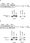
Exocytic SNARE complexes assemble only in vivo, but Sec1p can bind to SNARE complexes in lysates. For each experiment, a mixing protocol was used to test for the ability of epitope-tagged proteins expressed in two different strains to bind to each other. The strains that were mixed in each experiment are depicted schematically above the results, with the products of the relevant genes listed after the strain names. (A) Exocytic SNARE complexes assemble in vivo, not in yeast lysates. HA-SNC (NY1642) and MYC-SSO (NY1704) were mixed 1:1 before lysis (MIX). Sso proteins coprecipitated in the anti-HA IPs were detected with the Ssop antibody. Lysates shown represent ∼1% of the protein used for the IPs. (B) Sec1p binds to Ssop in yeast lysates. MYC-SEC1 (NY1689) and HA-SSO (NY1692) were mixed 1:1 before lysis (MIX). Sso proteins coprecipitated in the anti-MYC IPs were detected with the Ssop antibody. Lysates shown represent 1% of the protein used for the IPs. Note that in these mixing experiments HA-Ssop runs as a doublet, and there are two distinct cross-reacting bands detected in the Western blot, one in the lysate and one in the IPs.
Figure 4
Figure 4
The interaction between Sec1p and Ssop is limited by the abundance of SNARE complexes recovered from sec mutants. HA-SSO and MYC-SEC1 strains were mixed and lysed to look for coprecipitation of HA-Ssop with MYC-Sec1p as in Fig. 3 B, except that SEC+ cells were mixed with either sec4-8 or sec18-1 cells. For each pair of strains mixed, the sec genotypes are indicated in boxes between the MYC IPs and the lysates. In lanes 1–3, MYC-SEC1 SEC+ (NY1689) was mixed 1:1 with HA-SSO SEC+ (NY1692), HA-SSO sec4-8 (NY1693), and HA-SSO sec18-1 (NY1694), respectively. In lanes 4–6, HA-SSO SEC+ (NY1692) was mixed with MYC-SEC1 sec4-8 (NY1690), MYC-SEC1 sec18-1 (NY1691), and lysis buffer (—), respectively. Mixed cultures were shifted to 37°C for 10 min. Sso proteins coprecipitated with MYC-Sec1p were detected with the Ssop antibody. Lysates shown represent 1% of the protein used for the IPs.
Figure 5
Figure 5
Recombinant SNARE complexes bind Sec1p from yeast lysates. Amylose resin-bound MBP-Sso1 and MBP-Sso1:Sec9CT:Snc2 complex were used at 240 nM in 1-ml binding reactions with a lysate from a strain overexpressing Sec1p (NY13 transformed with pNB680). After washing, resin with MBP-Sso1 alone, MBP-Sso1 bound to lysate, MBP-Sso1:Sec9CT:Snc2 alone, and MBP-Sso1:Sec9CT:Snc2 bound to lysate were separated by 10% SDS-PAGE gels and blotted with the Sec1p antibody. A 15% SDS-PAGE Coomassie-stained gel indicates 25% of the resin-bound recombinant proteins used in each of the binding reactions. Note that carboxyl-terminal truncation products of MBP-Sso1 are also bound to the amylose resin and that Snc2 does not stain well with Coomassie (Nicholson et al. 1998).
Figure 6
Figure 6
The localization of GFP-Sec1p to sites of exocytosis is altered in sec mutants. SEC+ GFP-Sec1p (NY1699), SEC+; No GFP (NY 1700), sec4-8 GFP-Sec1p (NY1697), sec4-8; No GFP (NY1701) and sec18-1 GFP-Sec1p (NY1698) were grown to early log phase. 5 A600 units were concentrated and incubated at 37°C for 10 min (or incubated at 25°C), then diluted 10-fold into ice-cold wash buffer. Cells were fixed with methanol and acetone, washed with ice-cold PBS, and visualized by epifluorescence microscopy.

Similar articles

Cited by

References

    1. Aalto M.K., Ronne H., Keränen S. Yeast syntaxins Sso1p and Sso2p belong to a family of related membrane proteins that function in vesicular transport. EMBO (Eur. Mol. Biol. Organ.) J. 1993;12:4095–4104. - PMC - PubMed
    1. Aalto M.K., Jantti J., Ostling J., Keränen S., Ronne H. Mso1pa yeast protein that functions in secretion and interacts physically and genetically with Sec1p. Proc. Natl. Acad. Sci. USA. 1997;94:7331–7336. - PMC - PubMed
    1. Abeliovich H., Grote E., Novick P., Ferro-Novick S. Tlg2p, a yeast syntaxin homolog that resides on the Golgi and endocytic structures. J. Biol. Chem. 1998;273:11719–11727. - PubMed
    1. Barroso M., Nelson D.S., Sztul E. Transcytosis-associated protein (TAP)/p115 is a general fusion factor required for binding of vesicles to acceptor membranes. Proc. Natl. Acad. Sci. USA. 1995;92:527–531. - PMC - PubMed
    1. Bennet M.K., Scheller R.H. The molecular machinery for secretion is conserved from yeast to neurons. Proc. Natl. Acad. Sci. USA. 1993;90:2559–2563. - PMC - PubMed

Publication types

MeSH terms

Substances