. 1999 Aug;73(8):6370-9.
doi: 10.1128/JVI.73.8.6370-6379.1999.

The CC-chemokine RANTES increases the attachment of human immunodeficiency virus type 1 to target cells via glycosaminoglycans and also activates a signal transduction pathway that enhances viral infectivity

Affiliations

The CC-chemokine RANTES increases the attachment of human immunodeficiency virus type 1 to target cells via glycosaminoglycans and also activates a signal transduction pathway that enhances viral infectivity

A Trkola et al. J Virol. 1999 Aug.

Abstract

We have studied the mechanisms by which the CC-chemokine RANTES can enhance the infectivities of human immunodeficiency virus type 1 (HIV-1) and other enveloped viruses, when present at concentrations in excess of 500 ng/ml in vitro. Understanding the underlying mechanisms might throw light on fundamental processes of viral infection, in particular for HIV-1. Our principal findings are twofold: firstly, that oligomers of RANTES can cross-link enveloped viruses, including HIV-1, to cells via glycosaminoglycans (GAGs) present on the membranes of both virions and cells; secondly, that oligomers of RANTES interact with cell-surface GAGs to transduce a herbimycin A-sensitive signal which, over a period of several hours, renders the cells more permissive to infection by several viruses, including HIV-1. The enhancement mechanisms require that RANTES oligomerize either in solution or following binding to GAGs, since no viral infectivity enhancement is observed with a mutant form of the RANTES molecule that contains a single-amino-acid change (glutamic acid to serine at position 66) which abrogates oligomerization.


Figures

FIG. 1
FIG. 1
Effect of RANTES and RANTES variants on infection with MuLV pseudotypes. (a) CC-chemokines at the indicated concentrations were added to HeLa-CD4 cells for 24 h and then washed from the cells immediately before infection was initiated by the addition of HIV-1MuLV (3.2 ng of HIV-1 p24 antigen) for 2 h. No CC-chemokines were present during the infection period or subsequently. (b) CC-chemokines were added during the 2-h viral infection period but were not present before or after that time. In all experiments, unbound virus and CC-chemokines (if present) were washed away after the infection period and the cultures were replenished with fresh medium without CC-chemokine. The extent of viral infection was measured by determination of luciferase expression in quadruplicate cultures on day 3 postinfection; the data (mean ± standard deviation) are presented as percentages of control (no chemokine = 100%). The chemokines used were RANTES (■), RANTES(3-68) (▴), BB-10520 RANTES (▾), and MIP-1α (●). The untreated control values were 5,063 ± 948 RLU for both panels a and b. To test for statistical significance, we compared the data sets obtained in the presence and absence of CC-chemokine by the unpaired Student t test (95% confidence interval; two-tailed P values). Treatment of the cells for 24 h with 10, 5, and 1 μg of RANTES/ml (a) caused a significant increase in HIV-1MuLV infection compared to the untreated control (P < 0.0001). A significant increase in HIV-1MuLV infection was also caused by 10 μg of RANTES(3-68)/ml (P = 0.001) but not by MIP-1α (P = 0.299). Treatment with BB-10520 RANTES significantly decreased HIV-1MuLV infection (P < 0.001). In the experiment shown in panel b, all four compounds significantly increased HIV-1MuLV infection; even the moderate increases observed with BB-10520 RANTES and MIP-1α at 10 μg/ml achieved statistical significance (P < 0.001 and P = 0.011, respectively).
FIG. 2
FIG. 2
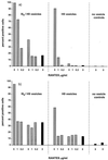
RANTES promotes virion adsorption to target cells. (a) A3.01 cells were incubated for 2 h on ice with preparations containing HIV-1IIIB virions (IIIB/H9 vesicles) or control microvesicles (H9 vesicles) in the presence of the indicated concentrations of RANTES (hatched bars) or BB-10520 RANTES (shaded bars) or with no CC-chemokine (solid bars). (b) A3.01 cells were treated for 24 h at 37°C with RANTES, BB-10520 RANTES, or no CC-chemokine, as described above. IIIB/H9 vesicles or control H9 vesicles were subsequently added for 2 h on ice in the absence of CC-chemokine. In both panels, cell-bound particles were detected by FACS after HLA-DR monoclonal antibody staining. The values shown are the percentages of positive cells that were gated.
FIG. 3
FIG. 3
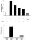
RANTES binds to virions. (a) RANTES was captured on magnetic beads via an anti-murine immunoglobulin G antibody and a murine anti-RANTES antibody (solid bars). HIV-1MuLV virions were then reacted with the beads for 2 h in the presence or absence of the indicated concentrations of soluble chondroitin sulfate (CS). The use of BB-10520 RANTES is indicated by the shaded bar, and the background binding in the absence of RANTES is represented by an open bar. The extent of virion capture was measured by p24 antigen determination and is expressed as the percentage of that achieved in the presence of RANTES but absence of CS (6.5 ng/sample, defined as 100%). The data shown are from one of two to three independent experiments. (b) Biotin-labeled antibodies to CC-chemokines were immobilized on streptavidin-coated magnetic beads and incubated with the appropriate CC-chemokines (solid bars, RANTES; shaded bars, MIP-1β) before the addition of HIV-1MuLV virions for 2 h. The extent of virion capture was measured by p24 antigen determination. The data shown are from one of three independent experiments.
FIG. 4
FIG. 4
RANTES and AOP-RANTES, but not BB-10520 RANTES, multimerize upon binding to heparin. Radiolabeled RANTES (■), AOP-RANTES (▴), or BB-10520 RANTES (●) was incubated with heparin-Sepharose beads in the presence of increasing amounts of the same, unlabeled chemokine, and the amount of radiolabeled chemokine bound to the beads was determined. The values shown are the means (± standard deviations) from five independent experiments.
FIG. 5
FIG. 5
RANTES-mediated infectivity enhancement is dependent upon GAG expression on target cells. CHO-K1 cells (■), heparan sulfate-deficient psgD-677 cells (●), or GAG-deficient psgA-745 cells (▴) were infected with HIV-1VSV (1.5 ng of HIV-1 p24 antigen) in the presence or absence of the indicated concentrations of RANTES. Unbound virus was removed after a 2-h incubation, and the cultures were replenished with fresh medium without RANTES. (a) RANTES was added to the cells for 24 h, and then the chemokine-containing medium was washed away immediately before the addition of virus. RANTES was absent during the 2-h infection period and subsequently. (b) RANTES was added during the 2-h infection period but was not present prior to or after that time. The extent of viral infection was measured by determination of luciferase expression in quadruplicate cultures on day 3 postinfection; the data (mean ± standard deviation) are presented as percentages of control (no RANTES = 100%). The untreated control values (in RLU) were as follows: (a) CHO-K1 cells, 71.4 ± 16.3; pgsD 677 cells, 19.9 ± 4.4; pgsA 745 cells, 71.8 ± 8.4; (b) CHO-K1, cells 47.4 ± 11.5; pgsD 677 cells, 22.9 ± 6.9; pgsA 745 cells, 37.8 ± 4.0.
FIG. 6
FIG. 6
RANTES-mediated infectivity enhancement is inhibited by soluble GAGs. HeLa-CD4 cells were infected with HIV-1MuLV as described in Materials and Methods. (a) The cells were pretreated for 24 h with RANTES (5 μg/ml) in the presence or absence of soluble GAGs, and then infection was initiated in the absence of both RANTES and GAGs. (b) RANTES and GAGs were both added simultaneously with the virus at the initiation of infection. The GAG used was heparan sulfate (■ and □) or chondroitin sulfate (▴ and ▵). Open symbols, no RANTES; closed symbols, plus RANTES. The extent of viral infection was measured by determination of luciferase expression in quadruplicate cultures on day 3 postinfection; the data (mean ± standard deviation) are presented as percentages of control (no RANTES = 100%). The untreated control values were (a) 27,290 ± 2,450 RLU and (b) 27,151 ± 5,468 RLU.
FIG. 7
FIG. 7
Effect of the tyrosine kinase inhibitor herbimycin A on RANTES-induced infectivity enhancement. HeLa-CD4 cells were incubated for 25 h with the indicated concentrations of herbimycin A. The cells were then infected with HIV-1MuLV (2.5 ng of HIV-1 p24 antigen) in the presence (■) or absence (●) of 10 μg of RANTES/ml. Unbound virus was removed after a 2-h incubation period, and the cultures were replenished with fresh medium without RANTES. (a) RANTES was added to the cells 24 h before the initiation of infection (i.e., 1 h after herbimycin A was added) and then washed away immediately before the addition of virus. Neither RANTES nor herbimycin A was present during the 2-h infection period or thereafter. (b) RANTES was added to the cells simultaneously with the viral inoculum so that both RANTES and herbimycin A were present during the 2-h infection period but neither agent was present after that period. In both experiments, the extent of viral infection was determined by measuring luciferase expression in quadruplicate cultures on day 3 postinfection; the data are presented as percentages of control (no chemokine = 100%). The untreated control values were (a) 183 ± 63 RLU and (b) 262 ± 53 RLU.
FIG. 8
FIG. 8
Effect of RANTES on HIV-1 Env-mediated cell-cell fusion. HeLa target cells expressing the HIV-1IIIB envelope glycoproteins were allowed to fuse with HeLa-CD4 effector cells in the presence of the indicated concentrations of RANTES (solid bars), MIP-1α (shaded bars), or medium (hatched bars). The designation 0 h means that the chemokine was only added to the mixed effector-target cell population during the fusion process; the designation 24 h indicates that the effector cells were pretreated with the chemokine for 24 h. The values shown are representative of those from four independent experiments.

Similar articles

Cited by

References

    1. Alkhatib G, Combadiere C, Broder C C, Feng Y, Kennedy P E, Murphy P M, Berger E A. CC CKR5: a RANTES, MIP-1α, MIP-1β receptor as a fusion cofactor for macrophage-tropic HIV-1. Science. 1996;272:1955–1958. - PubMed
    1. Amara A, Le Gall S, Schwartz O, Salamero J, Montes M, Loetscher P, Baggiolini M, Virelizier J-L, Arenzana-Seisdedos F. HIV coreceptor downregulation as antiviral principle: SDF-1α-dependent internalization of the chemokine receptor CXCR4 contributes to inhibition of HIV replication. J Exp Med. 1997;186:139–146. - PMC - PubMed
    1. Amzazi S, Ylisastigul Y L, Bakri Y, Rabehi L, Gattegno L, Parmentier M, Gluckman J C, Benjouad A. The inhibitory effect of RANTES on the infection of primary macrophages by R5 human immunodeficiency virus type-1 depends on the macrophage activation state. Virology. 1998;252:96–105. - PubMed
    1. Aramori I, Ferguson S S G, Bieniasz P D, Cullen B R, Caron M G. Molecular mechanism of desensitization of the chemokine receptor CCR-5: receptor signalling and internalization are dissociable from its role as an HIV-1 co-receptor. EMBO J. 1997;16:4606–4616. - PMC - PubMed
    1. Arenzana-Seisedos F, Virelizier J-L, Rousset D, Clark-Lewis I, Loetscher P, Moser B, Baggiolini M. HIV blocked by chemokine antagonist. Nature. 1996;383:400. - PubMed

Publication types

LinkOut - more resources