. 1999 Jul;104(1):123-33.
doi: 10.1172/JCI5844.

Der p 1 facilitates transepithelial allergen delivery by disruption of tight junctions

Affiliations

Der p 1 facilitates transepithelial allergen delivery by disruption of tight junctions

H Wan et al. J Clin Invest. 1999 Jul.

Abstract

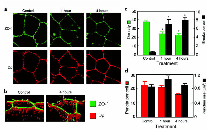
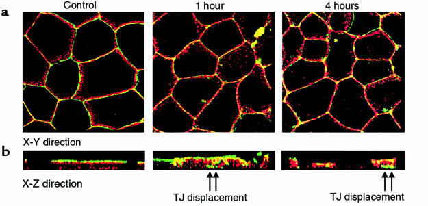
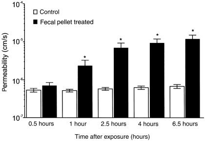
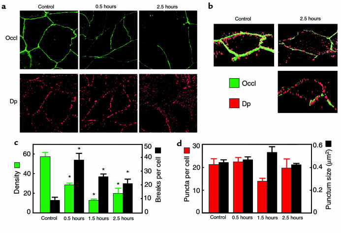
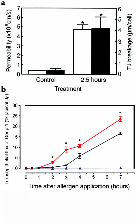
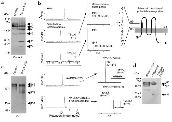
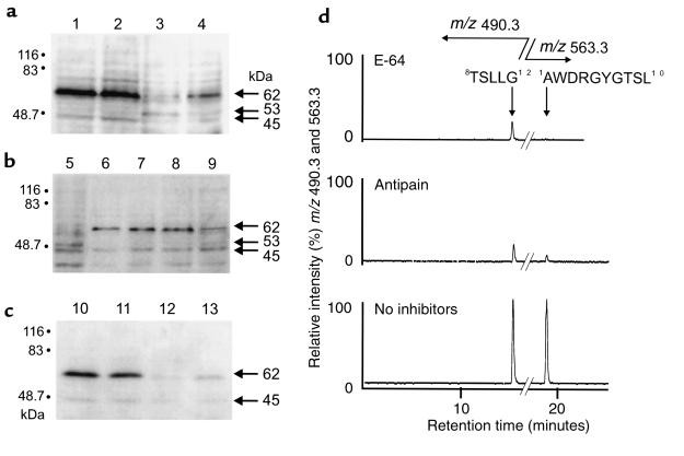
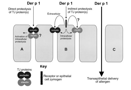
House dust mite (HDM) allergens are important factors in the increasing prevalence of asthma. The lung epithelium forms a barrier that allergens must cross before they can cause sensitization. However, the mechanisms involved are unknown. Here we show that the cysteine proteinase allergen Der p 1 from fecal pellets of the HDM Dermatophagoides pteronyssinus causes disruption of intercellular tight junctions (TJs), which are the principal components of the epithelial paracellular permeability barrier. In confluent airway epithelial cells, Der p 1 led to cleavage of the TJ adhesion protein occludin. Cleavage was attenuated by antipain, but not by inhibitors of serine, aspartic, or matrix metalloproteinases. Putative Der p 1 cleavage sites were found in peptides from an extracellular domain of occludin and in the TJ adhesion protein claudin-1. TJ breakdown nonspecifically increased epithelial permeability, allowing Der p 1 to cross the epithelial barrier. Thus, transepithelial movement of Der p 1 to dendritic antigen-presenting cells via the paracellular pathway may be promoted by the allergen's own proteolytic activity. These results suggest that opening of TJs by environmental proteinases may be the initial step in the development of asthma to a variety of allergens.


Figures

Figure 1
Figure 1
SDS-PAGE and immunoblot analysis of pure Der p 1. The electrophoresis gel was stained with Coomassie blue. Immunoblot detection was by ECL using mAb 5H8. Markers indicate the mobility of standards. Der p 1 migrated with an apparent mass of ∼24 kDa.
Figure 2
Figure 2
Effects of HDMFP on intercellular junctions of MDCK cells. (a) Sample through-focus images (2.8-μm thick) of ZO-1 and desmoplakin (DP) staining in control and after exposure to 40 solubilized fecal pellets apically applied for the periods indicated. (b) Isosurface-rendered images of the 3-dimensional distribution of ZO-1 (green) and desmoplakin (red) for a part of the image in a. Note the differential behavior of the 2 proteins. (c) Graphical analysis of immunoreactive ZO-1 concentration at the cell boundary and the number of breaks in the continuity of the ZO-1 staining belt per cell (*P < 0.05). Bars show mean ± SE of 10 cells. (d) Quantification of desmoplakin staining.
Figure 3
Figure 3
Displacement of ZO-1 in MDCK cells treated with HDMFP as described in Figure 2. The images shown in a are 2-color, extended-focus X-Y sections in cells immunostained for ZO-1 (green) and desmoplakin (red), with the corresponding X-Z sections shown in b. Arrows indicate the areas of TJ displacement seen in cells 1 hour and 4 hours after treatment.
Figure 4
Figure 4
Time-dependent effects of HDMFP on the mannitol permeability of MDCK cell monolayers cultured in Transwells. Forty HDMFPs solubilized in EMEM containing 0.5 mM of reduced glutathione were added to monolayers (filled bars). Open bars show monolayers sham treated with EMEM/glutathione alone. Data are mean ± SE of 4 experiments. *Significant differences from sham-treated monolayers (P < 0.05).
Figure 5
Figure 5
Effects of purified Der p 1 on intercellular junctions of 16HBE14o cells. (a) Sample through-focus images (3.2-μm thick) of occludin and desmoplakin staining in control and after exposure of the apical surface to Der p 1 for the periods indicated. The effects are similar to that seen in Figure 2a. (b) Isosurface-rendered images of the 3-dimensional distribution of occludin (Occl; green) and desmoplakin (Dp; red) for a part of the image in a. The left panel shows a typical untreated cell, whereas the 2 panels on the right show the heterogeneity of response after 2.5 hours. (c) Graphical analysis of occludin concentration and the number of breaks in continuity of the occludin ring per cell. Bars show mean and + SE of 8 cells (*P < 0.05). The method of measurement was the same as for Figure 2c. (d) Quantification of desmoplakin staining.
Figure 6
Figure 6
Effects of Der p 1 on permeability of MDCK cell monolayers. (a) Concordance between changes in [14C]mannitol permeability (open bars) and TJ breakage (i.e., the total length of breaks per cell; filled bars) in the same cell monolayer. Data are mean ± SE from 4 experiments (*P < 0.05). (b) The migration of immunoreactive Der p 1 across MDCK cell monolayers depends upon enzymatic activity. Data are expressed as the percentage of the starting concentration ([apical] t0) of Der p 1 in the apical compartment that was detected in the basal chamber at the indicated time points. Data in red denote monolayers treated with catalytically active allergen; black denotes cells treated with active allergen in the presence of E-64 (100 μM); and blue denotes cells treated with heat-inactivated allergen. Data are mean ± SE from 5 experiments. *Responses significantly different from both of the other treatments (P < 0.05). At 7 hours, the transepithelial flux of Der p 1 was 168-fold greater in monolayers exposed to active Der p 1 compared with those exposed to inactive Der p 1.
Figure 7
Figure 7
Proteolysis of occludin in situ, occludin extracellular loop peptide, ZO-1 in situ, and gel-enriched occludin by Der p 1. (a) Immunoblots of occludin from 16HBE14o cells prepared from sham-treated cells and after a 2.5-hour exposure to Der p 1. (b) Degradation by Der p 1 of peptide 88AWDRGYGTSLLG99 corresponding to residues 88–99 of human occludin (top right), with identified cleavage sites marked by arrows. The left section of b shows the HPLC A280 chromatogram and selected ion chromatograms for the 4-hour incubation products of Der p 1 with the peptide. Mass spectra for the peptides are shown. Major components correspond to unchanged 12-mer sequences (M+H+, m/z 1,295.6; M+H22+, m/z 648.5) and residues 1–10 (M+H+, m/z 1,125.5; M+H22+, m/z 563), 7–12 (M+H+, m/z 547), and 8–12 (M+H+, m/z 490). (c) Immunoblots showing degradation of ZO-1 after 2.5-hour exposure of 16HBE14o cells to active Der p 1. (d) Immunoblot showing degradation of gel-enriched human occludin by allergen. Lanes show whole-cell protein extract (Whole protein), and gel-enriched occludin without allergen treatment (Undigested) and with treatment (Allergen treated).
Figure 8
Figure 8
(a) Occludin immunoblots from MDCK cells. Lane 1: untreated cells; lane 2: cells treated with heat-inactivated Der p 1 for 1 hour; lane 3: following 1-hour treatment with active Der p 1 (activity 17 nmol/min); lane 4: active Der p 1 with 100 μM antipain. Note attenuation of degradation by antipain or heat inactivation. (b) Inhibition of occludin degradation in MDCK cells by occludin peptide 88AWDRGYGTSLLG99. Lane 5: cells treated for 1 hour with Der p 1; lane 6: peptide (100 μM) and cells alone; lanes 7–9: cells treated with peptide and Der p 1 for 0.25, 0.5, and 1 hour, respectively. (c) Lack of effect of mixed proteinase inhibitors on occludin degradation in MDCK cells treated with Der p 1. Lane 10: untreated cells; lane 11: mixed inhibitors (100 μM AEBSF, 1 μM BB-250, and 1 μM pepstatin) alone; lane 12: Der p 1 alone; lane 13: Der p 1 with mixed inhibitors. (d) HPLC/electrospray–selected ion mass chromatograms for m/z 490.3 (M+H+, GTSLLG) and m/z 563.3 (M+H22+, AWDRGYGTSL) are shown following Der p 1 treatment of AWDRGYGTSLLG in the presence of E-64 (100 μM), antipain (100 μM), or the absence of inhibitors. Both inhibitors reduced the formation of 2 major enzymic fragments, AWDRGYGTSL and GTSLLG.
Figure 9
Figure 9
Potential Der p 1 cleavage sites in claudin-1 extracellular loops. (a) Digestion of 64KVFDSLLNLS74 (peptide IV) for 2 hours resulted in 4 major peptides. The HPLC/electrospray–selected ion chromatograms are shown in the left section. I: LNLNS; II: KVFDSL; III: KVFDSLLN; and V: KVFDSLLNL. Peptides II–V were associated with A260 ultraviolet absorbance. Unchanged peptide was the major species in the total ion current (TIC) (not shown). Mass spectra for the peptides are shown on the right. Each peptide generated an M+H+ ion as shown. Larger peptides (III–V) produced intense doubly charged ions (M+H22+, bracketed); the molecular ion regions of these spectra were magnified 2- to 4-fold for display. (b) Digestion of 138WYGNRIVQ144 (peptide VI) generated 2 major fragments, WYG (VII) and NRIVQ (VIII). Small amounts of other fragments were observed. The HPLC A280 ultraviolet absorbance chromatogram is shown together with electrospray mass chromatograms for the TIC and ion channels m/z 1,035, 629, and 425. Mass spectra for VI–VIII are shown at the right; doubly charged ions are bracketed; and the molecular ion region of VI has been magnified for display. (c) Schematic representation of putative cleavage sites within claudin-1. The shaded boxes superimposed on the extracellular domains show approximate locations of the cleavable segments depicted. Arrows indicate cleavage sites identified in peptides. Residue numbering from murine claudin-1.
Figure 10
Figure 10
Recovery of occludin and ZO-1 in 16HBE14o epithelial cell monolayers following exposure to Der p 1. Cells were sham exposed (control) or treated with allergen for 4 hours (T 4 hours), and then changed to normal medium for recovery (R 1 hour, R 3 hours, R 16 hours). Images are shown as extended-focus X-Y sections with the corresponding X-Z images.
Figure 11
Figure 11
Schematic summary of how Der p 1 might open TJs in lung epithelium. In the simple case, cell A, Der p 1 acts upon extracellular loops of occludin, and possibly claudin-1. Extracellular cleavage of TJs initiates intracellular processing of junctional constituents. In cell B, Der p 1 is envisaged to operate indirectly on TJs by first activating a cell surface zymogen, which then proceeds to cleave the TJs. Intracellular processing arises from TJ perturbation as in A, or through a signal transduction pathway that ultimately affects TJs. Note that in A, we do not exclude the operation of a similar decoupled signal transduction pathway from contributing to the intracellular proteolysis. In both A and B, the result is the opening of the epithelial barrier and delivery of allergen (C).

Comment in

Similar articles

Cited by

References

    1. Tovey ER, Chapman MD, Platts-Mills TAE. Mite faeces are a major source of house dust allergens. Nature. 1981;289:592–593. - PubMed
    1. Sporik R, Holgate ST, Platts-Mills TA, Cogswell JJ. Exposure to house dust mite allergen (Der p 1) and the development of asthma in childhood. A prospective study. N Engl J Med. 1990;323:502–507. - PubMed
    1. Platts-Mills TA, Thomas WR, Aalberse RC, Vervolet D, Chapman MD. Dust mite allergens and asthma: report of a second international workshop. J Allergy Clin Immunol. 1992;89:1046–1060. - PubMed
    1. Peat JK, et al. House dust mite allergens. A major risk factor for childhood asthma in Australia. Am J Respir Crit Care Med. 1996;153:141–146. - PubMed
    1. Dowse GK, Turner KJ, Stewart GA, Alpers MP, Woolcock AJ. The association between Dermatophagoides mites and the increasing prevalence of asthma in village communities within the Papua New Guinea highlands. J Allergy Clin Immunol. 1985;75:75–83. - PubMed

Publication types

MeSH terms