

A brain sexual dimorphism controlled by adult circulating androgens
- PMID: 10377450
- PMCID: PMC22121
- DOI: 10.1073/pnas.96.13.7538
A brain sexual dimorphism controlled by adult circulating androgens
Abstract
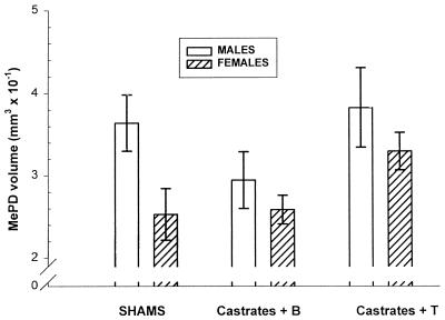
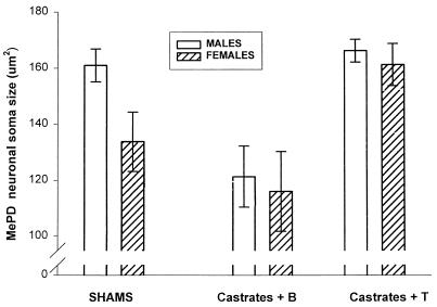
Reports of structural differences between the brains of men and women, heterosexual and homosexual men, and male-to-female transsexuals and other men have been offered as evidence that the behavioral differences between these groups are likely caused by differences in the early development of the brain. However, a possible confounding variable is the concentration of circulating hormones seen in these groups in adulthood. Evaluation of this possibility hinges on the extent to which circulating hormones can alter the size of mammalian brain regions as revealed by Nissl stains. We now report a sexual dimorphism in the volume of a brain nucleus in rats that can be completely accounted for by adult sex differences in circulating androgen. The posterodorsal nucleus of the medial amygdala (MePD) has a greater volume in male rats than in females, but adult castration of males causes the volume to shrink to female values within four weeks, whereas androgen treatment of adult females for that period enlarges the MePD to levels equivalent to normal males. This report demonstrates that adult hormone manipulations can completely reverse a sexual dimorphism in brain regional volume in a mammalian species. The sex difference and androgen responsiveness of MePD volume is reflected in the soma size of neurons there.
Figures

Comment in
-
Permanence of brain sex differences and structural plasticity of the adult brain.Proc Natl Acad Sci U S A. 1999 Jun 22;96(13):7128-30. doi: 10.1073/pnas.96.13.7128. Proc Natl Acad Sci U S A. 1999. PMID: 10377379 Free PMC article. No abstract available.
Similar articles
-
Sexual dimorphism in neuronal number of the posterodorsal medial amygdala is independent of circulating androgens and regional volume in adult rats.J Comp Neurol. 2008 Feb 10;506(5):851-9. doi: 10.1002/cne.21536. J Comp Neurol. 2008. PMID: 18076082
-
Steroid-dependent plasticity in the medial amygdala.Neuroscience. 2006;138(3):997-1005. doi: 10.1016/j.neuroscience.2005.06.018. Epub 2005 Dec 5. Neuroscience. 2006. PMID: 16330154 Review.
-
Sexual dimorphism and steroid responsiveness of the posterodorsal medial amygdala in adult mice.Brain Res. 2008 Jan 23;1190:115-21. doi: 10.1016/j.brainres.2007.11.005. Epub 2007 Nov 12. Brain Res. 2008. PMID: 18054901 Free PMC article.
-
Partial demasculinization of several brain regions in adult male (XY) rats with a dysfunctional androgen receptor gene.J Comp Neurol. 2005 Jun 27;487(2):217-26. doi: 10.1002/cne.20558. J Comp Neurol. 2005. PMID: 15880473
-
Morphological and functional features of the sex steroid-responsive posterodorsal medial amygdala of adult rats.Mini Rev Med Chem. 2012 Oct;12(11):1090-106. doi: 10.2174/138955712802762211. Mini Rev Med Chem. 2012. PMID: 22827219 Review.
Cited by
-
Kisspeptin in the medial amygdala and sexual behavior in male rats.Neurosci Lett. 2016 Aug 3;627:13-7. doi: 10.1016/j.neulet.2016.05.042. Epub 2016 May 24. Neurosci Lett. 2016. PMID: 27233219 Free PMC article.
-
Sex chromosomes and hormones independently influence healthy brain development but act similarly after cranial radiation.Proc Natl Acad Sci U S A. 2024 Sep 3;121(36):e2404042121. doi: 10.1073/pnas.2404042121. Epub 2024 Aug 29. Proc Natl Acad Sci U S A. 2024. PMID: 39207735
-
Sex Differences in the Development of the Rodent Corticolimbic System.Front Neurosci. 2020 Sep 30;14:583477. doi: 10.3389/fnins.2020.583477. eCollection 2020. Front Neurosci. 2020. PMID: 33100964 Free PMC article. Review.
-
Deletion of Bax eliminates sex differences in the mouse forebrain.Proc Natl Acad Sci U S A. 2004 Sep 14;101(37):13666-71. doi: 10.1073/pnas.0404644101. Epub 2004 Sep 1. Proc Natl Acad Sci U S A. 2004. PMID: 15342910 Free PMC article.
-
Anti-anxiety, cognitive, and steroid biosynthetic effects of an isoflavone-based dietary supplement are gonad and sex-dependent in rats.Brain Res. 2011 Mar 16;1379:164-75. doi: 10.1016/j.brainres.2010.12.025. Epub 2010 Dec 15. Brain Res. 2011. PMID: 21167133 Free PMC article.
References
-
- Swaab D F, Fliers E. Science. 1985;228:1112–1114. - PubMed
-
- Allen L S, Gorski R A. J Comp Neurol. 1990;302:697–706. - PubMed
-
- Schlaepfer T E, Harris G J, Tien A Y, Peng L, Lee S, Pearlson G D. Psychiatry Res. 1995;61:129–135. - PubMed
-
- Giedd J N, Vaituzis A C, Hamburger S D, Lange N, Rajapakse J C, Kaysen D, Vauss Y C, Rapoport J L. J Comp Neurol. 1996;366:223–230. - PubMed
Publication types
MeSH terms
Substances
Grants and funding
LinkOut - more resources
Full Text Sources
