. 1999 Jul;19(7):5143-54.
doi: 10.1128/MCB.19.7.5143.

Mitogen-activated protein kinase kinase kinase 1 activates androgen receptor-dependent transcription and apoptosis in prostate cancer

Affiliations

Mitogen-activated protein kinase kinase kinase 1 activates androgen receptor-dependent transcription and apoptosis in prostate cancer

M T Abreu-Martin et al. Mol Cell Biol. 1999 Jul.

Abstract

Mitogen-activated protein (MAP) kinases phosphorylate the estrogen receptor and activate transcription from estrogen receptor-regulated genes. Here we examine potential interactions between the MAP kinase cascade and androgen receptor-mediated gene regulation. Specifically, we have studied the biological effects of mitogen-activated protein kinase kinase kinase 1 (MEKK1) expression in prostate cancer cells. Our findings demonstrate that expression of constitutively active MEKK1 induces apoptosis in androgen receptor-positive but not in androgen receptor-negative prostate cancer cells. Reconstitution of the androgen receptor signaling pathway in androgen receptor-negative prostate cancer cells restores MEKK1-induced apoptosis. MEKK1 also stimulates the transcriptional activity of the androgen receptor in the presence or absence of ligand, whereas a dominant negative mutant of MEKK1 impairs activation of the androgen receptor by androgen. These studies demonstrate an unanticipated link between MEKK1 and hormone receptor signaling and have implications for the molecular basis of hormone-independent prostate cancer growth.


Figures
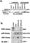
FIG. 1
FIG. 1
Biochemical characterization of LNCaP cells stably expressing mutant MEKK1. (a) MEKK1 immunoblot blot of LNCaP cells before and after G418 selection. LNCaP cells were infected with retrovirus pSRαMEKKΔ-DA or pSRαMEKKΔ-DN or Neo control virus. Whole-cell lysates were prepared from cells on the day after retroviral infection prior to G418 selection (unselected) or 2 weeks after G418 selection (G418-selected). The expression of truncated MEKK1 protein is similar in cells infected with pSRαMEKKΔ-DA or pSRαMEKK-DN immediately after infection, suggesting similar viral titers. After G418 selection, however, surviving cells express lower amounts of MEKKΔ-DA. Full-length MEKK1 is a 190-kDa protein not shown on this blot. This C-terminal-directed antibody recognizes a cleaved form of endogenous MEKK1 which runs at approximately 78-kDa and is the same in all lanes (5, 56). Equal protein loading was confirmed by protein assay and Ponceau S staining. (b) In vitro kinase assays of LNCaP sublines stably expressing MEKK isoforms as indicated. Cells expressing MEKKΔ-DA show approximately sixfold activation of JNK activity compared with control cells but only twofold activation of p38 kinase activity. A JNK1 immunoblot demonstrates the relative amounts of immunoprecipitated JNK1 in the different sublines.
FIG. 2
FIG. 2
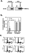
Effect of stable expression of MEKKΔ-DA in prostate cancer cell lines. (a) MEKK immunoblot of DU145 and PC3 cells after infection with pSRαMEKKΔ-DA retrovirus or Neo control virus. Whole-cell lysates were prepared from cells 2 weeks after G418 selection. Equal protein loading was confirmed by protein assay and Ponceau S staining. (b) Change in cell number of prostate cancer cell lines stably expressing MEKKΔ-DA. After antibiotic selection, DU145, PC3, and LNCaP sublines were plated at 100,000 cells per 60-mm-diameter plate, and the cell numbers were calculated after 5 days in culture. Data are expressed as the percentage of cells on day 5 in the sublines (Neo or MEKKΔ-DA) compared with the parental line. Experiments were performed in duplicate, and this is one representative of three independently derived stable cell lines. (c) Cell cycle analysis of LNCaP cells stably expressing mutant MEKK isoforms. Subconfluent LNCaP cells growing in 10% FCS were permeabilized, stained with propidium iodide, and analyzed on a Becton Dickinson flow cytometer. There are no differences between MEKKΔ-DA-expressing cells and Neo control cells with regard to G1, S, and G2 peaks.
FIG. 3
FIG. 3
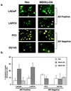
Effect of transient MEKKΔ-DA expression on apoptosis in androgen receptor-negative and androgen receptor-positive prostate cancer cell lines. (a) LNCaP cells were transiently transfected with GFP (0.4 μg) and cotransfected with Neo or MEKKΔ-DA (0.6 μg), and the total amount of transfected DNA (4 μg) was kept constant with Neo control vector. DU145, PC3, and LAPC4 cells were transfected with 3.6 μg of pCDNA3 Neo or MEKKΔ-DA and cotransfected with GFP (0.4 μg). Apoptotic cells demonstrate cytoplasmic blebbing (arrows). Cells were scored for apoptosis 48 h after transfection. The transfection efficiencies for each cell line are as follows: LNCaP Neo, 40 to 50%; MEKKΔ-DA, 40 to 50%; DU145 Neo, 20 to 30%; MEKKΔ-DA, 20 to 30%; PC3 Neo, 30 to 40%; MEKKΔ-DA, 30 to 40%; LAPC4 Neo, 20 to 30%; MEKKΔ-DA, 20 to 30%. (b) Graph represents three independent experiments in which 200 green fluorescent cells were counted and scored for cytoplasmic blebbing 48 h after transfection. For LNCaP cells, these experiments were also performed with transfected kinase-inactive MEKKΔ-DN (3 μg) which did not induce apoptosis. (c) LNCaP cells were transiently transfected with GFP (0.4 μg) and cotransfected with Neo or MEKKΔ-DA (0.6 μg), and the total amount of transfected DNA (4 μg) was kept constant with Neo control vector. DU145 cells were transfected with 3.6 μg of pCDNA3 Neo or MEKKΔ-DA and cotransfected with GFP (0.4 μg). At 48 h after transfection, cells were stained with the DNA dye Hoechst 33258, and GFP-positive cells were scored for chromatin condensation. There was no increase in chromatin condensation in DU145 cells transfected with Neo or MEKKΔ-DA. White arrows indicate GFP-positive cells, and yellow arrows indicate GFP-positive cells showing chromatin condensation.
FIG. 3
FIG. 3
Effect of transient MEKKΔ-DA expression on apoptosis in androgen receptor-negative and androgen receptor-positive prostate cancer cell lines. (a) LNCaP cells were transiently transfected with GFP (0.4 μg) and cotransfected with Neo or MEKKΔ-DA (0.6 μg), and the total amount of transfected DNA (4 μg) was kept constant with Neo control vector. DU145, PC3, and LAPC4 cells were transfected with 3.6 μg of pCDNA3 Neo or MEKKΔ-DA and cotransfected with GFP (0.4 μg). Apoptotic cells demonstrate cytoplasmic blebbing (arrows). Cells were scored for apoptosis 48 h after transfection. The transfection efficiencies for each cell line are as follows: LNCaP Neo, 40 to 50%; MEKKΔ-DA, 40 to 50%; DU145 Neo, 20 to 30%; MEKKΔ-DA, 20 to 30%; PC3 Neo, 30 to 40%; MEKKΔ-DA, 30 to 40%; LAPC4 Neo, 20 to 30%; MEKKΔ-DA, 20 to 30%. (b) Graph represents three independent experiments in which 200 green fluorescent cells were counted and scored for cytoplasmic blebbing 48 h after transfection. For LNCaP cells, these experiments were also performed with transfected kinase-inactive MEKKΔ-DN (3 μg) which did not induce apoptosis. (c) LNCaP cells were transiently transfected with GFP (0.4 μg) and cotransfected with Neo or MEKKΔ-DA (0.6 μg), and the total amount of transfected DNA (4 μg) was kept constant with Neo control vector. DU145 cells were transfected with 3.6 μg of pCDNA3 Neo or MEKKΔ-DA and cotransfected with GFP (0.4 μg). At 48 h after transfection, cells were stained with the DNA dye Hoechst 33258, and GFP-positive cells were scored for chromatin condensation. There was no increase in chromatin condensation in DU145 cells transfected with Neo or MEKKΔ-DA. White arrows indicate GFP-positive cells, and yellow arrows indicate GFP-positive cells showing chromatin condensation.
FIG. 4
FIG. 4
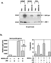
Role of JNK activation in MEKKΔ-DA-induced apoptosis. (a) Comparison of JNK activation in prostate cancer cell lines in response to MEKKΔ-DA. LNCaP, DU145, and PC3 cells were transfected with FLAG-tagged JNK1 (2 μg) and cotransfected with Neo or MEKKΔ-DA (2 μg). The top panel shows a JNK assay in which 100 μg of total cellular protein was immunoprecipitated with anti-FLAG antibody and reacted with GST–c-jun. The bottom panel shows an anti-FLAG immunoblot. LNCaP cells have approximately sixfold-higher amount of transfected JNK1 than DU145 cells as determined by densitometry analysis. When corrected for this difference in transfected protein, MEKKΔ-DA-induced JNK activation is approximately four- to sixfold in all three cell lines. (b) Effect of JNK inhibition on MEKKΔ-DA-induced apoptosis in LNCaP cells. LNCaP cells were cotransfected with MEKKΔ-DA (0.6 μg) or Neo and pCDNA3-JBD (JNK1 inhibitor), p35 (caspase inhibitor), or vector control. (Left panel) Effect of transfected JBD on MEKKΔ-DA-induced c-jun transcriptional activity as measured by a 5X-Gal-luciferase reporter (0.4 μg) and gal4-jun (0.4 μg). (Right panel) Transfected cells were scored for apoptosis 48 h after transfection.
FIG. 5
FIG. 5
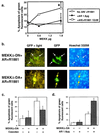
Modulation of androgen receptor function alters the sensitivity of prostate cancer cells to MEKKΔ-DA-induced apoptosis. Reconstitution of the androgen receptor signaling pathway in DU145 cells. DU145 cells were cotransfected with MEKKΔ-DA, as indicated, and androgen receptor (AR) (1.8 μg) in the presence or absence of androgen R1881 (10 nM). This graph is an average of the results from eight independent experiments. The P value for the combined experiments is 0.002 as determined by the paired Student t test for MEKKΔ-DA plus androgen receptor plus R1881 versus MEKKΔ-DA plus androgen receptor. (b) Morphology of DU145 reconstituted with androgen receptor and androgen R1881 and cotransfected with MEKKΔ-DN (top row) or MEKKΔ-DA (bottom row) 48 h after transfection. White arrows indicate GFP-positive cells, and yellow arrows indicate GFP-positive cells showing chromatin condensation. (c) Effect of the androgen receptor antagonist Casodex on MEKKΔ-DA-induced apoptosis. Graph of LNCaP transfected with 0.6 μg of MEKKΔ-DA or Neo control vector and treated with the androgen receptor antagonist Casodex (10 μM) as indicated. Graph represents results of four independent experiments in which 200 green fluorescent cells were counted and scored for cytoplasmic blebbing; P = 0.004 as determined by the paired Student t test for MEKKΔ-DA versus MEKKΔ-DA plus Casodex. Cells were scored for apoptosis at 48 h after transfection. (d) Graph of LNCaP transfected with 0.6 μg of pCDNA3 containing MEKKΔ-DA or the empty vector and cotransfected with androgen receptor (1.8 μg) as indicated. Graph represents three independent experiments in which 200 green fluorescent cells were counted and scored for cytoplasmic blebbing at 48 h after transfection; P = 0.01 as determined by the paired Student t test for MEKKΔ-DA versus MEKKΔ-DA plus androgen receptor.
FIG. 6
FIG. 6
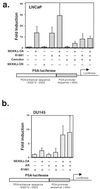
MEKKΔ-DA increases the transcriptional activity of androgen receptor-regulated promoters. (a) Graph of PSA P/E-luc transcriptional activity in LNCaP cells. LNCaP cells were transfected with MEKKΔ-DA (0.6 μg) or MEKKΔ-DN or Neo control vector (3.6 μg) and cotransfected with a PSA P/E-luc reporter construct (0.4 μg). R1881 was added to a final concentration of 10 nM, and Casodex was added at a final concentration of 10 μM. This graph represents an average of six independent experiments; P = 0.009 as determined by the paired Student t test for MEKKΔ-DA compared to the control. (b) Graph of PSA P/E-luc transcriptional activity in DU145. DU145 were cotransfected with MEKKΔ-DA or Neo control vector (1.6 μg), androgen receptor (1.8 μg), and PSA-luc reporter (0.4 μg). R1881 was added to a final concentration of 10 nM. Luciferase activity was measured 48 h after transfection. This graph represents an average of six experiments; P = 0.01 for MEKKΔ-DA versus MEKKΔ-DA plus androgen receptor.
FIG. 7
FIG. 7
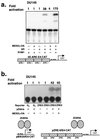
MEKKΔ-DA specifically increases the transcriptional activity of androgen receptor on a minimal promoter element. (a) Effect of MEKKΔ-DA on transcriptional activation of a promoter consisting of pure androgen response elements in DU145. A promoter consisting of four multimerized androgen response elements, 4X-ARE/E4-CAT (0.4 μg) was transfected into DU145 as in Fig. 6b. CAT production was analyzed by ELISA as described in Materials and Methods and by conventional CAT assay. ImageQuant software was used to analyze phosphorimager data for the conventional CAT assay. This is one representative experiment of four total. (b) Effect of MEKKΔ-DA on the transcriptional activation of a promoter consisting of ZEBRA response elements in DU145 cells. For these experiments, DU145 stably expressing androgen receptor or Neo-infected cells were transfected with the vectors as indicated: 0.8 μg of reporter plasmid, 0.8 μg of ZEBRA transcription factor, and 2.4 μg of MEKKΔ-DA or Neo vector control. The data shown were obtained with androgen receptor-expressing DU145 cells.
FIG. 8
FIG. 8
Expression of MEKKΔ-DN inhibits androgen-mediated activation of PSA P/E-luc. (Left panel) Effect of MEKKΔ-DN on MEKKΔ-DA-induced PSA P/E-luc activity. LNCaP cells were transfected with MEKKΔ-DA (0.6 μg) and cotransfected with 3.6 μg (6:1 ratio) of MEKKΔ-DN as indicated. This is one representative experiment of three total, all with similar results. (Right panel) Effect of increasing amounts of MEKKΔ-DN on R1881-induced PSA P/E-luc activity. LNCaP cells were transfected with increasing amounts of MEKKΔ-DN as indicated in the presence of R1881 (10 nM). This is one representative experiment of four total, all with similar results.

Similar articles

Cited by

References

    1. Abreu-Martin, M. T., and C. L. Sawyers. Unpublished data.
    1. Amati B, Land H. Myc-Max-Mad: a transcription factor network controlling cell cycle progression, differentiation and death. Curr Opin Genet Dev. 1994;4:102–108. - PubMed
    1. Brandstrom A, Westin P, Bergh A, Cajander S, Damber J E. Castration induces apoptosis in the ventral prostate but not in an androgen-sensitive prostatic adenocarcinoma in the rat. Cancer Res. 1994;54:3594–3601. - PubMed
    1. Bubulya A, Wise S C, Shen X Q, Burmeister L A, Shemshedini L. c-Jun can mediate androgen receptor-induced transactivation. J Biol Chem. 1996;271:24583–24589. - PubMed
    1. Bunone G, Briand P A, Miksicek R J, Picard D. Activation of the unliganded estrogen receptor by EGF involves the MAP kinase pathway and direct phosphorylation. EMBO J. 1996;15:2174–2183. - PMC - PubMed

Publication types

MeSH terms

Substances