. 1999 Jun;10(6):1957-72.
doi: 10.1091/mbc.10.6.1957.

Vesicle-associated membrane protein 4 is implicated in trans-Golgi network vesicle trafficking

Affiliations
Free PMC article

Vesicle-associated membrane protein 4 is implicated in trans-Golgi network vesicle trafficking

M Steegmaier et al. Mol Biol Cell. 1999 Jun.
Free PMC article

Abstract

The trans-Golgi network (TGN) plays a pivotal role in directing proteins in the secretory pathway to the appropriate cellular destination. VAMP4, a recently discovered member of the vesicle-associated membrane protein (VAMP) family of trafficking proteins, has been suggested to play a role in mediating TGN trafficking. To better understand the function of VAMP4, we examined its precise subcellular distribution. Indirect immunofluorescence and electron microscopy revealed that the majority of VAMP4 localized to tubular and vesicular membranes of the TGN, which were in part coated with clathrin. In these compartments, VAMP4 was found to colocalize with the putative TGN-trafficking protein syntaxin 6. Additional labeling was also present on clathrin-coated and noncoated vesicles, on endosomes and the medial and trans side of the Golgi complex, as well as on immature secretory granules in PC12 cells. Immunoprecipitation of VAMP4 from rat brain detergent extracts revealed that VAMP4 exists in a complex containing syntaxin 6. Converging lines of evidence implicate a role for VAMP4 in TGN-to-endosome transport.


Figures
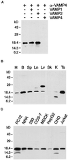
Figure 1
Figure 1
VAMP4 is broadly expressed. (A) Anti-VAMP4 antibodies preincubated with control buffer or recombinant VAMP1 or VAMP2 but not antibodies preincubated with recombinant VAMP4 recognize an 18-kDa band in brain postnuclear supernatant (PNS). (B) PNSs from rat heart (H), brain (B), spleen (Sp), lung (Ln), liver (Lv), skeletal muscle (Sk), kidney (K), and testis (Ts) (30 μg/lane) were analyzed by Western blot using affinity-purified rabbit polyclonal antibodies. A major band is detected at 18 kDa, with highest expression levels in brain and testis. The ∼25-kDa band in liver is recognized by polyclonal antibodies purified from two different rabbits immunized with VAMP4, suggesting that two isoforms of VAMP4 are expressed in liver. The lower-molecular-mass band recognized by the anti-VAMP4 antibodies in kidney PNS represents a degradation product of VAMP4. (C) PNSs of various cell lines (30 μg/lane) were analyzed by Western blot using affinity-purified rabbit polyclonal antibodies. VAMP4 was detected in all cell lines examined. The lower-molecular-mass band recognized by the anti-VAMP4 antibodies in PC12 and CHO cell lysate represents a degradation product of VAMP4.
Figure 2
Figure 2
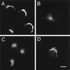
Perinuclear labeling for VAMP4 is detected in cell lines and primary cultures. Cells were fixed with 4% paraformaldehyde, permeabilized with saponin, and stained using affinity-purified anti-VAMP4 polyclonal antibodies and Texas Red–conjugated anti-rabbit IgG secondary antibody before processing for indirect immunofluorescence microscopy. (A) NRK; (B) COS-7; (C) NGF-differentiated PC12; (D) 12 d in vitro embryonic hippocampal cultures. Note that there is no VAMP4 staining observed in the processes of differentiated PC12 cells or embryonic hippocampal cultures. Bar: A–C, 20 μm; D, 25 μm.
Figure 3
Figure 3
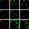
VAMP4 colocalizes with syntaxin 6. CHO cells were fixed with 4% paraformaldehyde, permeabilized with saponin, and costained using affinity-purified rabbit anti-VAMP4 antibodies (A, D, and G) and mouse mAbs against syntaxin 6 (B), p115 (E), and transferrin receptor (H). Texas Red–labeled anti-rabbit IgG and FITC-labeled anti-mouse IgG were used as secondary antibodies before processing for indirect immunofluorescence microscopy. In yellow are areas of overlap in merged images (C, F, and I). Bar, 20 μm.
Figure 4
Figure 4
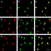
VAMP4 and syntaxin 6 both localize to the same compartment upon BFA treatment. CHO cells, treated for 15 min with 10 μg/ml BFA, were fixed with 4% paraformaldehyde, permeabilized with saponin, and costained using affinity-purified rabbit anti-VAMP4 antibodies (A, D, and G) and mouse mAbs against syntaxin 6 (B), p115 (E), and transferrin receptor (H). Texas Red–labeled anti-rabbit IgG and FITC-labeled anti-mouse IgG were used as secondary antibodies before processing for indirect immunofluorescence microscopy. In yellow are areas of overlap in merged images (C, F, and I). Bar, 20 μm.
Figure 5
Figure 5
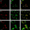
Nocodazole causes redistribution of VAMP4 and syntaxin 6 to the same compartment. CHO cells, treated for 30 min with 5 μg/ml nocodazole, were fixed with 4% paraformaldehyde, permeabilized with saponin, and costained using affinity-purified rabbit anti-VAMP4 antibodies (A, D, and G) and mouse mAbs against syntaxin 6 (B), p115 (E), and transferrin receptor (H). Texas Red-labeled anti-rabbit IgG and FITC-labeled anti-mouse IgG were used as secondary antibodies before processing for indirect immunofluorescence microscopy. In yellow are areas of overlap in merged images (C, F, and I). Bar, 20 μm.
Figure 6
Figure 6
VAMP4 localizes to the Golgi apparatus and the TGN. (A) Double-immunogold labeling of VAMP4 (10-nm gold) and negative control antibody 3C9.H6 (15-nm gold). Only VAMP4-representing 10-nm gold particles are seen. VAMP4 predominantly localizes to the Golgi stack (G) and vesicular and tubular TGN membranes in close vicinity to the Golgi stack. Lysosomes (L) are negative. (B and C) Double-immunogold labeling of VAMP4 (15-nm gold) and clathrin (10-nm gold). VAMP4 is present on clathrin-coated vesicles in the TGN (large arrowhead in B). Additional VAMP4 labeling (indicated by small arrowheads) is present at the limiting membranes of clathrin-positive ISGs (I in B) and of endosomal vacuoles (E in C). SGs that do not stain for clathrin are also negative for VAMP4. Bars, 200 nm.
Figure 7
Figure 7
VAMP4 colocalizes with syntaxin 6. (A) Double-immunogold labeling of VAMP4 (10-nm gold) and rbet1 (15-nm gold). The two SNARE proteins clearly reside in distinct membranes at either side of the Golgi stack. (B) Double-immunogold labeling of VAMP4 (15-nm gold) and syntaxin 6 (10-nm gold) showing colocalization to the same TGN membranes (large arrowheads). The extensive ER–Golgi intermediate compartment at the cis side of the Golgi is devoid of both VAMP4 and syntaxin 6. L, lysosome. Bars, 200 nm.
Figure 8
Figure 8
VAMP4 is a member of an ∼50- and ∼160-kDa protein complex. Rat brain membrane extract was fractionated by centrifugation through an 11–35% glycerol velocity gradient, and sequential fractions were analyzed by electrophoresis and immunobloting with antibodies against VAMP4, VAMP2, syntaxin 6, syntaxin 1, and synaptophysin. Arrows indicate the mobilities of marker proteins.
Figure 9
Figure 9
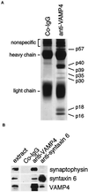
VAMP4 associates with at least six vesicle-trafficking proteins in two different protein complexes. (A) Crude membranes from rat brain were isolated and solubilized with 1% Triton X-100. The solubilized membranes were incubated with protein A-Sepharose beads loaded with either affinity-purified rabbit anti-VAMP4 antibodies or with nonspecific rabbit IgG. After the binding step the beads were washed, eluted with SDS protein sample dye, resolved by SDS-PAGE, and stained with Coomassie blue. The indicated bands were excised from the gel and subjected to internal amino acid sequencing (see Table 2). (B) Immunoprecipitations with control IgG, anti-VAMP4, and anti-syntaxin 6 antibodies were carriedout as detailed in MATERIALS AND METHODS, and the resulting precipitates were analyzed by electrophoresis and immunoblotting. Labels along the top correspond to the antibodies used for immunoprecipitation, whereas labels along the right indicate the antibodies used for immunobloting. Note that synaptophysin is precipitated with anti-VAMP4 but not with anti-syntaxin 6 antibodies.

Similar articles

Cited by

References

    1. Advani RJ, Bae HR, Bock JB, Chao DS, Doung YC, Prekeris R, Yoo JS, Scheller RH. Seven novel mammalian SNARE proteins localize to distinct membrane compartments. J Biol Chem. 1998;273:10317–10324. - PubMed
    1. Banker GA, Cowan WM. Rat hippocampal neurons in dispersed cell culture. Brain Res. 1977;126:397–442. - PubMed
    1. Bennett MK, Scheller RH. A molecular description of synaptic vesicle membrane trafficking. Annu Rev Biochem. 1994;63:63–100. - PubMed
    1. Bock JB, Klumperman J, Davanger S, Scheller RH. Syntaxin 6 functions in trans-Golgi network vesicle trafficking. Mol Biol Cell. 1997;8:1261–1271. - PMC - PubMed
    1. Bock JB, Lin RC, Scheller RH. A new syntaxin family member implicated in targeting of intracellular transport vesicles. J Biol Chem. 1996;271:17961–17965. - PubMed

MeSH terms

LinkOut - more resources