A novel mouse with B cells but lacking serum antibody reveals an antibody-independent role for B cells in murine lupus
- PMID: 10330443
- PMCID: PMC2193634
- DOI: 10.1084/jem.189.10.1639
A novel mouse with B cells but lacking serum antibody reveals an antibody-independent role for B cells in murine lupus
Abstract
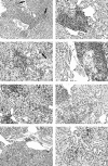
The precise role of B cells in systemic autoimmunity is incompletely understood. Although B cells are necessary for expression of disease (Chan, O., and M.J. Shlomchik. 1998. J. Immunol. 160:51-59, and Shlomchik, M.J., M.P. Madaio, D. Ni, M. Trounstine, and D. Huszar. 1994. J. Exp. Med. 180:1295-1306), it is unclear whether autoantibody production, antigen presentation, and/or other B cell functions are required for the complete pathologic phenotype. To address this issue, two experimental approaches were used. In the first, the individual contributions of circulating antibodies and B cells were analyzed using MRL/MpJ-Faslpr (MRL/lpr) mice that expressed a mutant transgene encoding surface immunoglobulin (Ig), but which did not permit the secretion of circulating Ig. These mice developed nephritis, characterized by cellular infiltration within the kidney, indicating that B cells themselves, without soluble autoantibody production, exert a pathogenic role. The results indicate that, independent of serum autoantibody, functional B cells expressing surface Ig are essential for disease expression, either by serving as antigen-presenting cells for antigen-specific, autoreactive T cells, or by contributing directly to local inflammation.
Figures


Similar articles
-
A new role for B cells in systemic autoimmunity: B cells promote spontaneous T cell activation in MRL-lpr/lpr mice.J Immunol. 1998 Jan 1;160(1):51-9. J Immunol. 1998. PMID: 9551955
-
Unexpected development of autoimmunity in BAFF-R-mutant MRL-lpr mice.Immunology. 2007 Feb;120(2):281-9. doi: 10.1111/j.1365-2567.2006.02500.x. Epub 2006 Oct 31. Immunology. 2007. PMID: 17073941 Free PMC article.
-
B and T cell immune response to small nuclear ribonucleoprotein particles in lupus mice: autoreactive CD4(+) T cells recognize a T cell epitope located within the RNP80 motif of the 70K protein.Eur J Immunol. 2000 Aug;30(8):2191-200. doi: 10.1002/1521-4141(2000)30:8<2191::AID-IMMU2191>3.0.CO;2-R. Eur J Immunol. 2000. PMID: 10940910
-
Dysregulated Lymphoid Cell Populations in Mouse Models of Systemic Lupus Erythematosus.Clin Rev Allergy Immunol. 2017 Oct;53(2):181-197. doi: 10.1007/s12016-017-8605-8. Clin Rev Allergy Immunol. 2017. PMID: 28500565 Review.
-
The cellular and genetic basis of murine lupus.Immunol Rev. 1981;55:121-54. doi: 10.1111/j.1600-065x.1981.tb00341.x. Immunol Rev. 1981. PMID: 7016728 Review. No abstract available.
Cited by
-
Belimumab and Rituximab in Systemic Lupus Erythematosus: A Tale of Two B Cell-Targeting Agents.Front Med (Lausanne). 2020 Jun 30;7:303. doi: 10.3389/fmed.2020.00303. eCollection 2020. Front Med (Lausanne). 2020. PMID: 32695790 Free PMC article. Review.
-
A Conversation with Cohn on the Activation of CD4 T Cells.Scand J Immunol. 2015 Aug;82(2):147-59. doi: 10.1111/sji.12315. Scand J Immunol. 2015. PMID: 25998043 Free PMC article.
-
Treating human autoimmune disease by depleting B cells.Ann Rheum Dis. 2002 Oct;61(10):863-6. doi: 10.1136/ard.61.10.863. Ann Rheum Dis. 2002. PMID: 12228152 Free PMC article. Review. No abstract available.
-
The regulation and activation potential of autoreactive B cells.Immunol Res. 2003;27(2-3):219-34. doi: 10.1385/IR:27:2-3:219. Immunol Res. 2003. PMID: 12857970 Review.
-
B cells, BAFF/zTNF4, TACI, and systemic lupus erythematosus.Arthritis Res. 2001;3(4):197-9. doi: 10.1186/ar299. Epub 2001 Mar 20. Arthritis Res. 2001. PMID: 11438034 Free PMC article.
References
-
- Kotzin B. Systemic lupus erythematosus. Cell. 1996;85:303–306. - PubMed
-
- Hewicker M, Trautwein G. Glomerular lesions in MRL mice. A light and immunofluorescence microscopic study. J Vet Med. 1986;33:727–739. - PubMed
-
- Hewicker M, Trautwein G. Sequential study of vasculitis in MRL mice. Lab Anim. 1987;21:335–341. - PubMed
-
- Alexander EL, Moyer C, Travlos GS, Roths JB, Murphy ED. Two histopathologic types of inflammatory vascular disease in MRL/Mp autoimmune mice. Arthritis Rheum. 1985;28:1146–1155. - PubMed
Publication types
MeSH terms
Substances
Grants and funding
LinkOut - more resources
Full Text Sources
Other Literature Sources
Molecular Biology Databases

