. 1999 Jun;19(6):4535-45.
doi: 10.1128/MCB.19.6.4535.

Identification of CHIP, a novel tetratricopeptide repeat-containing protein that interacts with heat shock proteins and negatively regulates chaperone functions

Affiliations

Identification of CHIP, a novel tetratricopeptide repeat-containing protein that interacts with heat shock proteins and negatively regulates chaperone functions

C A Ballinger et al. Mol Cell Biol. 1999 Jun.

Abstract

The chaperone function of the mammalian 70-kDa heat shock proteins Hsc70 and Hsp70 is modulated by physical interactions with four previously identified chaperone cofactors: Hsp40, BAG-1, the Hsc70-interacting protein Hip, and the Hsc70-Hsp90-organizing protein Hop. Hip and Hop interact with Hsc70 via a tetratricopeptide repeat domain. In a search for additional tetratricopeptide repeat-containing proteins, we have identified a novel 35-kDa cytoplasmic protein, carboxyl terminus of Hsc70-interacting protein (CHIP). CHIP is highly expressed in adult striated muscle in vivo and is expressed broadly in vitro in tissue culture. Hsc70 and Hsp70 were identified as potential interaction partners for this protein in a yeast two-hybrid screen. In vitro binding assays demonstrated direct interactions between CHIP and both Hsc70 and Hsp70, and complexes containing CHIP and Hsc70 were identified in immunoprecipitates of human skeletal muscle cells in vivo. Using glutathione S-transferase fusions, we found that CHIP interacted with the carboxy-terminal residues 540 to 650 of Hsc70, whereas Hsc70 interacted with the amino-terminal residues 1 to 197 (containing the tetratricopeptide domain and an adjacent charged domain) of CHIP. Recombinant CHIP inhibited Hsp40-stimulated ATPase activity of Hsc70 and Hsp70, suggesting that CHIP blocks the forward reaction of the Hsc70-Hsp70 substrate-binding cycle. Consistent with this observation, both luciferase refolding and substrate binding in the presence of Hsp40 and Hsp70 were inhibited by CHIP. Taken together, these results indicate that CHIP decreases net ATPase activity and reduces chaperone efficiency, and they implicate CHIP in the negative regulation of the forward reaction of the Hsc70-Hsp70 substrate-binding cycle.


Figures
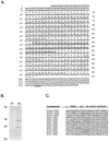
FIG. 1
FIG. 1
The hCHIP cDNA. (A) The complete nucleotide (upper line) and deduced amino acid (lower line) sequences of hCHIP. Residues comprising the TPR domains are singly underlined, a region rich in highly charged residues is doubly underlined, and a sequence similar to ubiquitin-proteasome-related proteins is dash underlined. Potential nuclear localization signals are in boldface. (B) In vitro transcription and translation of the hCHIP cDNA in the sense (T3) and antisense (T7) directions demonstrates a major band migrating with the 35-kDa molecular weight marker (position indicated on the left). The faster-migrating species likely represents a degradation product and was noted with in vitro but not in vivo protein expression. (C) Comparison of the TPR motifs of hCHIP with those of human Hip, protein phosphatase 5 (PP5), and CyP-40 (CYP) defines a consensus sequence for this class of TPR domains.
FIG. 2
FIG. 2
Comparison of the human, mouse, and Drosophila CHIP amino acid sequences. The deduced amino acid sequences derived from the human, mouse, and Drosophila open reading frames (ORFs) were aligned with GeneWorks 2.5.1. Similar or identical residues are boxed.
FIG. 3
FIG. 3
Tissue distribution of hCHIP. (A) Northern analysis was performed with a hCHIP cDNA probe on polyadenylated RNA from human tissues. A single hCHIP mRNA transcript of approximately 1.3 kb is visible. Filters were hybridized with β-actin to visualize RNA loading. (B) Northern analysis was performed as described above with 10 μg of total RNA from the cell lines indicated. HASMC, human aortic smooth muscle cells; HuSkMC, human skeletal muscle cells; HUVEC, human umbilical vein endothelial cells.
FIG. 4
FIG. 4
Cellular localization of hCHIP. COS-7 cells were transiently transfected with GFP fusion plasmids, and localization was assessed by direct epifluorescence 48 h after transfection. Upper left, expression of GFP-hCHIP fusions detected by epifluorescence with cytoplasmic localization; lower left, the same field analyzed by light microscopy; upper right, expression of GFP-GKLF fusion demonstrating nuclear expression; lower right, GFP alone demonstrating both nuclear and cytoplasmic localization. Original magnification, ×100.
FIG. 5
FIG. 5
CHIP interacts with both Hsp70 and Hsc70 in vitro and in vivo. (A) Binding assays were performed with 1 μg of Hsp70 or Hsc70 plus 15 μg of GST or GST-hCHIP bound to glutathione-Sepharose 4B beads. Western blots were probed with an anti-Hsp70-Hsc70 monoclonal antibody. The protein lane contains 1 μg of the specific protein indicated at the left. Sizes are indicated in kilodaltons. (B) Binding assays were performed with 1 μg of Hsp40 plus 15 μg of GST, GST-hCHIP, GST-Hsc70(540-650), or GST-Hsc70(1-540). Western blots were probed with an anti-Hsp40 polyclonal antibody. The protein lane contains 1 μg of Hsp40. (C) Human skeletal muscle whole cell lysates were immunoprecipitated with rabbit immune serum to hCHIP or rabbit preimmune serum and analyzed by Western blotting with an anti-Hsp70-Hsc70 monoclonal antibody. Human skeletal muscle whole-cell lysates (50 μg) were loaded as a control. The two bands between 47 and 73 kDa are nonspecific (NS); the lower band is the heavy chain of IgG. (D) Human skeletal muscle whole-cell lysates were immunoprecipitated with an anti-Hsp70-Hsc70 monoclonal antibody or a monoclonal antibody recognizing nitric oxide synthase (Nonspecific) and analyzed by Western blot with rabbit anti-hCHIP serum. Whole-cell lysates (50 μg) were loaded as a control. The specific 35-kDa band seen in whole-cell lysates was detected only in immunoprecipitates obtained with the anti-Hsp70-Hsc70 antibody.
FIG. 6
FIG. 6
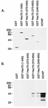
CHIP interacts with the carboxy-terminal domain of Hsc70 in vitro. Binding assays were performed with 3 μl of in vitro translation mixture containing 35S-labeled hCHIP with 3 μg of GST-Hsc70 deletion mutants. (A) Coomassie blue staining of the SDS-polyacrylamide gel demonstrates equivalent loading and appropriate folding of the fusion proteins. In vitro-translated 35S-labeled hCHIP is not visualized by Coomassie blue staining. Sizes are indicated in kilodaltons. (B) An autoradiogram of a duplicate gel shows that binding of the 35S-labeled hCHIP occurs to the carboxy-terminal GST-Hsc70 fusion proteins containing amino acids 373 to 650 and 540 to 650 of Hsc70 but not to those containing amino acids 1 to 540 or 373 to 540. An aliquot of 35S-labeled hCHIP was loaded into the last lane; lower-molecular weight species represent degradation of the in vitro-translated protein.
FIG. 7
FIG. 7
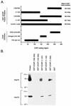
The TPR domain of CHIP is necessary but not sufficient for binding with Hsp70 and Hsc70. (A) Diagram depicting the location of the TPR, charged, and UFD2-like domains within the CHIP coding region. The diagram also shows the constructs of the GST-hCHIP fusion proteins that contain one or more of these domains. (B) Binding assays were performed with 1 μg of Hsp70 or Hsc70 and 15 μg of GST or GST-hCHIP fusion proteins bound to glutathione-Sepharose 4B beads. Western blots were probed with an anti-Hsp70-Hsc70 monoclonal antibody. The protein lane contains 1 μg of the specific protein indicated at the left. Sizes are indicated in kilodaltons.
FIG. 8
FIG. 8
Effects of CHIP on ATPase activities and nucleotide binding of heat shock proteins. (A) The ATPase activities of Hsc70 (black bars) and Hsp70 (white bars) were measured over 20 min in the presence or absence of CHIP, Hsp40, and/or Hsp90 as indicated. Data were normalized to the ATPase activity of Hsc70 alone. ∗ (or †) indicates a significant difference (P < 0.05) between the ATPase activity of Hsc70 alone (or Hsp70 alone) and Hsc70 plus Hsp40 (or Hsp70 plus Hsp40). ∗∗ (or ††) indicates a significant difference (P < 0.05) between the ATPase activity of Hsc70 plus Hsp40 (or Hsp70 plus Hsp40) and Hsc70 plus Hsp40 plus CHIP (or Hsp70 plus Hsp40 plus CHIP). Each condition was repeated for a total of six replicates. (B) The nucleotide species bound to Hsc70 in the absence or presence of CHIP, Hsp40, and/or BAG-1 was determined by thin-layer chromatography. A representative autoradiograph is shown.
FIG. 9
FIG. 9
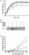
Effects of CHIP on chaperone functions of heat shock proteins. (A) The aggregation of rhodanese was measured at 340 nm over 5 min in the absence (⧫) or presence of CHIP (○), Hsp70-Hsp40 (□), or CHIP-Hsp70-Hsp40 (▴). The measured optical densities were normalized to the zero reading for each individual well, and the increase in absorbance was plotted as a percentage of the total increase of rhodanese alone. Each condition was repeated for a total of eight replicates, and points represent the mean ± standard error of the mean. (B) Binding assays were performed with denatured luciferase (Luc) and Hsc70 in the presence or absence of hCHIP and/or Hsp40. Binding of denatured luciferase was measured by coimmunoprecipitation using an anti-Hsc70 antibody, followed by blotting with an antiluciferase antibody. A representative experiment is shown. (C) Luciferase activity was measured as an indication of refolding after thermal denaturation. The refolding reactions were performed with Hsc70 (▵), Hsp40 (○), or both (□) in the absence (open symbols) and presence (closed symbols) of CHIP (⧫, CHIP alone). Luciferase activity was measured at various intervals between 0 and 120 min, and the activity for each reaction was normalized to luciferase in refolding buffer alone. Each condition was repeated for a total of 12 replicates, and points represent the mean ± standard error of the mean.
FIG. 10
FIG. 10
Model of the eukaryotic reaction cycle in the presence of CHIP, Hsp40, Hip, and BAG-1. The forward reaction, in which ATP is hydrolyzed to ADP and inorganic phosphate (Pi) is released, is enhanced by Hsp40. The biochemical data suggest that CHIP blocks this forward reaction. Hip stabilizes the ADP-bound, high-substrate-affinity conformation of Hsc70 and thus enhances chaperone activity. Conversely, BAG-1 accelerates nucleotide exchange, promoting substrate release and the formation of the low-substrate-affinity, ATP-bound conformation of Hsc70. In this model, both BAG-1 and CHIP would favor the low-affinity Hsc70 conformation, whereas Hip and Hsp40 would favor the high-affinity conformation. CTD, carboxy-terminal domain.

Similar articles

Cited by

References

    1. Angelier N, Moreau N, Rodriquez-Martin M L, Penrad-Mobayed M, Prudhomme C. Does the chaperone heat shock protein Hsp70 play a role in the control of developmental processes? Int J Dev Biol. 1996;40:521–529. - PubMed
    1. Ballinger, C. A., and C. Patterson. Unpublished observations.
    1. Benaroudj N, Fouchaq B, Ladjimi M M. The COOH-terminal peptide binding domain is essential for self-association of the molecular chaperone Hsc70. J Biol Chem. 1997;272:8744–8751. - PubMed
    1. Bercovich B, Stancovski I, Mayer A, Blumenfeld N, Laszlo A, Schwartz A L, Ciechanover A. Ubiquitin-dependent degradation of certain protein substrates in vitro requires the molecular chaperone Hsc70. J Biol Chem. 1997;272:9002–9010. - PubMed
    1. Boice J A, Hightower L E. A mutational study of the peptide-binding domain of Hsc70 guided by secondary structure prediction. J Biol Chem. 1997;272:24825–24831. - PubMed

Publication types

MeSH terms

Associated data