. 1999 Jun;73(6):4931-40.
doi: 10.1128/JVI.73.6.4931-4940.1999.

Feline leukemia virus long terminal repeat activates collagenase IV gene expression through AP-1

Affiliations

Feline leukemia virus long terminal repeat activates collagenase IV gene expression through AP-1

S K Ghosh et al. J Virol. 1999 Jun.

Abstract

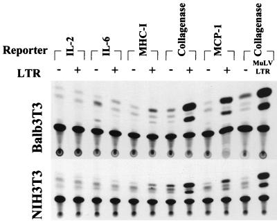
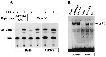
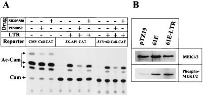
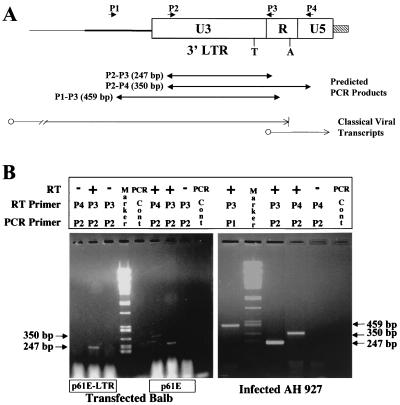
Leukemia and lymphoma induced by feline leukemia viruses (FeLVs) are the commonest forms of illness in domestic cats. These viruses do not contain oncogenes, and the source of their pathogenic activity is not clearly understood. Mechanisms involving proto-oncogene activation subsequent to proviral integration and/or development of recombinant viruses with enhanced replication properties are thought to play an important role in their disease pathogenesis. In addition, the long terminal repeat (LTR) regions of these viruses have been shown to be important determinants for pathogenicity and tissue specificity, by virtue of their ability to interact with various transcription factors. Previously, we have shown that, in the case of Moloney murine leukemia virus, the U3 region of the LTR independently induces transcriptional activation of specific cellular genes through an LTR-generated RNA transcript (S. Y. Choi and D. V. Faller, J. Biol. Chem. 269:19691-19694, 1994; S.-Y. Choi and D. V. Faller, J. Virol. 69:7054-7060, 1995). In this report, we show that the U3 region of exogenous FeLV LTRs can induce transcription from collagenase IV (matrix metalloproteinase 9) and monocyte chemotactic protein 1 (MCP-1) promoters up to 12-fold. We also show that AP-1 DNA-binding activity and transcriptional activity are strongly induced in cells expressing FeLV LTRs and that LTR-specific RNA transcripts are generated in those cells. Activation of mitogen-activated protein kinase kinases 1 and 2 (MEK1 and -2) by the LTR is an intermediate step in the FeLV LTR-mediated induction of AP-1 activity. These findings thus suggest that the LTRs of FeLVs can independently activate transcription of specific cellular genes. This LTR-mediated cellular gene transactivation may play an important role in tumorigenesis or preleukemic states and may be a generalizable activity of leukemia-inducing retroviruses.


Figures

FIG. 1
FIG. 1
Schematic representation of the FeLV LTR expression vectors. The open and partially shaded boxes represent viral sequences, whereas the hatched boxes represent flanking genomic sequences. Thick lines represent vector backbone sequences. Construction of these and similar clones is described in Materials and Methods. Sequence lengths are not to scale.
FIG. 2
FIG. 2
Transcriptional activation of a collagenase promoter-reporter by FeLV LTR and full-length FeLV proviral clone. (A) Induction of collagenase promoter by transiently expressed FeLV LTR. One microgram of −517/+62 collagenase-CAT reporter plasmid was cotransfected with 7.5 μg of individual LTR constructs into BALB/3T3 cells by the DEAE-dextran method, as described in Materials and Methods. The Mo-MuLV LTR construct XFUX was used as a positive control. Cotransfection with 7.5 μg of backbone vector plasmid pTZ19U was used to determine the constitutive basal expression of the collagenase promoter-reporter vector. Forty-eight hours after transfection, cells were washed with PBS and assessed microscopically for green fluorescence under UV light to normalize for transfection efficiency. Cells were harvested, and CAT assays were performed as described in Materials and Methods. Products were separated by thin-layer chromatography. (B) Induction of collagenase promoter-reporter by transient transfection of a full-length FeLV molecular clone. BALB/3T3 cells were cotransfected as described above with either 7.5 μg of FeLV or Mo-MuLV LTR constructs or 10 μg of full-length FeLV or Mo-MuLV proviral clones. These experiments were repeated three times. The thin-layer chromatogram of one representative experiment has been presented. The migration positions of chloramphenicol (Cam) and the acetylated products (Ac-Cam) are indicated. Fold activation for each sample shown at the bottom of each panel was calculated from quantitative data obtained from multiple experiments. Exposed X-ray films were photographed by using AlphaImager 3.4, and densitometric analysis of the image was carried out with the AlphaEase program (Alpha Innotech, San Leandro, Calif.). The error bars represent the standard errors of the means.
FIG. 3
FIG. 3
Collagenase promoter activation by cell lines stably expressing an FeLV LTR. G418-resistant stable BALB/3T3 cell lines BALB-LTR1, BALB-LTR6, and BALB-vector were transfected with 1.0 μg of −517/+62 collagenase-CAT reporter. Cell lines BALB-LTR1 (later shown to be PCR negative for the LTR) and BALB-LTR6 (later shown to be PCR positive for the LTR) were generated by transfecting BALB/3T3 cells with pCR3.1/61E-LTR-PK followed by G418 selection, whereas cell line BALB-vector was generated by transfecting the empty vector pCR3.1. In addition, normal BALB/3T3 cells were cotransfected with −517/+62 collagenase-CAT reporter and 7.5 μg of either pTZ19 (lane marked with minus sign) or 61E-LTR (lane marked with plus sign) as controls. Cells were harvested 48 h after transfection, and CAT assays were performed as described in the legend to Fig. 2. All these cell lines were also transfected separately with control plasmid CMV-Coll-CAT to compare their transfectability. These experiments were repeated four times. The thin-layer chromatogram of one representative experiment has been presented. The migration positions of chloramphenicol (Cam) and the acetylated products (Ac-Cam) are indicated.
FIG. 4
FIG. 4
Transcriptional activation of collagenase promoter-reporter by various FeLV LTR and full-length FeLV clones in the feline embryo fibroblast cell line AH927. Cotransfection experiments were carried out as described in the legend to Fig. 2, except that 0.5 μg of collagenase-CAT reporter was used for each plate and cells were treated with DEAE-dextran–DNA complex for only 3 h. For the full-length proviral clone, 10 μg of plasmid per plate was used, and for the LTR and backbone vector, 7.5 μg of plasmid per plate was used. In one plate, cells were also transfected with 1 μg of CMV-Coll-CAT construct alone (the control lane) to assess transfection efficiency in these cells compared to that in other cell lines. Forty-eight hours after transfection, CAT assays were performed as described in the legend to Fig. 2. An autoradiogram of a plate from a representative experiment is shown. The migration positions of chloramphenicol (Cam) and the acetylated products (Ac-Cam) are indicated. Fold induction calculations were performed as described in the legend to Fig. 2. Error bars indicate the standard errors of the means.
FIG. 5
FIG. 5
Analysis of transcriptional activation of promoters of various genes by FeLV LTR. Transient-cotransfection experiments were performed with either BALB/3T3 or NIH 3T3 cells as described in Materials and Methods, with p61E-LTR and CAT reporters driven by the promoter regions of various genes known to be important for cell growth or inflammation, plus an internal transfection control plasmid. Seven and one-half micrograms of pTZ19U (−) or p61E-LTR (+) plasmid was cotransfected with 7.5 μg of IL-2 (−585 IL-2-CAT), 7.5 μg of IL-6 (−225 IL-6-CAT) or MHC class I (KbHN-CAT), 1 μg of collagenase (−517/+62 Coll-CAT), or 4 μg of MCP-1 (−543 JE-CAT) reporter constructs. Collagenase gene induction by the Mo-MuLV LTR was used as control. Vector control for the Mo-MuLV LTR was 7.5 μg of pBR322 plasmid. Forty-eight hours after transfection, CAT assays were performed as described in the legend to Fig. 2. One representative autoradiogram for each cell line is shown. Experiments for both cell types were repeated three times with identical results.
FIG. 6
FIG. 6
Activation of the AP-1 complex by the FeLV LTR. (A) Transcriptional activation of a CAT reporter with 3×AP-1-binding-site-containing promoter element by the LTR. BALB/3T3 or AH927 cells were cotransfected with 3×AP-1-CAT and p61E-LTR(+) or vector pTZ19U(−) as detailed in the legends to Fig. 2 and 3. A transcriptional activation assay of the collagenase IV promoter by p61E-LTR(+) or vector pTZ19U(−) was also performed in the same set of experiments. Forty-eight hours after transfection, CAT assays were performed as described in the legend to Fig. 2. The migration positions of chloramphenicol (Cam) and the acetylated products (Ac-Cam) are indicated. (B) EMSAs with an oligonucleotide containing an AP-1-binding site. Nuclear extracts from cells were incubated with a radiolabeled double-stranded AP-1 consensus oligonucleotide probe as described in Materials and Methods, and DNA-protein complexes were separated on a 5% polyacrylamide gel. Before preparation of nuclear extracts, all cells were starved overnight in DMEM containing 0.5% appropriate serum. Extracts in lanes marked “61E-LTR” and “61E” came from transiently transfected BALB/3T3 cells 40 h after transfection. The lane marked “Mock” refers to normal BALB/3T3 cells. “Uninfected” and “Infected” AH927 cells refer to normal AH927 cells and AH927 cells that were transfected with full-length FeLV-A proviral clone p61E 3 months previously, respectively. Production of FeLV by this cell line has been confirmed with the Viracheck enzyme immunoassay kit (Synbiotics, Inc., San Diego, Calif.) for FeLV core protein antigen p27. The migration position of the DNA–AP-1 complex is indicated.
FIG. 7
FIG. 7
Effect of expression of the FeLV LTR on MAP kinase signal transduction pathways. (A) Action of MAP kinase-specific inhibitors PD98059 and SB203580 on transcriptional activation of 3×AP-1-binding-site-containing promoter and −517/+62 Coll-CAT reporter by FeLV LTR. The LTR expression plasmid p61E-LTR was cotransfected with either 3×AP-1-CAT reporter or −517/+62 Coll-CAT reporter. As a control, pTZ19U vector was also cotransfected with the above two reporters. In another set of experiments, BALB/3T3 cells were transfected with CMV-Coll-CAT reporter, as described in Materials and Methods. PD98059 and SB203580 (50 and 10 μM final concentrations, respectively) were added to the medium immediately after the chloroquine treatment step. Twenty-eight hours after transfection, the cells were harvested for CAT assays as described in the legend to Fig. 2. The migration positions of chloramphenicol (Cam) and the acetylated products (Ac-Cam) are indicated. This experiment was repeated twice with identical results. (B) Effect of expression of the FeLV LTR or full-length FeLV on phosphorylation of the Raf-MAPK pathway intermediates MEK1 and -2. BALB/3T3 cells were transiently transfected with vector pTZ19U, full-length clone p61E, or LTR clone p61E-LTR by the Lipofectamine Plus transfection method. Twenty-four hours after transfection, the medium was changed to DMEM with 0.5% DCS, and incubation was continued for another 24 h. Total cell lysates were then prepared, as detailed in Materials and Methods. Twenty micrograms of protein from each cell lysate was separated on a sodium dodecyl sulfate–10% polyacrylamide gel electrophoresis gel in duplicate and transferred to a nitrocellulose membrane. Total MEK1 and -2 protein and phosphorylated MEK1 and -2 protein on the membrane were detected by immunoblotting with pan-anti-MEK1 and -2 serum or phosphospecific anti-MEK1 and -2 serum (New England Biolabs) as described in Materials and Methods.
FIG. 8
FIG. 8
RT-PCR analysis of RNA isolated from cells expressing FeLV or the FeLV LTR. (A) Schematic diagram of the oligonucleotide primers and their locations on the viral genome. The origin and termination of the classical viral genomic transcripts are shown by thin lines. T, TATA box of classical genomic promoter; A, polyadenylation signal sequence. Predicted PCR products with various primer pair combinations and their sizes are also indicated. (B) (Right) RT-PCR with DNase-treated RNA isolated from BALB/3T3 cells transfected with either p61E-LTR or full-length virus 61E. (Left) RT-PCR with DNase-treated RNA isolated from FeLV 61E-infected AH927 cells. The origin of the infected AH927 cells has been described in the legend to Fig. 5. “PCR Cont” refers to an amplification product with all the primers and all other components of PCRs including Taq polymerase but no RNA or DNA template. This was done to test for contamination of the reagents used in the PCR. PCR products were separated on 2% agarose gels. PstI-cut lambda DNA was used as a marker in the gel. The migration positions and sizes of the amplified products are indicated.

Similar articles

Cited by

References

    1. Alessi D R, Cuenda A, Cohen P, Dudley D T, Saltiel A R. PD 098059 is a specific inhibitor of the activation of mitogen-activated protein kinase kinase in vitro and in vivo. J Biol Chem. 1995;270:27489–27494. - PubMed
    1. Andrews N C, Faller D V. A rapid micropreparation technique for extraction of DNA-binding proteins from limiting numbers of mammalian cells. Nucleic Acids Res. 1991;19:2499. - PMC - PubMed
    1. Angel P, Baumann I, Stein B, Delius H, Rahmsdorf H J, Herrlich P. 12-O-Tetradecanoyl-phorbol-13-acetate induction of the human collagenase gene is mediated by an inducible enhancer element located in the 5′-flanking region. Mol Cell Biol. 1987;7:2256–2266. - PMC - PubMed
    1. Angel P, Hattori K, Smeal T, Karin M. The jun proto-oncogene is positively autoregulated by its product, Jun/AP-1. Cell. 1988;55:875–885. - PubMed
    1. Angel P, Karin M. The role of Jun, Fos and the AP-1 complex in cell-proliferation and transformation. Biochim Biophys Acta. 1991;1072:129–157. - PubMed

Publication types

MeSH terms