. 1999 May;10(5):1367-79.
doi: 10.1091/mbc.10.5.1367.

Apg7p/Cvt2p: A novel protein-activating enzyme essential for autophagy

Affiliations
Free PMC article

Apg7p/Cvt2p: A novel protein-activating enzyme essential for autophagy

I Tanida et al. Mol Biol Cell. 1999 May.
Free PMC article

Abstract

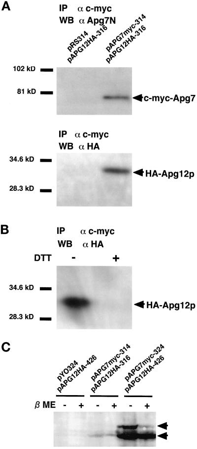
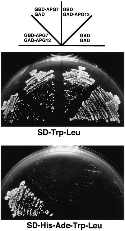
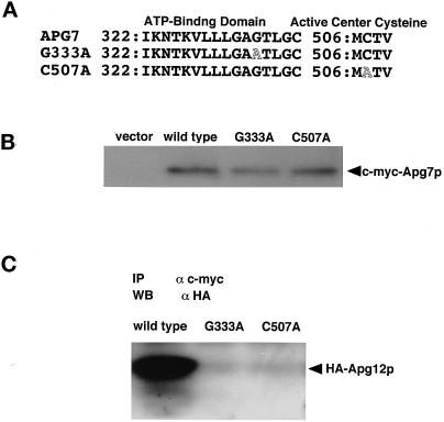
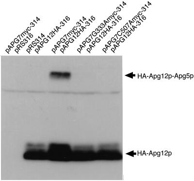
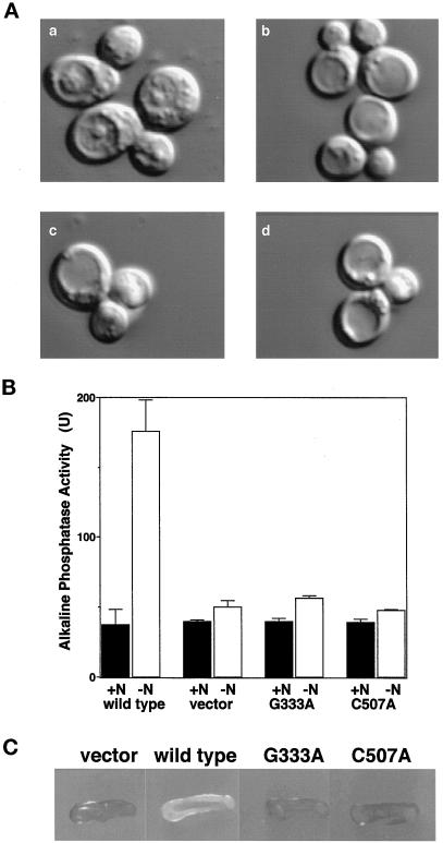
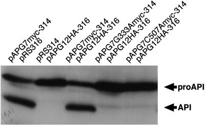
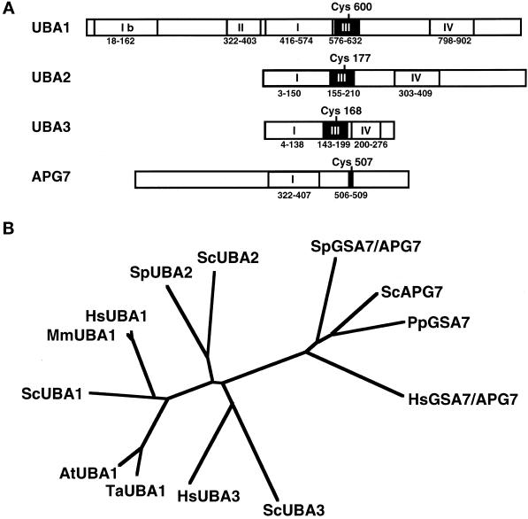
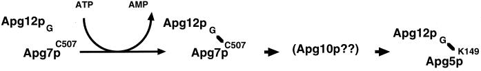
In the yeast Saccharomyces cerevisiae, the Apg12p-Apg5p conjugating system is essential for autophagy. Apg7p is required for the conjugation reaction, because Apg12p is unable to form a conjugate with Apg5p in the apg7/cvt2 mutant. Apg7p shows a significant similarity to a ubiquitin-activating enzyme, Uba1p. In this article, we investigated the function of Apg7p as an Apg12p-activating enzyme. Hemagglutinin-tagged Apg12p was coimmunoprecipitated with c-myc-tagged Apg7p. A two-hybrid experiment confirmed the interaction. The coimmunoprecipitation was sensitive to a thiol-reducing reagent. Furthermore, a thioester conjugate of Apg7p was detected in a lysate of cells overexpressing both Apg7p and Apg12p. These results indicated that Apg12p interacts with Apg7p via a thioester bond. Mutational analyses of Apg7p suggested that Cys507 of Apg7p is an active site cysteine and that both the ATP-binding domain and the cysteine residue are essential for the conjugation of Apg7p with Apg12p to form the Apg12p-Apg5p conjugate. Cells expressing mutant Apg7ps, Apg7pG333A, or Apg7pC507A showed defects in autophagy and cytoplasm-to-vacuole targeting of aminopeptidase I. These results indicated that Apg7p functions as a novel protein-activating enzyme necessary for Apg12p-Apg5p conjugation.


Figures

Figure 1
Figure 1
Apg12p conjugates with Apg7p via a thioester bond. (A) Apg12p was coimmunoprecipitated with Apg7p. pAPG7myc-314 plasmid (CEN) was transformed into apg7Δ cells producing HA-tagged Apg12p to express c-myc–tagged Apg7p. A transformant was designated as the YIT702 strain. The pRS314 vector was used as a control. Cell lysates were prepared as described in MATERIALS AND METHODS. A c-myc–tagged Apg7p was immunoprecipitated with Agarose beads conjugated with anti–c-myc mAb (9E10). The immunoprecipitates were subjected to SDS-PAGE on a 10% gel and transferred to a polyvinylidene difluoride membrane. Proteins were detected by Western blotting with αApg7N antibody (for Apg7p) and anti-HA mouse mAb (16B12; for HA-tagged Apg12p). (B) The coimmunoprecipitation of Apg12p with Apg7p is sensitive to DTT. A cell lysate of the YIT702 strain was prepared as described above. The lysate was boiled with (DTT+) or without 1 mM DTT (DTT−). Immunoprecipitation and Western blotting were performed as described above. (C) The higher molecular weight band of Apg7p in cells overexpressing Apg7p and Apg12p is sensitive to β-mercaptoethanol. Cells grown to early logarithmic phase in MVD medium were harvested and converted to spheroplasts in spheroplasting solution. The spheroplasts were harvested in 1.3 M sorbitol as a cushion, lysed with a 4× SDS sample buffer (Ausubel et al., 1995) with a protease-inhibitor mixture (Sigma). The lysate was boiled for 5 min in the presence (βME+) or absence (βME−) of 3% β-mercaptoethanol. SDS-PAGE on a 7% gel and Western blotting wereperformed as described above. pYO324/pAPG12HA-426: strain YIT704 cells; pAPG7myc-314/pAPG12HA-316: strain YIT702 cells; pAPG7myc-324/pAPG12HA-426: strain YIT703 cells.
Figure 2
Figure 2
Apg7p interacts with Apg12p in vivo. pGBD-APG7 (TRP1) and pGAD-APG12 (LEU2) were transformed into PJ69-4A strain (gal4Δ gal80Δ trp1 leu2 GAL2-ADE2 GAL1-HIS3) to express GAL4BD-Apg7p and GAL4AD-Apg12p, respectively. pGAD-C1 and pGBD-C1 were used as controls. Cells were plated on SC-Trp-Leu plate (positive control) and SC-Ade-His-Trp-Leu plate, and incubated at 30°C for 3 d. PJ69-4A strain counterclockwise from the bottom carried pGBD-C1 and pGAD-C1 (GBD GAD), pGBD-C1 and pGAD-APG12 (GBD GAD-APG12), pGBD-APG7 and pGAD-C1 (GBD-APG7 GAD), and pGBD-APG7 and pGAD-APG12 (GBD-APG7 GAD-APG12). A strain expressing both GAL4BD-Apg7p and GAL4AD-Apg12p grew well on SC-Ade-Trp-Leu plate, indicating that Apg7p interacts with Apg12p.
Figure 3
Figure 3
The ATP-binding domain and active site cysteine of Apg7p are essential for conjugation with Apg12p via a thioester bond. (A) Point mutation sites are schematically represented. Gly333 in a predicted ATP-binding domain of Apg7p was changed to Ala by site-directed mutagenesis, and Cys507 in Apg7p was also changed to Ala. (B) Apg7pG333A and Apg7pC507A are expressed in yeast cells at similar levels to Apg7p. The apg7Δ strain carrying pRS314 (vector), strain YIT702 (wild type), strain YIT7G333A (G333A), and strain YIT7C507A (C507A) cells were grown in MVD medium and lysed. c-myc–tagged Apg7p proteins were immunoprecipitated as described in Figure 1A. (C) No Apg12p is coimmunoprecipitated with Apg7pG333A and Apg7pC507A. c-myc–tagged Apg7p in the cell lysate of YIT702 (wild type), YIT7G333A (G333A), and YIT7C507A (C507A) strains were immunoprecipitated, and the coimmunoprecipitates were detected by Western blotting with anti-HA antibody as described above.
Figure 4
Figure 4
Apg7pG333A and Apg7pC507A show significant defects in the formation of the Apg12p–Apg5p conjugate. The conjugate in cell lysates expressing HA-tagged Apg12p was detected by Western blotting using αHA antibody as described previously (Mizushima et al. 1998a). pAPG7myc-314/pRS316: apg7Δ cells expressing c-myc–tagged wild-type Apg7p only; pRS314/pAPG12HA-316: apg7Δ cells expressing HA-tagged Apg12p only; pAPG7myc-314/pAPG12HA-316: strain YIT702 cells; pAPG7G333Amyc-314/pAPG12HA-316: strain YIT7G333A cells; pAPG7C507Amyc-314/pAPG12HA-316: strain YIT7C507A cells.
Figure 5
Figure 5
The ATP-binding domain and active site cysteine of Apg7p are essential for autophagy. (A) Gly333 and Cys507 in Apg7p are essential for the accumulation of autophagic bodies under nitrogen-starvation conditions. Cells grown to early-logarithmic phase in MVD+Ura medium were transferred to nitrogen-starvation medium in the presence of PMSF and incubated for 8 h at 30°C. Representative Nomarski images of the cells are shown. Autophagic bodies accumulated in vacuoles of apg7Δ cells carrying pAPG7myc-314 (a). Few autophagic bodies accumulated in vacuoles of apg7Δ cells carrying pRS314 (b), pAPG7G333Amyc-314 (c), and pAPG7C507Amyc-314 (d). (B) Autophagy in apg7Δ cells expressing Apg7pG333A and Apg7pC507A was monitored by an alkaline phosphatase processing assay. We constructed strain YTS2 (pho8::pho8Δ60 apg7::HIS3 trp1) and transformed it with plasmids pRS314 (vector), pAPG7myc-314 (wild type), pAPG7G333Amyc-314 (G333A), and pAPG7C507Amyc-314 (C507A). Cells growing logarithmically in MVD+Ade+Ura medium (N+) were transferred to nitrogen-starvation medium and incubated for 4 h at30°C (N−). Alkaline phosphatase activity in cells under rich or nitrogen-starvation conditions was measured as described by Noda et al. (1998). (C) Gly333 and Cys507 in Apg7p are essential for cell viability under nitrogen-starvation conditions. Cells were plated on nitrogen-starvation medium containing 10 μg/ml phloxine B and incubated at 30°C for 3 d. Nonviable cells were stained red (gray in monochrome), whereas viable cells were not stained (white in monochrome).
Figure 6
Figure 6
The ATP-binding domain and active center cysteine of Apg7p are essential for cytoplasm-to-vacuole targeting of API. The processing of API in cells growing logarithmically was measured as described by Mizushima et al. (1998a). Lanes correspond to those in Figure 4.
Figure 7
Figure 7
Apg7p is present in the cytosol. A wild-type strain (YW5–1B) was cultured in MVD+Ura+Trp medium, transferred to nitrogen-starvation medium, and incubated for 30 min. Cells growing vegetatively were converted to spheroplasts in rich buffer (1% yeast extract, 2% polypeptone, 0.5% glucose, and 0.7 M sorbitol), whereas nitrogen-starved cells were converted to spheroplasts in nitrogen-starvation buffer (0.15% yeast nitrogen base without amino acids and ammonium sulfate, 2% glucose, and 0.9 M sorbitol). Cells were lysed and fractionated by centrifugation as described by Huang and Chiang (1997). LSP, 15,000 × g pellet; HSP, 100,000 × g pellet; HSS, 100,000 × g supernatant.
Figure 8
Figure 8
Apg7p is distantly related to Uba1p and its relatives. (A) Schematic representation of the similarity domains among UBA1, UBA2, UBA3, and APG7. The putative active site cysteines are located within similarity box III (UBA domain), as indicated. The essential ATP-binding domains are located within similarity box I. UBA1 contains another ATP-binding motif within similarity box Ib, which is nonessential for function. The similarity boxes correspond to those of Johnson et al. (1997) and Liakopoulos et al. (1998), except box Ib. (B) The phylogenetic tree of E1 enzymes. The homologous regions of E1-like enzymes containing the ATP-binding domain and active site cysteine were multi-aligned and analyzed by a CLUSTAL W multiple alignment program (Thompson et al., 1994). Shown are ScAPG7 (residues 321 to 520 of S. cerevisiae Apg7p; PID: g731742), PpAPG7 (residues 322 to 521 of P. pastoris Gsa7p), HsAPG7/GSA7 (residues 235 to 434 of Homo sapiens APG′7/GSA7), SpAPG7/GSA7 (residues 332 to 531 of Schizosaccharomyces pombe Apg7p/Gsa7p; PID g2894280), HsUBA1 (residues 465 to 664 of H. sapiens UBA1; PID: g340072), MmUBA1 (residues 465 to 664 of Mus musculus UBA1; PID: g220629), ScUBA1 (residues 431 to 630 of S. cerevisiae Uba1p; PID: g4715), AtUBA1 (residues 489 to 688 of A. thaliana UBA1; PID: g1750376), TaUBA1 (residues 459 to 658 of Triticum aestivum UBA1; PID g136632), ScUBA2 (residues 18 to 219 of S. cerevisiae Uba2p; PID: g1717852), SpUBA2 (residues 22 to 221 of S. pombe Uba2p; PID: g2956755), HsUBA3 (residues 45 to 244 of H. sapiens UBA3; PID: g3342564), and ScUBA3 (residues 1 to 200 of S. cerevisiae Uba3p; PID: g2980755) (Hatfield et al., 1990; Handley et al., 1991; McGrath et al., 1991; Imai et al., 1992; Dohmen et al., 1995; Hatfield et al., 1997; Liakopoulos et al., 1998; Mizushima et al., 1998a; Osaka et al., 1998, Yuan et al., 1999).
Figure 9
Figure 9
Working hypothesis for the function of Apg7p in the Apg12p conjugation system. Apg12p associates with Apg7p. The C-terminal Gly of Apg12p conjugates with Cys507 in Apg7p via a thioester bond in an ATP-dependent manner. The reaction occurs in the cytoplasm or on the cytoplasmic side of an Apg12p-associated compartment(s). Although the E2 enzyme has not been identified, one candidate is Apg10p.

Similar articles

Cited by

References

    1. Ausubel F, Brent R, Kingston RE, Moore DD, Seidman JG, Smith JA, Struhl K. Short Protocols in Molecular Biology. 3rd ed. New York: John Wiley & Sons; 1995.
    1. Baba M, Ohsumi M, Ohsumi Y. Analysis of the membrane structures involved in autophagy in yeast by freeze-replica method. Cell Struct Funct. 1995;20:465–471. - PubMed
    1. Baba M, Takeshige K, Baba N, Ohsumi Y. Ultrastructural analysis of the autophagic process in yeast: detection of autophagosomes and their characterization. J Cell Biol. 1994;124:903–913. - PMC - PubMed
    1. Bartel B, Wunning I, Varshavsky A. The recognition component of the N-end rule pathway. EMBO J. 1990;9:3179–3189. - PMC - PubMed
    1. Boddy MN, Howe K, Etkin LD, Solomon E, Freemont PS. PIC1, a novel ubiquitin-like protein which interacts with the PML component of a multiprotein complex that is disrupted in acute promyelocytic leukemia. Oncogene. 1996;13:971–982. - PubMed

Publication types

MeSH terms