. 1999 Apr 15;13(8):935-45.
doi: 10.1101/gad.13.8.935.

Control of the nuclear localization of Extradenticle by competing nuclear import and export signals

Affiliations

Control of the nuclear localization of Extradenticle by competing nuclear import and export signals

M Abu-Shaar et al. Genes Dev. .

Abstract

The Drosophila PBC protein Extradenticle (Exd) is regulated at the level of its subcellular distribution: It is cytoplasmic in the absence of Homothorax (Hth), a Meis family member, and nuclear in the presence of Hth. Here we present evidence that, in the absence of Hth, Exd is exported from nuclei due to the activity of a nuclear export signal (NES). The activity of this NES is inhibited by the antibiotic Leptomycin B, suggesting that Exd is exported by a CRM1/exportin1-related export pathway. By analyzing the subcellular localization of Exd deletion mutants in imaginal discs and cultured cells, we identified three elements in Exd, a putative NES, a nuclear localization sequence (NLS), and a region required for Hth-mediated nuclear localization. This latter region coincides with a domain in Exd that binds Hth protein in vitro. When Exd is uncomplexed with Hth, the NES dominates over the NLS. When Exd is expressed together with Hth, or when the NES is deleted, Exd is nuclear. Thus, Hth is required to overcome the influence of the NES, possibly by inducing a conformational change in Exd. Finally, we provide evidence that Hth and Exd normally interact in the cytoplasm, and that Hth also has an NLS. We propose that in Exd there exists a balance between the activities of an NES and an NLS, and that Hth alters this balance in favor of the NLS.


Figures

Figure 1
Figure 1
Schematic diagram of Exd protein variants and summary of their subcellular localization. Full-length Exd(1–376) contains three major domains of conservation with vertebrate Pbx1 and C. elegans Ceh-20; PBC-A, PBC-B, and the homeodomain (HD) as indicated. In addition, these proteins are similar for an additional 13 residues carboxy-terminal to the homeodomain (dark gray shading). A classical NLS (RRKRR) is present in the amino-terminal arm of the homeodomain. NLS–myc–Exd has the NLS sequence from the SV40 large T antigen inserted amino-terminal to the myc tag and NLS–β gal–Exd has the same NLS and the nearly full-length β-galactosidase ORF inserted in place of the myc tag. All proteins except for NLS–β gal–Exd were myc-tagged at their amino termini (small broken boxes). Protein domains are shown to scale with the exception of the myc epitope, which is 86 amino acids. The columns at right summarize the subcellular localization of these proteins in the presence (+HTH) or absence (−HTH) of Homothorax, and when expressed at high levels in the absence of Hth. (N) Nuclear; (C) cytoplasmic; (mixed) nuclear and cytoplasmic; (N > C) more nuclear than cytoplasmic; (nd) not determined. Class I proteins are nuclear in the presence of Hth but generally cytoplasmic in its absence; class II proteins are not regulated by Hth and generally cytoplasmic; class III proteins are not regulated by Hth and generally nuclear.
Figure 2
Figure 2
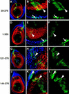
Subcellular localization of Exd(1–376) in leg imaginal discs. Shown are leg imaginal discs in which Exd(1–376) expression was induced by ptc–Gal4 and monitored by staining with an anti-myc antibody (green). These discs were costained for Hth (blue) and Dac (red), which, in these focal planes, serves as a nuclear marker for non-Hth-expressing cells. (A–C) A leg disc grown at 22°C; (D–F) a leg disc grown at 29°C, which results in higher levels of Exd(1–376). (A,D) Low magnification views of the entire disc; the ptc–Gal4 induced expression of Exd(1–376) can be seen as a green stripe extending across the entire disc. (B,C,E,F) High magnification views showing regions of these discs with Hth+, Dac cells next to Hth, Dac+ cells; (C,F) show only the signal from the myc staining. At 22°C, nearly all of the Exd(1–376) is in the cytoplasm in the absence of Hth (A–C) whereas at 29°C (D–F), Exd(1–376) is partially localized to nuclei in the absence of Hth (arrows in F). In this and subsequent figures, arrows point to examples of nuclear localization and arrowheads point to examples of cytoplasmic localization.
Figure 3
Figure 3
Subcellular localization of Exd truncations in leg imaginal discs. Antibody stains and views of leg discs are as described in Fig. 2. Exd(39–376) (A–C); Exd(1–300) (D–F); Exd(121–376) (G–I); Exd(144–376) (J–L); Exd(178–376) (M–O); Exd(220–376) (P–R); Exd(257–376) (S–U). All discs were grown at 22°C. See Fig. 1 for schematic illustrations of these mutants. (Arrows) Examples of nuclear localization; (arrowheads) examples of cytoplasmic localization.
Figure 3
Figure 3
Subcellular localization of Exd truncations in leg imaginal discs. Antibody stains and views of leg discs are as described in Fig. 2. Exd(39–376) (A–C); Exd(1–300) (D–F); Exd(121–376) (G–I); Exd(144–376) (J–L); Exd(178–376) (M–O); Exd(220–376) (P–R); Exd(257–376) (S–U). All discs were grown at 22°C. See Fig. 1 for schematic illustrations of these mutants. (Arrows) Examples of nuclear localization; (arrowheads) examples of cytoplasmic localization.
Figure 4
Figure 4
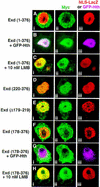
Exd is excluded from nuclei because of an LMB-sensitive NES. Shown are S2 cells immunostained for proteins expressed from transiently transfected plasmids; all Exd derivatives are myc tagged and shown in green. Expression of NLS–βgal served as a nuclear marker except for B and G, in which GFP–Hth was a nuclear marker. (A) Exd(1–376) + NLS–βgal (red); (B) Exd(1–376) + GFP–Hth (fuchsia); (C) Exd(1–376) + NLS–βgal (red) + 10 nm LMB; (D) Exd(220–376) + NLS–βgal (red); (E) Exd(Δ179–219) + NLS–βgal (red); (F) Exd(178–376) + NLS–βgal (red); (G) Exd(178–376) + GFP–Hth (fuchsia); (H) Exd(178–376) + NLS–βgal (red) + 10 nm LMB. (i,ii,iii) The merged, green, and red or fuchsia channels, respectively.
Figure 5
Figure 5
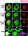
The endogenously expressed Exd protein in S2 cells is only partially sensitive to LMB. Shown are S2 cells, transfected with NLS–β gal or GFP–Hth, stained for Exd (green) and β gal (red) or GFP–Hth (fuchsia) proteins. Prior to fixation, the cells were incubated with either no LMB (A,D), 10 nm LMB (B), or 25 nm LMB (C). Without LMB (A) Exd was in the cytoplasm. In the presence of LMB (B,C), although most of the Exd was in nuclei, some remained in the cytoplasm. Similar results were obtained with concentrations of LMB up to 400 nm (not shown). When GFP–Hth was expressed in S2 cells, all of the endogenous Exd protein became nuclear (D, arrow), whereas in untransfected cells, Exd was cytoplasmic (D, arrowhead). (i,ii,iii) The merged, green, and red/fuchsia channels, respectively.
Figure 6
Figure 6
The PBC-A domain of Exd binds Hth. (A) Schematic illustration of the his-tagged 35S-labeled-Exd proteins used in this experiment. (B) GST pull-down experiment. Crude Escherichia coli extracts containing GST–Hth (lanes 2,5,8) or GST–Exd(144–376) (lanes 3,6,9) were incubated with 35S-labeled-Exd(1–376) (lanes 2,3), 35S-labeled-Exd(144–376) (lanes 5,6), or 35S-labeled-Exd(1–143) (lanes 8,9). Bound proteins were analyzed by SDS-PAGE and autoradiography. For each of the three 35S-labeled Exd proteins, 25% of the amount used in the binding reactions was directly loaded in lanes 1, 4, and 7, respectively.
Figure 7
Figure 7
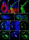
The subcellular localization of Exd(1–223) in the presence and absence of endogenous Exd. (A–C) ptc–Gal4 driven expression of Exd(1–223) in otherwise wild-type (exd+) leg discs; views and stains are as in Fig. 2 [Hth, blue; Dac, red; and Exd(1–223), green]. Exd(1–223), which includes the Hth-interaction domain and the NES, but excludes the NLS, is cytoplasmic in Hth-expressing and nonexpressing cells (arrowheads). (D–K) Embryos expressing Exd(1–223) in stripes with the ptc–Gal4 driver line. These embryos were stained for Hth (blue) and the myc epitope of Exd(1–223) (green). (D,F–H) An embryo that had no maternal exd expression but had wild-type zygotic (paternal) exd expression; (E,I–K) a sibling embryo that had no maternal or zygotic exd expression. In these exd embryos, the stripes of ptc–Gal4-induced Exd(1–223) expression are disorganized because of a breakdown in engrailed expression (Peifer and Wieschaus 1990). (D,E) Low magnification views of the entire embryos (anterior is left); (F–K) higher magnification views of the anterior regions of these embryos. In the presence of zygotic Exd (F–H), Hth protein is detected in many nuclei (G) and Exd(1–223) is predominantly cytoplasmic (H, arrowhead). In the absence of maternal and zygotic Exd (I–K), Hth was not detected in most cells (asterisk) but could be detected in cells that also express Exd(1–223) (J,K; arrow). In these cells, both proteins are predominantly nuclear.
Figure 8
Figure 8
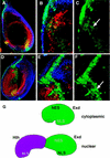
The regulated nuclear import of Exd depends on a balance between nuclear export and localization signals. (A–C) The subcellular localization of NLS–myc–Exd in leg discs; (D–F) the subcellular localization of NLS–β gal–Exd in leg discs. The stains and views of these discs are as described in Fig. 2 (Hth, blue; Dac, red; and NLS–myc–Exd or NLS–β gal–Exd, green). In the absence of Hth, NLS–myc–Exd is weakly nuclear (C, arrow), whereas NLS–β gal–Exd is predominantly nuclear (F, arrow). The nuclear localization of NLS–β gal–Exd is incompatible with Dac expression (Abu-Shaar and Mann 1998); therefore, these nuclei appear green (E). (G) A model summarizing these findings. We propose that Exd contains both an NES and an NLS and that, due to its conformation, the activity of the NES dominates when Hth is not present. In the presence of Hth, we suggest that Hth binds to a region within the PBC-A domain of Exd (striped region) and induces a conformational change, resulting in the NLS dominating.

Similar articles

Cited by

References

    1. Abu-Shaar M, Mann RS. Generation of multiple antagonistic domains along the proximodistal axis during Drosophila leg development. Development. 1998;125:3821–3830. - PubMed
    1. Aspland SE, White RA. Nucleocytoplasmic localisation of extradenticle protein is spatially regulated throughout development in Drosophila. Development. 1997;124:741–747. - PubMed
    1. Blank V, Kourilsky P, Israel A. NF-kappa B and related proteins: Rel/dorsal homologies meet ankyrin-like repeats. Trends Biochem Sci. 1992;17:135–140. - PubMed
    1. Brand A, Perrimon N. Targeted gene expression as a means of altering cell fates and generating dominant phenotypes. Development. 1993;118:401–415. - PubMed
    1. Briggs LJ, Stein D, Goltz J, Corrigan VC, Efthymiadis A, Hubner S, Jans DA. The cAMP-dependent protein kinase site (Ser312) enhances dorsal nuclear import through facilitating nuclear localization sequence/importin interaction. J Biol Chem. 1998;273:22745–22752. - PubMed

Publication types

LinkOut - more resources