. 1999 Apr 19;145(2):255-64.
doi: 10.1083/jcb.145.2.255.

CRM1-mediated recycling of snurportin 1 to the cytoplasm

Affiliations

CRM1-mediated recycling of snurportin 1 to the cytoplasm

E Paraskeva et al. J Cell Biol. .

Abstract

Importin beta is a major mediator of import into the cell nucleus. Importin beta binds cargo molecules either directly or via two types of adapter molecules, importin alpha, for import of proteins with a classical nuclear localization signal (NLS), or snurportin 1, for import of m3G-capped U snRNPs. Both adapters have an NH2-terminal importin beta-binding domain for binding to, and import by, importin beta, and both need to be returned to the cytoplasm after having delivered their cargoes to the nucleus. We have shown previously that CAS mediates export of importin alpha. Here we show that snurportin 1 is exported by CRM1, the receptor for leucine-rich nuclear export signals (NESs). However, the interaction of CRM1 with snurportin 1 differs from that with previously characterized NESs. First, CRM1 binds snurportin 1 50-fold stronger than the Rev protein and 5,000-fold stronger than the minimum Rev activation domain. Second, snurportin 1 interacts with CRM1 not through a short peptide but rather via a large domain that allows regulation of affinity. Strikingly, snurportin 1 has a low affinity for CRM1 when bound to its m3G-capped import substrate, and a high affinity when substrate-free. This mechanism appears crucial for productive import cycles as it can ensure that CRM1 only exports snurportin 1 that has already released its import substrate in the nucleus.


Figures
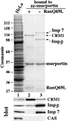
Figure 1
Figure 1
Identification of potential snurportin 1 export factors. A HeLa cell extract was subjected to binding to immobilized snurportin 1 in the presence or absence of 1.3 μM RanQ69L (GTP-bound form). Starting material and bound fractions were analyzed by SDS-PAGE followed by Coomassie staining and Western blotting. The load in the bound fractions corresponds to 20 times the starting material. Note that CRM1 preferentially bound to snurportin 1 in the presence of RanQ69L, but importin β and importin 7 in its absence.
Figure 2
Figure 2
Snurportin 1 interacts directly with CRM1 and importin β in a RanGTP-regulated manner. (A) Immobilized snurportin 1 was used to bind recombinant CRM1 from a total E. coli lysate. Where indicated, 1.3 μM RanQ69L (GTP) was also added. Starting material and bound fractions were analyzed by SDS-PAGE followed by Coomassie staining. The load in the bound fractions corresponded to 20 times the starting material. (B) Immobilized RanQ69L was incubated with total E. coli lysates expressing either recombinant CRM1 (lanes 1 and 2), importin β (lanes 3 and 4), or a control lysate (lane 5). Where indicated, (+) 300 μl of total E. coli lysate expressing a GST-snurportin 1 fusion protein was also included (lanes 1, 3, and 5). The interaction between snurportin 1 and CRM1 in the presence of RanQ69L is direct and a trimeric snurportin/CRM1/RanQ69L complex is formed.
Figure 3
Figure 3
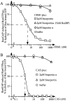
Kinetic characterization of the snurportin/CRM1/ RanGTP interaction. (A) The kinetic assay to measure complex formation is based on the observation that binding of RanGTP to an importin β–like factor prevents GTPase activation by RanGAP. 50 pM Ran-[γ-32P]GTP was preincubated at 15°C with the indicated concentrations of CRM1 in the absence or presence of 2 μM snurportin 1 or 2 μM importin α (human Rch1p). After 30 min, a 30-s GTPase reaction was started by addition of 40 nM Rna1p, the S. pombe RanGAP. Hydrolysis of Ran-bound GTP was determined as released [32P]phosphate. Note that Ran and snurportin 1 bound to CRM1 in a highly cooperative manner, with snurportin 1 increasing the apparent affinity of CRM1 for RanGTP roughly 1,000-fold. The apparent constant for dissociation of RanGTP from the trimeric complex is 4–5 nM. The presence of 15 nM RanBP1 relieved the GAP resistance of the complex completely. (B) Measurements were performed exactly as in A except that CAS was added instead of CRM1. Note that snurportin 1 bound selectively to CRM1, but not to CAS, while importin α bound to CAS but not to CRM1.
Figure 4
Figure 4
CRM1 promotes the export of snurportin 1 from the nucleus. Nuclear import of 1.6 μM fluorescein-labeled snurportin 1 and 1.6 μM Texas red–labeled importin α was allowed for 10 min in the presence of importin β (0.7 μM) and nucleoplasmin (1.4 μM) as described in Materials and Methods. After 10 min, one aliquot of the sample was fixed. The remaining sample was split in three. Either 3 μM CRM1, 3 μM CAS, or buffer was added and the incubation was continued for another 10 min before fixation. Nuclei were spun onto coverslips and fluorescent proteins were detected by confocal microscopy (63× oil objective). Note that addition of CRM1 specifically leads to a depletion of the intranuclear snurportin 1 and the appearance of an NPC staining, but had no effect on importin α distribution. In contrast, CAS promoted export of importin α, but not of snurportin.
Figure 5
Figure 5
Binding of m3G-cap and of CRM1 to snurportin 1 are mutually exclusive. (A) Immobilized snurportin 1 was used to bind recombinant CRM1 out of total E. coli lysate either in the presence (lanes 3 and 5) or absence (lanes 2 and 4) of a 10-fold molar excess of a m3G-cap oligonucleotide (5 μM). Where indicated, 11 μM RanQ69L was also added (lanes 4 and 5). In the absence of Ran-GTP, m3G-cap prevented binding of CRM1 to snurportin, while in the presence of Ran-GTP binding of CRM1 to snurportin 1 is reduced but not abolished by m3G-cap. (B) Formation of the trimeric snurportin/CRM1/ RanGTP complex was measured as in Fig. 2 A, with the modification that the concentration of CRM1 was kept constant at 300 nM and the concentration of snurportin 1 was varied. Where indicated, snurportin 1 had been preincubated with either 5 μM m3G-capped oligonucleotide or m7GpppG dinucleotide. Note that the m3G-cap RNA oligonucleotide specifically inhibited trimeric complex formation, while the m7G-cap analogue had no effect. (C) Measurements were performed exactly as in B except that a Rev-NES-BSA conjugate was used instead of snurportin. Note that the m3G-cap RNA oligonucleotide had no effect on the Rev-NES/CRM1/RanGTP interaction. (D) Measurements were performed exactly as in B, varying the concentrations of the following export substrates: snurportin 1; HIV Rev protein; Rev-NES peptide 1, which is a synthetic peptide (cys-LPPLERLTL) corresponding to the minimum Rev activation domain, Rev-NES peptide 2, which is a slightly larger peptide (cys-PVPLQLPPLERLTLD) that also includes NH2- and COOH-terminally flanking residues, Mut. NES peptide (cys-LPPDLRLTL), which corresponds to a loss-of-function mutant of the activation domain, was used as a negative control.
Figure 6
Figure 6
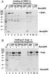
Binding properties of snurportin 1 deletion mutants. Immobilized full-length snurportin 1 (lanes 2 and 3) or deletion mutants (lanes 4–11) were tested for binding of (A) CRM1 and (B) importin β from total E. coli lysates. Where indicated, 1.5 μM RanQ69L GTP had also been added. Analysis was as in Fig. 2. Note that deletion of the 26 NH2-terminal residues of snurportin 1 abolished binding to CRM1. Deletion of >74 residues from the COOH terminus of snurportin 1 also resulted in the loss of the CRM1 interaction. All fragments that contained the IBB domain (residues 25–65) bound importin β.
Figure 7
Figure 7
Effects of nuclear injection of snurportin 1 on RNA export. Xenopus laevis oocyte nuclei were coinjected with a mixture of 32P-labeled RNAs and, where indicated, with purified recombinant GST-snurportin 1 fusion protein at the concentrations indicated above the lanes. The mixture of RNAs consisted of DHFR mRNA, histone H4 mRNA, U1ΔSm snRNA, U5ΔSm snRNA, U6Δss snRNA, and human initiator methionyl tRNA. The ΔSm U snRNAs lack the Sm binding site required for reimport into the nucleus. U6Δss does not leave the nucleus and is an internal control for nuclear integrity and proper nuclear injection. Synthesis of DHFR and histone H4 mRNA, and U1ΔSm and U5ΔSm, was primed with the m7GpppG-cap dinucleotide and synthesis of U6Δss was primed with γ-mGTP. RNA samples from total oocytes (T) or cytoplasmic (C) and nuclear (N) fractions were collected immediately after injection in lanes 1–3 or 180 min after injection in lanes 4–15. RNAs were resolved on 8% acrylamide/7 M urea denaturing gels. Note that coinjection of snurportin 1 specifically competed export of U1 and U5 snRNA but had only a negligible effect on mRNA export.
Figure 8
Figure 8
The snurportin 1 nucleocytoplasmic transport cycle. (1) In the cytoplasm, snurportin 1 binds m3G-capped U snRNAs and (2) is translocated by importin β into the nucleus. Translocation is terminated by direct binding of nuclear RanGTP to importin β (3), which results in the dissociation of importin β from snurportin. (4) Release of the m3G-capped U snRNA from snurportin 1 is prerequisite for (5) incorporation of snurportin 1 into a trimeric export complex with CRM1 and RanGTP. The trimeric complex is exported to the cytoplasm where (6) RanGTP is removed from CRM1 through the action of RanBP1 and RanGAP. (7) CRM1 is displaced from snurportin 1 by the next m3G-capped import substrate and a new transport cycle can proceed. β stands for importin β, Snp for snurportin 1; Sm for the Sm core domain; and m3G for the m3G-cap import signal of U snRNPs.

Similar articles

Cited by

References

    1. Adam SA, Sterne R, Marr, Gerace L. Nuclear protein import in permeabilized mammalian cells requires soluble cytoplasmic factors. J Cell Biol. 1990;111:807–816. - PMC - PubMed
    1. Arts GJ, Fornerod M, Mattaj IW. Identification of a nuclear export receptor for tRNA. Curr Biol. 1998;8:305–314. - PubMed
    1. Bischoff FR, Görlich D. RanBP1 is crucial for the release of RanGTP from importin β-related nuclear transport factors. FEBS Lett. 1997;419:249–254. - PubMed
    1. Bischoff FR, Krebber H, Kempf T, Hermes I, Ponstingl H. Human RanGTPase activating protein RanGAP1 is a homolog of yeast RNA1p involved in messenger RNA processing and transport. Proc Natl Acad Sci USA. 1995a;92:1749–1753. - PMC - PubMed
    1. Bischoff FR, Krebber H, Smirnova E, Dong WH, Ponstingl H. Coactivation of RanGTPase and inhibition of GTP dissociation by Ran GTP binding protein RanBP1. EMBO (Eur Mol Biol Organ) J. 1995b;14:705–715. - PMC - PubMed

Publication types

MeSH terms